The Influence of the Sympathetic Nervous System on the Development and Progression of Cancer
Shin-Hyung Park1,2*, Gyoo-Yong Chi1,2 and Yung Hyun Choi3,4*
1Department of Pathology, College of Korean Medicine, Dongeui University, Busan 47227, Korea
2Research Institute of Korean Medicine, Dongeui University, Busan 47227, Korea
3Department of Biochemistry, College of Korean Medicine, Dongeui University, Busan 47227, Korea
4Anti-Aging Research Center, Dongeui University, Busan 47340, Korea
Received September 26, 2017 /Revised December 5, 2017 /Accepted December 7, 2017
Living creatures possess long-conserved mechanisms to maintain homeostasis in response to various stresses. However, chronic and continuous exposure to stress can result in the excessive production of stress hormones, including catecholamines, which have harmful effects on health. Studies on the relationship between the sympathetic nervous system (SNS) and cancer have been conducted based on the traditional hypothesis that stress can promote cancer progression. Many preclinical and epi- demiological studies have suggested that the regulation of β-adrenergic signaling, which mediates SNS activity, can suppress the progression of solid tumors. SNS activation has highly pleiotropic ef- fects on tumor biology, as it stimulates oncogenes, survival pathways, the epithelial–mesenchymal transition, and invasion. Moreover, it inhibits DNA repair and programmed cell death and regulates the tumor microenvironment, including immune cells, endothelial cells, the extracellular matrix, mes- enchymal cells, and adipocytes. Although targeted therapies on the molecular basis of tumor pro- liferation are currently receiving increased attention, they have clinical limitations, such as the com- pensatory activation of other signaling pathways, emergence of drug resistance, and various side ef- fects, which raise the need for pleiotropic cancer regulation. This review summarizes the effects of the SNS on the development and progression of cancer and discusses the clinical perspectives of β -blockade as a novel therapeutic strategy for this disease.
Key words : β-adrenergic signaling, cancer progression, catecholamines, stress, sympathetic nervous system
*Corresponding authors
*Tel : +82-51-850-8646, Fax : +82-51-853-4036
*E-mail : [email protected] (Shin-Hyung Park)
*Tel : +82-51-850-7413, Fax : +82-51-853-4036
*E-mail : [email protected] (Yung Hyun Choi)
This is an Open-Access article distributed under the terms of the Creative Commons Attribution Non-Commercial License (http://creativecommons.org/licenses/by-nc/3.0) which permits unrestricted non-commercial use, distribution, and reproduction in any medium, provided the original work is properly cited.
Journal of Life Science 2018 Vol. 28. No. 1. 116~129 DOI : https://doi.org/10.5352/JLS.2018.28.1.116
서 론
스트레스와 암의 연관성은 오래 전부터 흥미로운 연구 주제 였다. 수십년 전 이미 동물모델에서 만성적 스트레스가 암의 성장과 전이를 촉진시키고 생존율을 감소시킨다는 것이 보고 되었으며[127, 128], 임상적으로도 정신적 혹은 사회적 스트레 스가 발암여부 및 예후와 밀접한 연관이 있음이 알려져 있다 [56, 111]. 그 기전으로서 스트레스에 의해 시상하부-뇌하수체- 부신피질 축(hypothalamic-pitutary-adrenal axis; HPA axis) 이 활성화되어 코티솔과 같은 glucocorticoid의 혈중 분비가 증가하면 T 세포의 불활성화를 야기함으로써 암세포를 공격
하는 면역계의 기능이 억제된다는 주장이 있다[56, 115]. 그러 나 만성적인 스트레스를 받은 마우스의 혈중에서 오히려 호중 구, 단핵구, 림프구가 증가되어 있음이 보고되어 단순히 HPA axis에 의한 면역억제가 암의 개시와 발전의 주요한 원인이 된다고 보기는 어렵다[65].
또 다른 기전으로서 교감신경-부신수질 축(sympathetic- adrenal medullary axis; SAM axis)에 의한 교감신경계 활성화 를 들 수 있다. 일반적으로 교감신경계는 스트레스에 반응하 여 ‘fight-or-flight’ 반응의 일환으로서 일시적인 근력과 기동 성 및 지각력을 향상시키는 것으로 알려져 있다[140]. 그러나 만성적 혹은 주기적인 교감신경계의 활성은 다양한 조직 장기 의 유전자 발현을 보다 지속적으로 조절하는 효과를 가진다 [60, 74, 114, 118]. 이러한 만성적 조절의 일환으로서 최근 몇 년간 교감신경계가 암의 발전과 전이를 촉진한다는 연구결과 가 축적되고 있다. 아드레날린과 노르아드레날린을 포함한 교 감신경계 신경전달물질은 β-adrenergic signaling pathway를 통해 암을 형성하고, 만성적인 스트레스에 노출된 mouse의 혈중에서 아드레날린계 신경전달물질이 증가하면서 종양 내 신생혈관 형성을 촉진한다는 보고도 있다[64, 122, 136]. 본 리 뷰에서는 교감신경계가 암의 성장과 전이에 미치는 영향과
- Review -
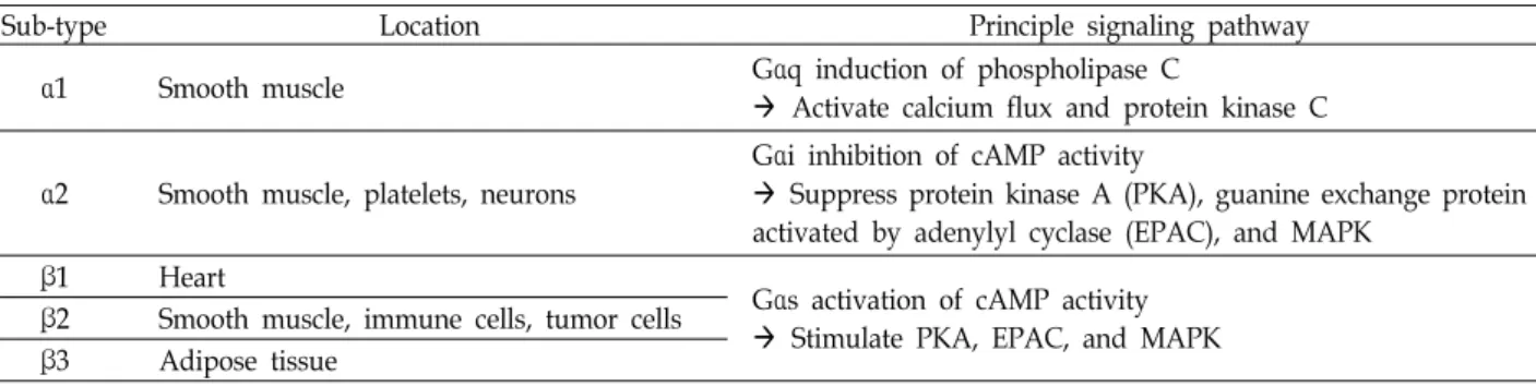
Table 1. Five types of adrenergic receptor
Sub-type Location Principle signaling pathway
α1 Smooth muscle Gαq induction of phospholipase C
Activate calcium flux and protein kinase C
α2 Smooth muscle, platelets, neurons
Gαi inhibition of cAMP activity
Suppress protein kinase A (PKA), guanine exchange protein activated by adenylyl cyclase (EPAC), and MAPK
β1 Heart
Gαs activation of cAMP activity Stimulate PKA, EPAC, and MAPK β2 Smooth muscle, immune cells, tumor cells
β3 Adipose tissue
그 조절 기전을 분석하고, 임상적으로 교감신경계 조절을 통 한 암 치료의 가능성에 대해 고찰해보고자 한다.
본 론 교감신경계의 인체조절 기전
교감신경계는 노르아드레날린과 아드레날린의 두 가지 카 테콜아민을 통해 인체를 조절한다. 노르아드레날린은 표적 장 기에 직접 분포하는 교감신경섬유 말단에서 주로 분비되어 국소적 조절을 담당하며, 아드레날린은 부신수질에서 혈중으 로 분비되어 전신적 조절에 관여한다. 이들은 서로 다른 조직 에 분포하는 다섯 종류의 adrenergic receptor (AR)에 의해 신호를 전달하는데, 이 중 β2-AR은 다양한 암세포에서 발현되 는 것으로 알려져 교감신경계 조절을 통한 항암제 개발의 주 요 타겟이 되고 있다[30](Table 1).
β-adrenergic signaling pathway
β-AR이 노르아드레날린 혹은 아드레날린과 결합하면 Gαs 를 활성화시켜 adenylyl cyclase (AC)를 통해 cyclic AMP (cAMP)를 합성한다. cAMP는 2개의 주요한 downstream ef- fector를 통해 신호를 전달하는데, protein kinase A (PKA)와 guanine exchange protein activated by adenylyl cyclase (EPAC)가 여기에 해당한다. 먼저 PKA는 표적 단백질의 ser- ine과 threonine 잔기를 인산화시켜 세포의 형태, 분화, 이동 능, 분비, 신경전달 등 다양한 부분을 조절한다. PKA의 표적 단백질로서 약 20%에 달하는 인간 유전자를 조절한다고 알려 져 있는 cAMP-responsive element binding protein (CREB) family와 β-receptor signaling을 탈감작시키는 β-AR kinase (BARK)가 있다[101, 149]. BARK는 Src/Ras/mitogen-acti- vated protein kinase (MAPK) pathway를 활성화시킨다고 알 려져 있다[93]. cAMP의 두번째 주요한 downstream effector 인 EPAC은 Ras-related protein-1a (Rap1A)/B-Raf/ERK pathway를 활성화시켜 세포의 형태, 이동능, 분비 등을 조절 한다[41](Fig. 1).
만성적 교감신경계 활성화에 의한 유전자 발현 변화 사회적 스트레스 인자로 인한 만성적 혹은 반복적인 교감신 경계 활성은 혈중으로 분비되는 아드레날린보다 교감신경섬 유 말단에서 분비되는 노르아드레날린을 더욱 현저히 증가시 키는 것으로 알려져 있다[140, 141]. 동물모델에서도 만성적인 사회적 스트레스가 교감신경섬유의 성장과 분지형성을 촉진 시켜 AR을 통한 신호를 활성화시키는 것이 보고되어 있다 [129, 130]. 급성 스트레스에 대처하기 위한 ‘fight-or-flight’ 반 응이 번역 후 조절과정에 의해 빠른 속도로 일어나는 반면 만성적인 교감신경계 활성은 표적기관의 유전자 발현을 조절 하거나 세포 구조를 변화시켜 인체의 다양한 생리적 요구와 스트레스 상황에 대처한다[26, 28, 60, 71, 118].
교감신경계 신호전달에 의한 유전자 전사조절의 예로서 면 역계 조절을 들 수 있는데, 교감신경 활성은 골수 내에서 림프 구계 세포나 적혈구계 세포보다 단백구(monocyte)나 과립구 (granulocyte)와 같은 골수구계 면역세포의 발달을 촉진하여 염증반응을 활성화시킨다[65, 114]. 비장과 림프절을 포함한 2차 림프조직에 분포한 교감신경은 interferon의 전사를 저해 하고 type 2 및 type 17 helper T 세포에서 분비되는 cytokine 들의 전사를 조절함으로써 세포매개 면역반응(cellular im- mune response)을 약화시키는 대신 체액성 면역반응(humoral immune response)을 촉진시킨다[45, 78, 130]. 또한 adrener- gic signaling을 통해 단백구, 대식세포(macrophage), 자연살 생세포(natural killer cell, NK cell)와 같은 innate immune cell 에서 pro-inflammatory cytokine의 전사를 활성화시킨다[45, 71, 114]. 이 밖에도 교감신경계를 통한 신호전달은 epithelial- mesenchymal transition (EMT), 섬유아세포(fibroblast) 활성 화 및 혈관신생(angiogenesis)과 관련된 유전자 전사를 조절함 으로써 상처 치유(wound-healing)에도 관여한다고 알려져 있 다[18, 67, 136]. 이와 같이 만성적, 반복적 스트레스에 의한 교감신경계 활성은 다양한 조직기관 내 유전자 전사를 조절함 으로써 불행히도 암의 진행과 발전에도 영향을 미치게 되었 다.
교감신경계의 암세포 유전자 발현 조절
교감신경계의 활성화는 SAM axis를 통해 부신수질로부터
Fig. 1. β-adrenergic receptor signaling pathway in cancer.
혈중으로 아드레날린을 분비시켜 ‘fight-or-flight’ 반응을 유도 하지만, 이것이 고형 종양(solid tumor)에 미치는 영향은 아직 명확하지 않다. 임상적으로 혈중 카테콜아민 수치와 종양 내 유전자 발현과의 관계는 일관되지 않으며, 이들이 어떻게 특 정 종양 속으로 침투하여 조절효과를 나타내는지도 미지수이 다[30, 90, 112]. 또한 일반적으로 종양 조직 내 노르아드레날린 의 수치가 혈중 노르아드레날린이나 아드레날린 수치와 일관 되지 않은 것으로 알려져 있어 카테콜아민 신호전달에 의한 종양의 조절은 호르몬을 통한 조절보다 종양 주변의 국소적인 교감신경섬유 분포에 의한 조절일 가능성이 높다[90, 91]. 그 동안 교감신경계가 종양 진행에 영향을 미치는 기전에 대해 지속적인 분석이 이뤄졌으며, 크게 암 유전자(oncogene) 조절, DNA repair 관련 유전자 조절, 전이 관련 유전자 조절, 세포 생존 및 사멸 유전자 조절로 나눠볼 수 있다.
암 유전자 조절
β-adrenergic signaling에 의한 암 유전자 조절은 크게 두 가지 방면으로 이루어진다. 첫 번째는 Src와 HER2와 같은 세 포 종양 유전자(cellular oncogene)을 활성화하는 것이다. β- adrenergic signaling은 protein kinase A (PKA)를 통해 Src의 Y419 residue를 인산화시켜 결과적으로 tumor growth, mi- gration, invasion 등을 촉진하며[5], 전사인자인 signal trans- ducer and activator of transcription 3 (STAT3)를 활성화시켜
HER2를 encoding하는 ERBB2 유전자의 전사를 촉진한다고 알려져 있다[126]. 두 번째는 종양발생 바이러스(oncogenic vi- rus)를 활성화시키는 것이다. 예를 들어 카포시육종을 일으키 는 human herpesvirus 8 (HHV8)에 감염된 B 림프구에서 β- adrenergic signaling이 활성화되면 PKA를 통해 RTA와 같은 바이러스성 전사인자를 과발현 혹은 활성화시켜 바이러스성 암유전자(viral oncogene)의 전사를 촉진시키므로 B 림프구의 악성종양을 일으키는 것으로 알려져 있다[22, 150].
DNA repair 관련 유전자 조절
β-adrenergic signaling은 DNA repair를 억제하여 염색체 의 불안정성을 야기함으로써 암의 개시에 영향을 미칠 수 있 다. 특히 DNA repair와 apoptosis를 담당하는 종양억제 유전 자 p53을 조절할 수 있는데, 예를 들어 β-arrestin/AKT path- way를 통해 p53의 E3 ubiquitin ligase인 murine double mi- nute 2 (MDM2)를 활성화시켜 p53의 분해를 촉진하는 것 등이 다[61]. 이러한 현상은 β-adrenergic antagonist인 propranolol 에 의해 현저히 억제되는데[61, 62], 신경아세포종(neuroblas- toma)에서 propronolol은 p53 발현을 높여 topoisomerase in- hibitor인 SN-38에 대한 감수성을 높인다는 보고가 있다[142].
그러나 아직까지 β-adrenergic signaling에 의한 DNA repair 저해만으로 암이 발생한다는 in vivo 데이터는 없는 실정이므 로 향후 연구가 필요한 부분이다[30].
Fig. 2. Regulation of sympathetic nervous system in the tumor microenvironment.
전이 관련 유전자 조절
β-adrenergic signaling은 암의 전이를 촉진하는 유전자의 발현을 조절함으로써 암의 진행과 발전에 영향을 미칠 수 있 다. 노르아드레날린이 PKA를 통해 Src (Y419)나 CREB을 인산 화시킴으로써 대장암, 유방암 및 전립선암 등의 전이를 일으 킨다는 것이 보고되었으며, 이러한 현상은 β-adrenergic an- tagonist에 의해 공통적으로 억제되었다[5, 43, 84, 107]. 또한 β-adrenergic signaling은 matrix metalloproteinases (MMPs) 의 발현을 높여 암세포가 주변 조직에 침윤하는 것을 용이하 게 하는데, STAT3를 통한 MMP-2와 MMP-9의 발현 증가가 대표적이다[85, 133, 144]. 노르아드레날린에 의한 β-AR의 활 성화는 암세포의 EMT도 유도할 수 있다. 이 때 transforming growth factor-β (TGF-β)와 hypoxia-inducible factor 1-α (HIF- 1α) 등이 전사인자인 Snail의 발현을 증가시킴으로써 EMT 과 정을 매개하는 것으로 알려져 있다[124, 148]. 최근 연구에 따 르면 β-adrenergic signaling은 암세포가 이동하기 위해 세포 막의 일부가 길게 늘어나는 invadopodia를 증가시킴으로써 유방암의 전이를 촉진하기도 하는데, 이는 암세포의 형태를 변화시킴으로써 전이를 돕는 대표적인 예이다[34].
세포 생존 및 사멸 유전자 조절
β-adrenergic signaling은 세포 생존 및 사멸과 관련된 다양 한 신호전달경로를 조절할 수 있다. 예를 들어, focal adhesion 에 관련된 focal adhesion kinase (FAK)를 조절함으로써 암세 포가 전이를 위해 extracellular matrix (ECM)에서 떨어져 나 올 때 세포사멸을 일으키는 anoikis 과정을 저해한다[134]. 그 리고 BCL-2-associated agonist of cell death (BAD)나 p53 등
을 조절함으로써 apoptosis에 저항하기 때문에 β-AR antago- nist를 이용하여 암세포의 항암화학요법(chemotherapy)에 대 한 감수성을 높일 수 있다[109, 117, 142]. 또한 노르아드레날린 은 β-AR/cAMP/PKA signaling pathway를 통해 vascular endothelial growth factor (VEGF), interleukin (IL)-8, IL-6를 포함한 성장인자의 발현을 높임으로써 sunitinib과 같은 ty- rosine kinase inhibitor에 대한 저항성을 획득할 수 있다고 알 려져 있다[40].
교감신경계의 종양미세환경 조절
종양미세환경은 매우 복잡하면서 역동적이다. 암의 발달과 정 동안 혈관내피세포, 면역세포, 섬유아세포를 포함한 다양 한 기질세포와 암세포 간의 긴밀한 상호작용으로 암의 성장과 전이가 촉진된다. 교감신경계 역시 종양이 위치하는 국소부위 에 신경분포를 통해 종양미세환경을 직접적으로 조절하거나 종양과 멀리 떨어진 조직장기를 조절함으로써 간접적으로 종 양미세환경에 영향을 미칠 수 있다(Fig. 2).
면역계 조절
암세포는 면역세포를 종양미세환경으로 유인하는 주화성 인자(chemotactic factor)를 분비한다[77]. 미세환경 속에는 암 을 촉진하거나 억제하는 다양한 면역세포들이 존재하는데, M2 대식세포, 골수유래 억제세포(myeloid derived sup- pressor cell, MDSC), 조절 T 세포(regulatory T cell, Treg)는 대표적으로 암을 촉진하는 면역세포에 해당하며, 자연살해세 포와 세포독성 T 세포(cytotoxic T cell)는 암을 억제하는 역할 을 한다[51]. 교감신경계는 이러한 heterogenous immune cell
들을 다양한 방식으로 조절하여 암의 성장과 전이를 촉진하게 된다.
먼저 교감신경계는 대식세포를 종양 주변으로 유인하는 역 할을 한다. 대식세포는 염증반응을 매개하고 종양미세환경을 조절하여 암의 전이를 촉진하는 핵심적인 면역세포이다. 암세 포는 β-adrenergic signaling을 통해 집락자극인자(colony- stimulating factor 1, CSF1)나 CCL2와 같은 주화성인자를 분 비하여 대식세포를 종양미세환경으로 유인한다[6, 132]. 유인 된 대식세포 역시 β-AR을 발현하므로 β-adrenergic signaling 을 통해 transforming growth factor-β (TGF-β), VEGF, IL- 6, MMP-9, PTGS2 등의 유전자 발현을 증가시켜 궁극적으로 종양의 전이를 돕는 혈관신생 촉진, ECM 리모델링, 염증성 환경 조성, 면역세포 유인 및 항암 면역반응 억제 등을 일으킨 다[96, 132, 136]. CSF1이나 monocyte chemotactic protein 1 (MCP1) 억제제에 의해 스트레스성 암 전이가 감소된다는 보 고는 종양으로 유인된 대식세포가 β-adrenergic signaling에 의한 암의 전이 과정에서 핵심적인 역할을 담당함을 보여준다 [6, 132].
다음으로 교감신경계는 자연살해세포와 T 세포를 조절할 수 있는데, 이들은 대식세포와 마찬가지로 β-AR을 발현하고 있다. β-adrenergic signaling은 자연살해세포의 활성을 억제 하며[123], 세포독성 T 세포의 생성 및 종양으로의 접근을 차 단한다[33, 73, 103]. 또한 type ⅠⅡ interferon의 전사를 억제 하여 세포매개 면역반응을 저해한다[29, 32]. 그러나 면역결핍 마우스 모델인 severe combined immunodeficiency disease (SCID) 마우스나 누드 마우스에서도 교감신경계 활성에 의해 암의 전이가 촉진될 수 있다고 보고됨에 따라 스트레스로 인 한 암의 진행에 세포매개 면역반응 조절이 영향을 미칠 수는 있으나 반드시 필수적인 인자는 아닌 것으로 보인다[64, 95, 136].
마지막으로 골수에 분포된 교감신경계는 단핵구와 호중구 (neutrophil)와 같은 골수구계 면역세포의 생성을 촉진하여 이 들이 tumor-associated macrophage (TAM)와 같이 종양미세 환경으로 유인되어 종양의 전이를 돕도록 유도한다[114, 132].
때로는 면역세포들이 다른 면역세포들과 소통하기 위해서 스 스로 카테콜아민을 합성하여 분비하기도 한다[48, 108]. 이러 한 보고들은 결국 교감신경계 활성으로 인한 β-adrenergic signaling이 항암 면역반응을 회피하고 종양으로의 진행을 촉 진함을 보여주는 예이다.
혈관신생 조절
β-adrenergic signaling은 VEGF나 IL-6와 같은 혈관성장인 자의 분비를 촉진하여 tumor의 성장과 전이를 돕는다[21, 88, 105, 136, 144, 145]. VEGF 항체나 VEGFR 저해제 처리로 혈관 신생을 억제했을 때 스트레스로 인한 암의 성장 및 전이가 억제된다는 보고는 교감신경계 활성에 의한 암의 진행에 혈관 신생이 핵심적인 역할을 함을 보여준다[136].
간엽세포 및 세포외기질 조절
Adrenergic signaling은 종양 주변을 둘러싸고 있는 섬유아 세포, 주피세포(pericyte), 중배엽 중기세포(mesenchymal stem cell)와 같은 간엽세포를 활성화시켜 pro-inflammatory cytokine 분비를 증가시키거나 혈관신생을 촉진하여 암의 성 장과 전이를 돕는다. 이 과정에서 간엽세포가 발현하는 α2-, β2- 및 β3-AR을 경유하여 신호가 전달된다고 알려져 있다[16, 18, 35, 49, 67].
또한 β-adrenergic signaling은 ECM을 재구성할 수 있는 다양한 성장인자와 cytokine 분비를 조절할 수 있다. 예를 들 어 노르아드레날린은 TGF-β signaling을 활성화시켜 fibro- nectin이나 콜라겐과 같은 ECM 단백질들의 발현을 증가시킴 으로써 조직의 섬유화를 일으킬 수 있다[10, 125]. 섬유화되어 단단해진 ECM은 암세포의 integrin과 성장인자 수용체를 활 성화하여 암의 생성과 전이를 촉진할 뿐만 아니라 항암제에 대한 물리적 장벽으로 작용하기도 한다[11, 69, 89, 100, 110, 120]. 또한 ECM의 재구성은 면역세포의 이동을 조절할 수 있 다. 예를 들어 저산소 환경에서 유방암세포가 분비하는 lysyl oxidase는 콜라겐을 단단하게 결합시키는 작용을 하는데, 종 양 성장을 촉진하는 면역세포(CD11b+ myeloid cells)는 콜라 겐과 결합하기 때문에 종양 기질(tumor stroma)로 쉽게 침윤 할 수 있는 반면, 종양 증식을 억제하는 T 세포는 단단히 결합 된 ECM 속에서 이동하기가 어려워진다[11, 47, 116]. 이와 같 이 adrenergic signaling은 간엽세포와 ECM을 암의 진행에 유리하도록 변화시킨다.
지방세포 조절
지방세포에서 분비되는 leptin 호르몬은 뇌에 대사정보를 전달하는 동시에 조직 유형에 따라 성장인자, 대사조절자, 혈 관신생인자, 생존인자로서 작용하기도 한다[139]. 임상적으로 혈청 내 leptin 수치가 전립선암, 유방암 및 피부암의 발생과 밀접한 관련이 있음이 보고되어 있다[24, 52, 54]. 지방조직에 분포된 교감신경섬유는 β-adrenergic signaling을 통해 지방 세포에서 분비되는 leptin의 농도를 낮춰 leptin이 성장인자로 작용하는 leptin-sensitive tumor의 성장을 억제한다. 반대로 암의 성장을 억제하는 adiponectin의 발현과 분비는 증가시킨 다고 알려져 있다[19, 20].
교감신경계와 암의 상호작용
다양한 고형암이 신경섬유의 직접적 분포 아래 교감신경계 의 지배를 받고 있다. 가장 일반적인 유형으로서 교감신경섬 유가 혈관과 함께 암 실질 속으로 분지를 내는 경우가 있으며, 때로는 암 주변의 정상조직에 분포하던 교감신경섬유가 암세 포가 분비하는 신경성장인자 및 축삭안내물질(axon guidance molecule)에 의해 암으로 이동하는 경우도 있다[7, 60, 95]. 여 러 연구결과에 따르면 암은 신경섬유의 성장과 분지형성을 촉진하며 심지어 새로운 신경섬유를 형성하기도 하는데, nerve
growth factor (NGF), brain-derived growth factor (BDNF), semaphorins 및 netrins을 포함한 신경성장인자와 축삭안내 물질이 이 과정을 매개한다고 알려져 있다[7, 60, 137]. 흥미롭 게도 이러한 인자들은 신경섬유뿐만 아니라 암세포의 생존과 성장을 촉진하기도 한다. 예를 들어 NGF는 유방암과 전립선 암 등에서 발견되며, 암세포의 apoptosis와 혈관신생을 조절 하는 역할을 하는데, NGF 저해에 의해 암 주변 신경섬유의 분지형성과 신생혈관형성이 동시에 억제됨이 보고되었다[1, 72, 79, 102]. BDNF 역시 정상 조직보다 암조직에서 잘 관찰되 며, BDNF signaling 저해에 의해 암세포의 성장이 억제되는 것을 볼 수 있다[15, 81, 146, 147]. Neurotrophin은 PI3K/AKT 경로나 Ras/MEK/MAPK 경로를 조절하여 다양한 암의 생존 과 증식을 돕는다[17, 98, 106]. 축삭안내물질인 netrin은 암세 포의 apoptosis를 억제하고 신생혈관 형성을 촉진하는데[44, 75, 87], 대표적 혈관성장인자인 VEGF가 신경계 발달에 영향 을 미친다는 보고[44]와 말초에서 동맥이 분비하는 artemin과 endothelin을 따라 교감신경섬유가 분포한다는 보고[36, 97]와 더불어 신경성장과 혈관형성이 상호 밀접하게 연관되어 있음 을 보여준다. 신경과 혈관의 발달과정에 나타나는 이러한 접 점들은 VEGF나 VEGFR을 표적으로 하는 치료들이 신경형성 에도 영향을 미쳐 암을 치료하는 데 더욱 효과적일 것임을 시사하지만, 많은 암들이 기존의 VEGF 요법을 회피한다고 알 려져 있으므로 향후 그 기전을 분석할 필요가 있다. 이와 같이 암에서 분비되는 다양한 신경성장인자와 축삭안내물질 등이 암 주변을 둘러싼 신경과 혈관의 형성과 분포를 조절하기도 하지만, 역으로 교감신경계 활성화는 말초신경에서 CXCL12 와 같은 chemokine을 분비시켜 이에 대한 수용체를 가진 암세 포를 신경섬유 쪽으로 유인할 수도 있다[7, 59, 137, 143]. 따라 서 암과 신경 간의 상호작용에 대해 구체적인 기전을 규명하 는 것이 새로운 암 치료법 개발에 필수적이다.
암 치료 타겟으로서 β-adrenergic signaling 조절의 임상 적 의의
상기한 바와 같이 교감신경계는 원발성 암과 그것을 둘러싼 종양미세환경 및 교감신경의 지배를 받고 있는 원장기에까지 영향을 미쳐 암의 성장과 발전과정을 광범위하게 조절할 수 있으므로 β-adrenergic signaling 억제제는 암을 효과적으로 치료할 수 있는 새로운 대안으로 대두되고 있다. 현재 임상에 서 adrenergic antagonist는 대부분 심장질환을 치료하려는 목 적으로 사용되고 있다(Table 2). β-adrenergic signaling과 암 과의 관련을 분석하는 연구는 애초에 스트레스와 암의 발전 사이에 연관성이 있을 거라는 가능성 때문에 시작되었다[2, 4, 23]. 또한 최근에 약물-역학 데이터에서 β-adrenergic antag- onist가 암의 발전을 막는다는 보고와 함께 더욱 촉발되었다 [9, 31, 39, 42, 57, 58, 99, 113, 138]. 동물 모델에서도 행동적 스트레스가 유방암, 전립선암, 난소암, 신경아종세포, 악성 흑
색종, 췌장암 및 혈액암 등을 가속화시킬 수 있음이 밝혀졌다 [46, 55, 63, 64, 70, 76, 82, 94, 107, 109, 132, 136, 142]. 이러한 현상은 β-adrenergic antagonist에 의해 대부분 저해되었으며, 반대로 β-adrenergic agonist에 의해 스트레스 없이 mimic 되 었다[31].
전임상 데이터를 바탕으로 한 약리학적 분석에 따르면 교감 신경계가 암에 미치는 영향은 β2-와 β3-AR에 의해 대부분 매 개된다[19, 25, 80, 136]. 몇몇 전임상 연구에서 α-adrenergic antagonist의 생리활성을 조사하였으나 결과는 일관되지 않는 다[83, 135]. 역학연구 결과 역시 α-adrenergic antagonist가 암 의 발생률이나 진행률에 주목할만한 효과를 나타내지 않으며, 심지어 미약하게 암을 진행시킨다고 보고하였다[50]. 또한 동 물모델에서 β-AR antagonist만으로도 스트레스로 인한 암의 진행을 충분히 차단할 수 있으므로 α-adrenergic signaling이 핵심적인 역할을 담당한다고 보기 어렵다.
β-adrenergic antagonist는 비교적 저렴하고 인체에 안전하 다고 알려져 있지만, 암 치료에 활용하기 위해서는 몇 가지 해결해야 할 과제가 있다. 첫 번째는 암 치료효과가 가장 뛰어 난 최적의 약물을 선정하는 것이다. 예를 들어 β2-AR을 비선 택적으로 저해하는 propranolol은 atenolol과 같이 심장질환 에 주로 사용하는 β1-AR 특이적 억제제보다 항암작용이 우수 한 것으로 알려져 있다[9, 31, 53, 109, 136, 138, 142]. 두 번째는 β-adrenergic antagonist를 적용할 수 있는 최적의 질병 환경 을 설정하는 것이다. 일부 약물-역학 연구에 따르면 β-adre- nergic antagonist는 estrogen receptor (ER), progesterone re- ceptor (PR), HER2를 모두 발현하지 않는 triple-negative 유방 암에서 이들 중 하나 이상을 발현하는 유방암보다 더욱 탁월 한 효과를 보인다[12, 99]. 세 번째는 최적의 투약 시점과 기간 을 설정하는 것이다. 역학 연구에 따르면 스트레스가 기존의 암을 가속화시킨다는 것은 일관되게 밝혀지고 있지만, 암을 유발한다는 증거는 상대적으로 부족하다. 동물모델에 있어서 도 이종이식(xenograft)과 syngeneic 종양 모델에 스트레스를 부과했을 경우 암이 악화되는 일관된 결과를 얻을 수 있었으 나[80, 107], 스트레스가 원발성 암을 유발한다는 보고는 몇 가지 연구[14, 19, 66, 119]를 제외하면 상대적으로 적은 편이 다. 또한 수술 전에 β-adrenergic antagonist를 투약하면 교감 신경계 활성으로 인한 수술 후 전이를 감소시킬 수 있다는 보고가 있다[68, 104]. 이러한 연구결과들은 암을 예방하거나 이미 전이가 광범위하게 일어난 말기 단계의 암보다 암의 전 이능을 비교적 조절할 수 있는 초기 단계의 암이 β-adrenergic antagonist를 더욱 효과적으로 활용할 수 있는 적응증이 된다 는 것을 시사한다. 따라서 유방암이나 전립선암과 같이 일반 적으로 초기 단계에 발견되며 β-adrenergic signaling을 통해 전이를 일으킨다고 알려진 암종에 β-adrenergic antagonist를 활용할 수 있을 것이다[31]. 마지막으로 β-AR과 downstream 을 포함한 교감신경계 관련 유전자의 발현 정도와 환자의 스
Table 2. Pharmacological characteristics of β-blockers
Name Major current therapeutic indications
Non-selective β-blocking agents Alprenolol
Oxprenolol Pindolol Propronolol Timolol
Sotalol Nadolol Carteolol Bupranolol Penbutolol
Angina pectoris
Angina, hypertension and cardiac arrhythmias Angina pectoris and hypertension
Hypertension, angina and hemangiomas
Hypertension, congestive heart failure, acute myocardiac Infarction, and open-angle glaucoma
Cardiac arrhythmias and ventricular arrhythmias Hypertension and angina pectoris
Hypertension and reduce intraocular pressure Hypertension, tachycardia and glaucoma Hypertension
Selective β-blocking agents Practolol
Metoprolol Atenolol Acebutolol Betaxolol Bisoprolol Celiprolol Esmolol Nebivolol
Cardiac arrhythmias
Hypertension, angina pectoris, tachycardia, heart failure, vasovagal syncope Hypertension, coronary heart disease, arrhythmias and angina pectoris Hypertension, cardiac arrhythmias, acute myocardial infarction Hypertension, angina pectoris and glaucoma
Hypertension
Hypertsnsion and angina Tachycardia
Hypertension α and β blocking agents
Labetalol Carvedilol
Hypertension
Hypertension, congestive heart failure
트레스 지수를 바탕으로 최적의 환자군을 설정하는 것이다[2, 3, 27, 90, 91, 132]. 주의할 점은 환자의 암세포에서 β-AR 발현 정도가 높다고 해서 반드시 β-adrenergic antagonist에 민감하 게 반응하는 것은 아니라는 점이다[13, 142]. 이는 교감신경계 에 의한 암의 진행이 암세포뿐만 아니라 암세포를 둘러싸고 있는 종양미세환경과 혈관 및 골수를 구성하는 다양한 세포에 서 발현하는 AR에 의해 매개될 수 있기 때문이다. 또한 교감신 경계에 의해 광범위한 신호전달경로가 조절되므로 이들 중 β-adrenergic antagonist를 적용할 수 있는 명확한 biomarker 를 규명할 필요가 있다.
이미 다수의 연구를 통해 β-adrenergic antagonist가 암의 진행을 억제한다는 사실이 밝혀졌으나[9, 39, 86, 99, 113, 121], 비대조 관찰 연구라는 방법론적인 한계로 인해 임상적인 활용 도를 보장하기 어렵다. 전통적으로 β-adrenergic antagonist를 사용해온 심혈관질환은 흡연, 비만, 전신적 염증과 같이 암과 공통적인 악화요인을 가지고 있으며, 심혈관질환 치료를 위해 β-adrenergic antagonist를 투약하는 동안 안지오텐신 전환효 소 저해제와 같은 다른 약물에 노출된 경우 암의 진행에 영향 을 미칠 수 있다. 또한 심혈관질환을 대상으로 한 연구는 암의 진행률이나 사망률에 대한 정보가 부족하며, 암을 대상으로 한 연구는 β-adrenergic antagonist 투약 여부에 대한 정보가
부족하다. 게다가 β-adrenergic antagonist를 투약하는 방식에 따라 암생존율 추이 역시 달라지는 경향성을 보인다[31]. 이러 한 문제점들은 기존의 관찰 연구가 β-adrenergic antagonist의 항암작용을 증명함에 있어서 해석상의 오류를 남길 수 있음을 의미하며, 이를 해결하기 위해 β-adrenergic antagonist가 암 의 진행을 실제로 차단하는 효과가 있는지 무작위 대조 임상 시험으로 확인할 필요가 있다.
결 론
일반적으로 암은 상피세포의 유전적 혹은 후성유전적 변이 로 인해 발생하는 것으로 알려져 있으나, 최근에는 암세포 자 체의 변이뿐만 아니라 종양미세환경을 구성하고 있는 다양한 기질세포와의 상호작용이 중요하다는 사실이 여러 연구를 통 해 밝혀지고 있다. 교감신경계는 암세포 내의 다양한 유전자 발현과 활성을 조절할 뿐만 아니라 면역세포, 혈관세포, 간엽 세포, 세포외기질, 지방세포 및 골수와 같은 광범위한 대상을 조절함으로써 암의 진행과 발전에 영향을 미칠 수 있다. 따라 서 교감신경계로부터 비롯된 신호를 전달하는 핵심적 신호전 달경로인 β-adrenergic signaling을 차단하는 것이 새로운 암 치료법으로 대두되고 있으며, 여러 전임상 데이터와 관찰연구
를 통해 실제로 그 가능성을 인정받고 있다. β-adrenergic an- tagonist는 이미 FDA 승인된 안전하고 비교적 저렴한 약물이 므로 여기에 명확한 약리학적 기전 분석과 항암 작용에 대한 무작위 대조 임상시험 결과가 더해진다면 새로운 암 치료제로 서 활용가치가 높을 것이다.
감사의 글
본 연구는 한국연구재단의 일반연구자지원사업(신진연구 지원사업)에 의해 수행되었음(과제번호 NRF-2016R1C1B201 5076).
References
1. Adriaenssens, E., Vanhecke, E., Saule, P., Mougel, A., Page, A., Romon, R., Nurcombe, V., Le Bourhis, X. and Honder- marck, H. 2008. Nerve growth factor is a potential ther- apeutic target in breast cancer. Cancer Res. 68, 346-351.
2. Antoni, M. H., Lutgendorf, S. K., Cole, S. W., Dhabhar, F.
S., Sephton, S. E., McDonald, P. G., Stefanek, M. and Sood, A. K. 2006. The influence of bio-behavioural factors on tu- mour biology: pathways and mechanisms. Nat. Rev. Cancer 6, 240-248.
3. Antoni, M. H., Lutgendorf, S. K., Blomberg, B., Carver, C.
S., Lechner, S., Diaz, A., Stagl, J., Arevalo, J. M. and Cole, S. W. 2012. Cognitive-behavioral stress management re- verses anxiety-related leukocyte transcriptional dynamics.
Biol. Psychiatry 71, 366-372.
4. Armaiz-Pena, G. N., Allen, J. K., Cruz, A., Stone, R. L., Nick, A. M., Lin, Y. G., Han, L. Y., Mangala, L. S., Villares, G.
J., Vivas-Mejia, P., Rodriguez-Aguayo, C., Nagaraja, A. S., Gharpure, K. M., Wu, Z., English, R. D., Soman, K. V., Shahzad, M. M., Zigler, M., Deavers, M. T., Zien, A., Soldatos, T. G., Jackson, D. B., Wiktorowicz, J. E., Torres- Lugo, M., Young, T., De Geest, K., Gallick, G. E., Bar-Eli, M., Lopez-Berestein, G., Cole, S. W., Lopez, G. E., Lutgen- dorf, S. K. and Sood, A. K. 2013. Src activation by β-adrenor- eceptors is a key switch for tumour metastasis. Nat.
Commun. 4, 1403.
5. Armaiz-Pena, G. N., Cole, S. W., Lutgendorf, S. K. and Sood, A. K. 2013. Neuroendocrine influences on cancer progression. Brain Behav. Immun. 30, S19-S25.
6. Armaiz-Pena, G. N., Gonzalez-Villasana, V., Nagaraja, A. S., Rodriguez-Aguayo, C., Sadaoui, N. C., Stone, R. L., Matsuo, K., Dalton, H. J., Previs, R. A., Jennings, N. B., Dorniak, P., Hansen, J. M., Arevalo, J. M., Cole, S. W., Lutgendorf, S.
K., Sood, A. K. and Lopez-Berestein, G. 2015. Adrenergic regulation of monocyte chemotactic protein 1 leads to en- hanced macrophage recruitment and ovarian carcinoma growth. Oncotarget 6, 4266-4273.
7. Ayala, G. E., Dai, H., Powell, M., Li, R., Ding, Y., Wheeler, T. M., Shine, D., Kadmon, D., Thompson, T., Miles, B. J., Ittmann, M. M. and Rowley, D. 2008. Cancer-related axono-
genesis and neurogenesis in prostate cancer. Clin. Cancer Res. 14, 7593-7603.
8. Aydiner, A., Ciftci, R., Karabulut, S. and Kilic, L. 2013. Does β-blocker therapy improve the survival of patients with metastatic non-small cell lung cancer? Asian Pac. J. Cancer Prev. 14, 6109-6114.
9. Barron, T. I., Connolly, R. M., Sharp, L., Bennett, K. and Visvanathan, K. 2011. β blockers and breast cancer mortal- ity: a population-based study. J. Clin. Oncol. 29, 2635-2644.
10. Boluyt, M. O., Long, X., Eschenhagen, T., Mende, U., Schmitz, W., Crow, M. T. and Lakatta, E. G. 1995. Isoproterenol in- fusion induces alterations in expression of hypertrophy-as- sociated genes in rat heart. Am. J. Physiol. 2, H638-647.
11. Bonnans, C., Chou, J. and Werb, Z. 2014. Remodelling the extracellular matrix in development and disease. Nat. Rev.
Mol. Cell Biol. 12, 786-801.
12. Botteri, E., Munzone, E., Rotmensz, N., Cipolla, C., De Giorgi, V., Santillo, B., Zanelotti, A., Adamoli, L., Colleoni, M., Viale, G., Goldhirsch, A. and Gandini, S. 2013. Therapeutic effect of β-blockers in triple-negative breast cancer postmeno- pausal women. Breast Cancer Res. Treat. 140, 567-575.
13. Boucek, R. J. Jr., Kirsh, A. L., Majesky, M. W. and Perkins, J. A. 2013. Propranolol responsiveness in vascular tumors is not determined by qualitative differences in adrenergic receptors. Otolaryngol. Head Neck Surg. 149, 772-776.
14. Boyd, A. L., Salleh, A., Humber, B., Yee, J., Tomes, L. and Kerr, L. R. 2010. Neonatal experiences differentially infl uence mammary gland morphology, estrogen receptor a protein levels, and carcinogenesis in BALB/c mice. Cancer Prev. Res. (Phila) 3, 1398-1408.
15. Bronzetti, E., Artico, M., Forte, F., Pagliarella, G., Felici, L.
M., D’Ambrosio, A., Vespasiani, G. and Bronzetti, B. 2008.
A possible role of BDNF in prostate cancer detection. Oncol.
Rep. 19, 969-974.
16. Bruzzone, A. Piñero, C. P., Rojas, P., Romanato, M., Gass, H., Lanari, C. and Lüthy, I. A. 2011. α2-adrenoceptors en- hance cell proliferation and mammary tumor growth acting through both the stroma and the tumor cells. Curr. Cancer Drug Targets 11, 763-774.
17. Burger, R., Bakker, F., Guenther, A., Baum, W., Schmidt Arras, D., Hideshima, T., Tai, Y. T., Shringarpure, R., Catley, L., Senaldi, G. Gramatzki, M. and Anderson, K. C. 2003.
Functional significance of novel neurotrophin-1/B cell-stim- ulating factor-3 (cardiotrophin-like cytokine) for human myeloma cell growth and survival. Brit. J. Haematol. 123, 869-878.
18. Calvani, M., Pelon, F., Comito, G., Taddei, M. L., Moretti, S., Innocenti, S., Nassini, R., Gerlini, G., Borgognoni, L., Bambi, F., Giannoni, E., Filippi, L. and Chiarugi, P. 2015.
Norepinephrine promotes tumor microenvironment re- activity through β3-adrenoreceptors during melanoma progression. Oncotarget 6, 4615-4632.
19. Cao, L. Liu, X., Lin, E. J., Wang, C., Choi, E. Y., Riban, V., Lin, B. and During, M. J. 2010. Environmental and genetic activation of a brain–adipocyte BDNF/leptin axis causes cancer remission and inhibition. Cell 142, 52-64.
20. Cao, L. and During, M. J. 2012. What is the brain–cancer connection? Annu. Rev. Neurosci. 35, 331-345.
21. Chakroborty, D., Sarkar, C., Basu, B., Dasgupta, P. S. and Basu, S. 2009. Catecholamines regulate tumor angiogenesis.
Cancer Res. 69, 3727-3730.
22. Chang, M., Brown, H. J., Collado-Hidalgo, A., Arevalo, J.
M., Galic, Z., Symensma, T. L., Tanaka, L., Deng, H., Zack, J. A., Sun, R. and Cole, S. W. 2005. β-adrenoreceptors re- activate Kaposi’s sarcoma-associated herpesvirus lytic repli- cation via PKA-dependent control of viral RTA. J. Virol. 79, 13538-13547.
23. Chida, Y., Hamer, M., Wardle, J. and Steptoe, A. 2008. Do stress-related psychosocial factors contribute to cancer in- cidence and survival? Nat. Clin. Pract. Oncol. 5, 466-475.
24. Cirillo, D., Rachiglio, D. A. M., la Montagna R, Giordano, A. and Normanno, N. 2008. Leptin signaling in breast can- cer: an overview. J. Cell. Biochem. 105, 956-964.
25. Cole, S. W. 2011. Beta-adrenergic regulation of gene ex- pression in cancer. In: Proceeding of the 102nd Annual Meetingofthe American Association for Cancer Research.
26. Cole, S. W. 2013. Social regulation of human gene ex- pression: mechanisms and implications for public health.
Am. J. Publ. Health 103, S84-S92.
27. Cole, S. W. 2013. Nervous system regulation of the cancer genome. Brain Behav. Immun. 30, S10-S18.
28. Cole, S. W. 2014. Human social genomics. PLoS Genet. 10, e1004601.
29. Cole, S. W., Korin, Y. D., Fahey, J. L. and Zack, J. A. 1998.
Norepinephrine accelerates HIV replication via protein kin- ase A-dependent effects on cytokine production. J. Immunol.
161, 610-616.
30. Cole, S. W., Nagaraja, A. S., Lutgendorf, S. K., Green, P.
A. and Sood, A. K. 2015. Sympathetic nervous system regu- lation of the tumour microenvironment. Nat. Rev. Cancer.
15, 563-572.
31. Cole, S. W. and Sood, A. K. 2012. Molecular pathways: be- ta-adrenergic signaling in cancer. Clin. Cancer Res. 5, 1201- 1206.
32. Collado-Hidalgo, A., Sung, C. and Cole, S. 2006. Adrenergic inhibition of innate anti-viral response: PKA blockade of type I interferon gene transcription mediates catecholamine support for HIV-1 replication. Brain Behav. Immun. 20, 552- 563.
33. Cook-Mills, J. M., Mokyr, M. B., Cohen, R. L., Perlman, R.
L. and Chambers, D. A. 1995. Neurotransmitter suppression of the in vitro generation of a cytotoxic T lymphocyte re- sponse against the syngeneic MOPC-315 plasmacytoma.
Cancer Immunol. Immunother. 2, 79-87.
34. Creed, S. J., Le, C. P., Hassan, M., Pon, C. K., Albold, S., Chan, K. T., Berginski, M. E., Huang, Z., Bear, J. E., Lane, J. R., Halls, M. L., Ferrari, D., Nowell, C. J. and Sloan, E.
K. 2015. β2-adrenoceptor signaling regulates invadopodia formation to enhance tumor cell invasion. Breast Cancer Res.
17, 145.
35. Dal Monte, M., Casini, G., Filippi, L., Nicchia, G. P., Svelto, M. and Bagnoli, P. 2013. Functional involvement of β3-adre-
nergic receptors in melanoma growth and vascularization.
J. Mol. Med. 91, 1407-1419.
36. Damon, D. H., Teriele, J. A. and Marko, S. B. 2007. Vascular- derived artemin: a determinant of vascular sympathetic in- nervation? Am. J. Physiol. 293, H266-273
37. De Giorgi, V., Gandini, S., Grazzini, M., Benemei, S., Mar- chionni, N. and Geppetti, P. 2013. Effect of β-blockers and other antihypertensive drugs on the risk of melanoma re- currence and death. Mayo Clin. Proc. 88, 1196-1203.
38. De Giorgi, V., Grazzini, M., Gandini, S., Benemei, S., Lotti, T., Marchionni, N. and Geppetti, P. 2011. Treatment with b-blockers and reduced disease progression in patients with thick melanoma. Arch. Intern. Med. 171, 779-781.
39. Deng, G. H., Liu, J., Zhang, J., Wang, Y., Peng, X. C., Wei, Y. Q. and Jiang, Y. 2014. Exogenous norepinephrine attenu- ates the efficacy of sunitinib in a mouse cancer model. J.
Exp. Clin. Cancer Res. 33, 21.
40. De Rooij, J., Zwartkruis, F. J., Verheijen, M. H., Cool, R.
H., Nijman, S. M., Wittinghofer, A. and Bos, J. L. 1998. Epac is a Rap1 guanine-nucleotide-exchange factor directly acti- vated by cyclic AMP. Nature 396, 474-477.
41. Diaz, E. S., Karlan, B. Y. and Li, A. J. 2012. Impact of β blockers on epithelial ovarian cancer survival. Gynecol.
Oncol. 127, 375-378.
42. Drell, T. L., Joseph, J., Lang, K., Niggemann, B., Zaenker, K. S. and Entschladen, F. 2003. Effects of neurotransmitters on the chemokinesis and chemotaxis of MDA-MB-468 hu- man breast carcinoma cells. Breast Cancer Res. Treat. 80, 63- 70.
43. Eichmann, A. and Thomas, J. L. 2013. Molecular parallels between neural and vascular development. Cold Spring Harb. Perspect. Med. 3, a006551.
44. Elenkov, I. J., Wilder, R. L., Chrousos, G. P. and Vizi, E.
S. 2000. The sympathetic nerve - an integrative interface be- tween two supersystems: the brain and the immune system.
Pharmacol. Rev. 52, 595-638.
45. Eng, J. W., Reed, C. B., Kokolus, K. M., Pitoniak, R., Utley, A., Bucsek, M. J., Ma, W. W., Repasky, E. A. and Hylander, B. L. 2015. Housing temperature-induced stress drives ther- apeutic resistance in murine tumour models through β2- adrenergic receptor activation. Nat. Commun. 6, 6426.
46. Erler, J. T., Bennewith, K. L., Cox, T. R., Lang, G., Bird, D., Koong, A., Le, Q. T. and Giaccia, A. J. 2009. Hypoxia-in- duced lysyl oxidase is a critical mediator of bone marrow cell recruitment to form the premetastatic niche. Cancer Cell 1, 35-44.
47. Flierl, M. A., Rittirsch, D., Nadeau, B. A., Chen, A. J., Sarma, J. V., Zetoune, F. S., McGuire, S. R., List, R. P., Day, D. E., Hoesel, L. M., Gao, H., Van Rooijen, N., Huber-Lang, M.
S., Neubig, R. R. and Ward, P. A. 2007. Phagocyte-derived catecholamines enhance acute inflammatory injury. Nature 449, 721-725.
48. Flint, M. S., Baum, A., Episcopo, B., Knickelbein, K. Z., Liegey Dougall, A. J., Chambers, W. H. and Jenkins, F. J.
2013. Chronic exposure to stress hormones promotes trans- formation and tumorigenicity of 3T3 mouse fibroblasts.
Stress 16, 114-121.
49. Friedman, G. D., Udaltsova, N. and Habel, L. A. 2011.
Norepinephrine antagonists and cancer risk. Int. J. Cancer 128, 737-738.
50. Fridman, W. H., Pages, F., Sautes-Fridman, C. and Galon, J. 2012. The immune contexture in human tumours: impact on clinical outcome. Nat. Rev. Cancer 4, 298-306.
51. Garofalo, C. and Surmacz, E. 2006. Leptin and cancer. J. Cell.
Physiol. 207, 12-22.
52. Ganz, P. A. and Cole, S. W. 2011. Expanding our therapeutic options: β blockers for breast cancer? J. Clin. Oncol. 29, 2612-2616.
53. Gogas, H., Trakatelli, M., Dessypris, N., Terzidis, A., Katsambas, A., Chrousos, G. P. and Petridou, E. T. 2008.
Melanoma risk in association with serum leptin levels and lifestyle parameters: a case-control study. Ann. Oncol. 19, 384-389.
54. Goldfarb, Y., Sorski, L., Benish, M., Levi, B., Melamed, R.
and Ben-Eliyahu, S. 2011. Improving postoperative immune status and resistance to cancer metastasis: a combined peri- operative approach of immunostimulation and prevention of excessive surgical stress responses. Ann. Surg. 253, 798- 810.
55. Glaser, R. and Kiecolt-Glaser J. K. 2005. Stress-induced im- mune dysfunction: implications for health. Nat. Rev.
Immunol. 5, 243-251.
56. Grytli, H. H., Fagerland, M. W., Fossa, S. D., Tasken, K.
A. and Haheim, L. L. 2013. Use of β-blockers is associated with prostate cancer-specific survival in prostate cancer pa- tients on androgen deprivation therapy. Prostate 73, 250-260.
55. Grytli, H. H., Fagerland, M. W., Fossa, S. D. and Tasken, K. A. 2014. Association between use of β-blockers and pros- tate cancer-specific survival: a cohort study of 3561 prostate cancer patients with high-risk or metastatic disease. Eur.
Urol. 65, 635-641.
58. Guo, K. Ma, Q., Li, J., Wang, Z., Shan, T., Li, W., Xu, Q.
And Xie, K. 2013. Interaction of the sympathetic nerve with pancreatic cancer cells promotes perineural invasion through the activation of STAT3 signaling. Mol. Cancer Ther. 12, 264- 273.
59. Hanoun, M., Maryanovich, M., Arnal-Estape, A. and Fre- nette, P. S. 2015. Neural regulation of hematopoiesis, in- flammation, and cancer. Neuron 86, 360-373.
60. Hara, M. R., Kovacs, J. J., Whalen, E. J., Rajagopal, S., Strachan, R. T., Grant, W., Towers, A. J., Williams, B., Lam, C. M., Xiao, K., Shenoy, S. K., Gregory, S. G., Ahn, S., Duckett, D. R. and Lefkowitz, R. J. 2011. A stress response pathway regulates DNA damage through β2-adrenoreceptors and β-arrestin-1. Nature 477, 349-353.
61. Hara, M. R., Sachs, B. D., Caron, M. G. and Lefkowitz, R.
J. 2013. Pharmacological blockade of a β2AR-βarrestin-1 sig- naling cascade prevents the accumulation of DNA damage in a behavioral stress model. Cell Cycle 12, 219-224.
62. Hasegawa, H. and Saiki, I. 2002. Psychosocial stress aug- ments tumor development through β-adrenergic activation in mice. Jpn. J. Cancer Res. 93, 729-735.
63. Hassan, S., Karpova, Y., Baiz, D., Yancey, D., Pullikuth, A., Flores, A., Register, T., Cline, J. M., D’Agostino, R. Jr., Danial, N. Datta, S. R. and Kulik, G. 2013. Behavioral stress accelerates prostate cancer development in mice. J. Clin.
Invest. 123, 874-886.
64. Heidt, T., Sager, H. B., Courties, G., Dutta, P., Iwamoto, Y., Zaltsman, A., von Zur Muhlen C., Bode, C., Fricchione, G.
L., Denninger, J., Lin, C. P., Vinegoni, C., Libby, P., Swirski, F. K., Weissleder, R. and Nahrendorf, M. 2014. Chronic vari- able stress activates hematopoietic stem cells. Nat. Med. 20, 754-758.
65. Hermes, G. L., Delgado, B., Tretiakova, M., Cavigelli, S. A., Krausz, T., Conzen, S. D. and McClintock, M. K. 2009. Social isolation dysregulates endocrine and behavioral stress while increasing malignant burden of spontaneous mammary tumors. Proc. Natl. Acad. Sci. USA 106, 22393-22398.
66. Hori, Y., Ishii, K., Kanda, H., Iwamoto, Y., Nishikawa, K., Soga, N., Kise, H., Arima, K. and Sugimura, Y. 2011. Nafto- pidil, a selective α1-adrenoceptor antagonist, suppresses hu- man prostate tumor growth by altering interactions between tumor cells and stroma. Cancer Prev. Res. 4, 87-96.
67. Horowitz, M., Neeman, E., Sharon, E. and Ben-Eliyahu, S.
2015. Exploiting the critical perioperative period to improve long-term cancer outcomes. Nat. Rev. Clin. Oncol. 12, 213- 226.
68. Huang, S. and Ingber, D. E. 2005. Cell tension, matrix me- chanics, and cancer development. Cancer Cell 3, 175-176.
69. Inbar, S., Neeman, E., Avraham, R., Benish, M., Rosenne, E. and Ben-Eliyahu, S. 2011. Do stress responses promote leukemia progression? An animal study suggesting a role for epinephrine and prostaglandin-E2 through reduced NK activity. PLoS ONE 6, e19246.
70. Irwin, M. R. and Cole, S. W. 2011. Reciprocal regulation of the neural and innate immune systems. Nat. Rev. Immunol.
11, 625-632.
71. Jimenez-Andrade, J. M., Bloom, A. P., Stake, J. I., Mantyh, W. G., Taylor, R. N., Freeman, K. T., Ghilardi, J. R., Kuskowski, M. A. and Mantyh, P. W. 2010. Pathological sprouting of adult nociceptors in chronic prostate cancer-in- duced bone pain. J. Neurosci. 30, 14649-14659.
72. Kalinichenko, V. V., Mokyr, M. B., Graf, L. H. Jr., Cohen, R. L. and Chambers, D. A. 1999. Norepinephrine-mediated inhibition of antitumor cytotoxic T lymphocyte generation involves a beta-adrenergic receptor mechanism and de- creased TNF-alpha gene expression. J. Immunol. 5, 2492- 2499.
73. Katayama, Y., Battista, M., Kao, W. M., Hidalgo, A., Peired, A. J., Thomas, S. A. and Frenette, P. S. 2006. Signals from the sympathetic nervous system regulate hematopoietic stem cell egress from bone marrow. Cell 124, 407-421.
74. Keino-Masu, K., Masu, M., Hinck, L., Leonardo, E. D., Chan, S. S., Culotti, J. G. and Tessier-Lavigne, M. 1996. Deleted in colorectal cancer (DCC) encodes a netrin receptor. Cell 87, 175-185.
75. Kim-Fuchs, C., Le, C. P., Pimentel, M. A., Shackleford, D., Ferrari, D., Angst, E., Hollande, F. and Sloan, E. K. 2014.
Chronic stress accelerates pancreatic cancer growth and in- vasion: a critical role for β-adrenergic signaling in the pan- creatic microenvironment. Brain Behav. Immun. 40, 40-47.
76. Kitamura, T., Qian, B. Z. and Pollard, J. W. 2015. Immune cell promotion of metastasis. Nat. Rev. Immunol. 2, 73-86.
77. Kohm, A. P. and Sanders, V. M. 2001. Norepinephrine and β 2-adrenergic receptor stimulation regulate CD4+ T and B lymphocyte function in vitro and in vivo. Pharmacol. Rev.
53, 487-525.
78. Kruttgen, A., Schneider, I. and Weis, J. 2006. The dark side of the NGF family: neurotrophins in neoplasias. Brain Pathol.
16, 304-310.
79. Kulik, G. A., Hassan, S., Karpova, Y. and Baurin, V. 2011.
Behavioral stress protects prostate cancer cells from apopto- sis. In: Proceedings of the 102nd Annual Meeting of the American Association for Cancer Research.
80. Lai, P. C., Chiu, T. H. and Huang, Y. T. 2010. Overexpression of BDNF and TrkB in human bladder cancer specimens.
Oncol. Rep. 24, 1265-1270.
81. Lamkin, D. M., Sloan, E. K., Patel, A. J., Chiang, B. S., Pimentel, M. A., Ma, J. C., Arevalo, J. M., Morizono, K. and Cole, S. W. 2012. Chronic stress enhances progression of acute lymphoblastic leukemia via β-adrenergic signaling.
Brain Behav. Immun. 26, 635-641.
82. Lamkin, D. M., Sung, H. Y., Yang, G. S., David, J. M., Ma, J. C., Cole, S. W. and Sloan, E. K. 2015. α2-adrenergic block- ade mimics the enhancing effect of chronic stress on breast cancer progression. Psychoneuroendocrinology 51, 262-270.
83. Lang, K., Drell, T. L. 4th., Lindecke, A., Niggemann, B., Kaltschmidt, C., Zaenker, K. S. and Entschladen, F. 2004.
Induction of a metastatogenic tumor cell type by neuro- transmitters and its pharmacological inhibition by estab- lished drugs. Int. J. Cancer 112, 231-238.
84. Landen, C. N. Jr., Lin, Y. G., Armaiz Pena, G. N., Das, P.
D., Arevalo, J. M., Kamat, A. A., Han, L. Y., Jennings, N.
B., Spannuth, W. A., Thaker, P. H., Lutgendorf, S. K., Savary, C. A., Sanguino, A. M., Lopez-Berestein, G., Cole, S. W. and Sood, A. K. 2007. Neuroendocrine modulation of signal transducer and activator of transcription-3 in ovar- ian cancer. Cancer Res. 67, 10389-10396.
85. Lemeshow, S. Sørensen, H. T., Phillips, G., Yang, E. V., Antonsen, S., Riis, A. H., Lesinski, G. B., Jackson, R. and Glaser, R. 2011. β-blockers and survival among Danish pa- tients with malignant melanoma: a population-based cohort study. Cancer Epidemiol. Biomarkers Prev. 20, 2273-2279.
86. Leonardo, E. D., Hinck, L., Masu, M., Keino-Masu, K., Ackerman, S. L. and Tessier-Lavigne, M. 1997. Vertebrate homologues of C. elegans UNC-5 are candidate netrin receptors. Nature 386, 833-838.
87. Liu, J., Deng, G. H., Zhang, J., Wang, Y., Xia, X. Y., Luo, X. M., Deng, Y. T., He, S. S., Mao, Y. Y., Peng, X. C., Wei, Y. Q. and Jiang, Y. 2015. The effect of chronic stress on anti- angiogenesis of sunitinib in Lu colorectal cancer models.
Psychoneuroendocrinology 52, 130-142.
88. Lu, P., Weaver, V. M. and Werb, Z. 2012. The extracellular matrix: a dynamic niche in cancer progression. J. Cell Biol.
4, 395-406.
89. Lutgendorf, S. K., DeGeest, K., Sung, C. Y., Arevalo, J. M., Penedo, F., Lucci, J., Goodheart, M., Lubaroff, D., Farley, D. M., Sood, A. K. and Cole, S. W. 2009. Depression, social support, and β-adrenergic transcription control in human ovarian cancer. Brain Behav. Immun. 23, 176-183.
90. Lutgendorf, S. K., DeGeest, K., Dahmoush, L., Farley, D., Penedo, F., Bender, D., Goodheart, M., Buekers, T. E., Mendez, L., Krueger, G., Clevenger, L., Lubaroff, D. M., Sood, A. K. and Cole, S. W. 2011. Social isolation is asso- ciated with elevated tumor norepinephrine in ovarian car- cinoma patients. Brain Behav. Immun. 25, 250-255.
91. Lutgendorf, S. K., Sood, A. K. and Antoni, M. H. 2010.
Host factors and cancer progression: biobehavioral signal- ing pathways and interventions. J. Clin. Oncol. 28, 4094- 4099.
92. Luttrell, L. M., Ferguson, S. S., Daaka, Y., Miller, W. E., Maudsley, S., Della Rocca, G. J., Lin, F., Kawakatsu, H., Owada, K., Luttrell, D. K., Caron, M. G. and Lefkowitz, R. J. 1999. Beta-arrestin-dependent formation of beta2 adre- nergic receptor-Src protein kinase complexes. Science 283, 655-661.
93. Madden, K. S., Szpunar, M. J. and Brown, E. B. 2011. β- adrenergic receptors (β-AR) regulate VEGF and IL-6 pro- duction by divergent pathways in high β-AR-expressing breast cancer cell lines. Breast Cancer Res. Treat. 130, 747- 758.
94. Magnon, C., Hall, S. J., Lin, J., Xue, X., Gerber, L., Freedland, S. J. and Frenette, P. S. 2013. Autonomic nerve develop- ment contributes to prostate cancer progression. Science 341, 1236361.
95. Mantovani, A., Allavena, P., Sica, A. and Balkwill, F. 2008.
Cancer-related inflammation. Nature 7203, 436-444.
96. Makita, T., Sucov, H. M., Gariepy, C. E., Yanagisawa, M.
and Ginty, D. D. 2008. Endothelins are vascular-derived axonal guidance cues for developing sympathetic neurons.
Nature 452, 759-763.
97. McGregor, L. M., McCune, B. K., Graff, J. R., McDowell, P. R., Romans, K. E., Yancopoulos, G. D., Ball, D. W., Baylin, S. B. and Nelkin, B. D. 1999. Roles of trk family neurotrophin receptors in medullary thyroid carcinoma de- velopment and progression. Proc. Nat. Acad. Sci. USA 96, 4540-4545.
98. Melhem-Bertrandt, A., Chavez-Macgregor, M., Lei, X., Brown, E. N., Lee, R. T., Meric-Bernstam, F., Sood, A. K., Conzen, S. D., Hortobagyi, G. N. and Gonzalez-Angulo, A. M. 2011. β-blocker use is associated with improved re- lapse-free survival in patients with triple-negative breast cancer. J. Clin. Oncol. 29, 2645-2652.
99. Minchinton, A. I. and Tannock, I. F. 2006. Drug penetration in solid tumours. Nat. Rev. Cancer 8, 583-592.
100. Montminy, M. 1997. Transcriptional regulation by cyclic AMP. Annu. Rev. Biochem. 66, 807-822.
101. Mantyh, W. G., Jimenez-Andrade, J. M., Stake, J. I., Bloom, A. P., Kaczmarska, M. J., Taylor, R. N., Freeman, K. T., Ghilardi, J. R., Kuskowski, M. A. and Mantyh, P. W. 2010.
Blockade of nerve sprouting and neuroma formation mark- edly attenuates the development of late stage cancer pain.
Neuroscience 171, 588-598.
102. Nakai, A., Hayano, Y., Furuta, F., Noda, M. and Suzuki, K. 2014. Control of lymphocyte egress from lymph nodes through beta2-adrenergic receptors. J. Exp. Med. 13, 2583- 2598.
103. Neeman, E., Zmora, O. and Ben-Eliyahu, S. 2012. A new approach to reducing postsurgical cancer recurrence: peri- operative targeting of catecholamines and prostaglandins.
Clin. Cancer Res. 18, 4895-4902.
104. Nilsson, M. B., Armaiz-Pena, G., Takahashi, R., Lin, Y. G., Trevino, J., Li, Y., Jennings, N., Arevalo, J., Lutgendorf, S.
K., Gallick, G. E., Sanguino, A. M., Lopez-Berestein, G., Cole, S. W. and Sood, A. K. 2007. Stress hormones regulate interleukin-6 expression by human ovarian carcinoma cells through a Src-dependent mechanism. J. Biol. Chem. 282, 29919-29926.
105. Ohta, T., Numata, M., Tsukioka, Y., Futagami, F., Kayahara, M., Kitagawa, H., Nagakawa, T., Yamamoto, M., Wakayama, T., Kitamura, Y., Terada, T. and Nakanuma, Y. 1997.
Neurotrophin-3 expression in human pancreatic cancers.
J. Pathol. 181, 405-412.
106. Palm, D., Lang, K., Niggemann, B., Drell, T. L., Masur, K., Zaenker, K. S. and Entschladen, F. 2006. The norepine- phrine-driven metastasis development of PC-3 human prostate cancer cells in BALB/c nude mice is inhibited by β-blockers. Int. J. Cancer 118, 2744-2749.
107. Pongratz, G. and Straub, R. H. 2014. The sympathetic nerv- ous response in inflammation. Arthritis Res. Ther. 6, 504.
108. Pasquier, E., Street, J., Pouchy, C., Carre, M., Gifford, A.
J., Murray, J., Norris, M. D., Trahair, T., Andre, N. and Kavallaris, M. 2013. β-blockers increase response to chemo- therapy via direct antitumour and antiangiogenic mecha- nisms in neuroblastoma. Br. J. Cancer 108, 2485-2494.
109. Paszek, M. J., Zahir, N., Johnson, K. R., Lakins, J. N., Rozenberg, G. I., Gefen, A. Reinhart-King, C. A., Margulies, S. S., Dembo, M., Boettiger, D., Hammer, D. A. and Weaver, V. M. 2005. Tensional homeostasis and the malignant phenotype. Cancer Cell 3, 241-254.
110. Penninx, B. W., Guralnik, J. M., Pahor, M., Ferrucci, L., Cerhan, J. R., Wallace, R. B. and Havlik, R. J. 1998.
Chronically depressed mood and cancer risk in older persons. J. Nat. Cancer Inst. 90, 1888-1893.
111. Peters, L. J. and Kelly, H. 1977. The influence of stress and stress hormones on the transplantability of a non-immuno- genic syngeneic murine tumor. Cancer 39, 14828.
112. Powe, D. G., Voss, M. J., Z€anker, K. S., Habashy, H. O., Green, A. R., Ellis, I. O. and Entschladen, F. 2010. Beta- blocker drug therapy reduces secondary cancer formation in breast cancer and improves cancer specific survival.
Oncotarget 1, 628-638.
113. Powell, N. D., Sloan, E. K., Bailey, M. T., Arevalo, J. M., Miller, G. E., Chen, E., Kobor, M. S., Reader, B. F., Sheridan, J. F. and Cole, S. W. 2013. Social stress up-regulates in- flammatory gene expression in the leukocyte transcriptome
via β-adrenergic induction of myelopoiesis. Proc. Natl.
Acad. Sci. USA 110, 16574-16579.
114. Reiche, E. M., Nunes, S. O. and Morimoto, H. K. 2004.
Stress, depression, the immune system, and cancer. Lancet Oncol. 5, 617-625.
115. Salmon, H., Franciszkiewicz, K., Damotte, D., Dieu-Nosjean, M. C., Validire, P., Trautmann, A. Mami-Chouaib, F. and Donnadieu, E. 2012. Matrix architecture defines the prefer- ential localization and migration of T cells into the stroma of human lung tumors. J. Clin. Invest. 3, 899-910.
116. Sastry, K. S., Karpova, Y., Prokopovich, S., Smith, A. J., Essau, B., Gersappe, A., Carson, J. P., Weber, M. J., Register, T. C., Chen, Y. Q., Penn, R. B. and Kulik, G. 2007. Epine- phrine protects cancer cells from apoptosis via activation of cAMP-dependent protein kinase and BAD phosphor- ylation. J. Biol. Chem. 282, 14094-14100.
117. Scheiermann, C., Kunisaki, Y. and Frenette, P. S. 2013.
Circadian control of the immune system. Nat. Rev. Immunol.
13, 190-198.
118. Schuler, L. A. and Auger, A. P. 2010. Psychosocially infl uenced cancer : diverse early life stress experiences and links to breast cancer. Cancer Prev. Res. (Phila) 3, 1365-1370.
119. Seo, B. R., Bhardwaj, .P., Choi, S., Gonzalez, J., Andresen Eguiluz, R. C., Wang, K., Mohanan, S., Morris, P. G., Du, B., Zhou, X. K., Vahdat, L. T., Verma, A., Elemento, O., Hudis, C. A., Williams, R. M., Gourdon, D., Dannenberg, A. J. and Fischbach, C. 2015. Obesitydependent changes in interstitial ECM mechanics promote breast tumorigenesis.
Sci. Transl. Med. 7, 301ra130.
120. Shah, S. M., Carey, I. M., Owen, C. G., Harris, T., Dewilde, S. and Cook, D. G. 2011. Does b-adrenoceptor blocker ther- apy improve cancer survival? Findings from a population- based retrospective cohort study. Br. J. Clin. Pharmacol. 72, 157-161.
121. Shahzad, M. M., Arevalo, J. M., Armaiz-Pena, G. N., Lu, C., Stone, R. L., Moreno-Smith, M., Nishimura, M., Lee, J. W., Jennings, N. B., Bottsford-Miller, J. Vivas-Mejia, P., Lutgendorf, S. K., Lopez-Berestein, G., Bar-Eli, M., Cole, S. W. and Sood, A. K. 2010. Stress effects on FosB- and interleukin-8 (IL8)-driven ovarian cancer growth and meta- stasis. J. Biol. Chem. 285, 35462-35470.
122. Shakhar, G. and Ben-Eliyahu, S. 1998. In vivo beta-adrener- gic stimulation suppresses natural killer activity and com- promises resistance to tumor metastasis in rats. J. Immunol.
7, 3251-3258.
123. Shan, T., Cui, X., Li, W., Lin, W., Li, Y., Chen, X. and Wu, T. 2014. Novel regulatory program for norepinephrine-in- duced epithelial-mesenchymal transition in gastric ad- enocarcinoma cell lines. Cancer Sci. 105, 847-856.
124. Shanley, C. J., Gharaee-Kermani, M., Sarkar, R., Welling, T. H., Kriegel, A., Ford, J. W., Stanley, J. C. and Phan, S.
H. 1997. Transforming growth factor-beta 1 increases lysyl oxidase enzyme activity and mRNA in rat aortic smooth muscle cells. J. Vasc. Surg. 3, 446-452.
125. Shi, M., Liu, D., Duan, H., Qian, L., Wang, L., Niu, L., Zhang, H., Yong, Z., Gong, Z., Song, L., Yu, M., Hu, M.,