Short-term comparative study of three-dimensional and Short-term comparative study of three-dimensional and two-dimensional laparoscopic surgery for
two-dimensional laparoscopic surgery for
total extraperitoneal primary inguinal hernia repair total extraperitoneal primary inguinal hernia repair
Ah Yoon Kim, Sung Il Choi, Jung Hyun Yeom
Department of Surgery, Kyung Hee University Hospital at Gangdong, Seoul, Korea
Purpose: The aim of this study was to compare the short-term outcomes of two-dimensional (2D) and three-dimensional (3D) laparoscopic surgery for total extraperitoneal (TEP) primary inguinal hernia repair.
Methods: This was a single-center, retrospective, observational database study of 38 patients who underwent laparoscopic TEP inguinal hernia repair from March 1, 2019 to August 30, 2019 at Kyung Hee University Hospital at Gangdong in Seoul, Korea.
Results: There was no significant difference in sex ratio, age, or body mass index between the two groups.
The 2D group had two patients with direct hernia and 18 patients with indirect hernia. The 3D group had five patients with direct hernia, 11 patients with indirect hernia, and two patients with femoral hernia. The mean operation time was 38.2 minutes in the 2D group compared with 37.2 minutes in the 3D group. There was no severe intraoperative bleeding in either group. During the operation, peritoneal tearing occurred in 12 out of 20 patients in the 2D group compared with five out of 18 patients in the 3D group (p = 0.02). The average length of hospital stay was 1.3 days in both groups. The numeric rating scale score was 3.3 and 3 in the 2D group and the 3D group, respectively. In the 2D group, two patients revisited the outpatient clinic because of the postoperative occurrence of seroma and varicocele.
Conclusion: A 3D laparoscopic surgery is feasible and safe for inguinal hernia repair and showed less peritoneal tearing compared with 2D laparoscopic surgery for primary inguinal hernia repair.
Keywords: Herniorrhaphy, Laparoscopy, Three-dimensional image, Medical imaging, Image enhancement
Received February 16, 2021 Revised April 21, 2021 Accepted May 23, 2021 Corresponding author Sung Il Choi
Department of Surgery, Kyung Hee University Hospital at Gangdong, 892 Dongnam-ro, Gangdong-gu, Seoul 05278, Korea
Tel: +82-2-440-6136 Fax: +82-2-440-8197 E-mail: [email protected] ORCID:
https://orcid.org/0000-0002-0662-0951
This is an Open Access article distributed under the terms of the Creative Commons Attribution Non-Commercial License (http://
creativecommons.org/licenses/by-nc/4.0/) which permits unrestricted non-commercial use, distribution, and reproduction in any medium, provided the original work is properly cited.
Copyright © The Korean Society of Endo- scopic and Laparoscopic Surgeons.
Journal of Minimally Invasive Surgery Journal of Minimally Invasive Surgery
J Minim Invasive Surg 2021;24(2):98-103
INTRODUCTION
With more than 20 million patients undergoing inguinal her- nia repair annually, it is one of the most frequently performed surgical procedures worldwide [1]. Over the last two centuries, a number of procedures have been described for the repair of inguinal hernias, starting from Marcy repair and the milestone Bassini repair up to laparoscopic inguinal hernia repair. There are two standardized techniques for laparoscopic inguinal hernia
repair; transabdominal preperitoneal (TAPP) repair, which was described by Arregui et al. [2] in 1992, and total extraperitoneal (TEP) repair, as described by McKernan and Laws [3] in 1993.
In the early 1990s, laparoscopic techniques entered the field of general surgery; the first cases of minimally invasive inguinal hernia repair were performed in 1992 [3]. Laparoscopic surgery has revolutionized clinical practice. Over the last decades, tech- nological advances, such as high-definition (HD) cameras, dedi- cated instruments, and articulating staplers have improved the
safety and feasibility of laparoscopic procedures [4,5].
Nevertheless, laparoscopic surgery is particularly challenging for beginners. The main challenge is that the three-dimensional (3D) working space is projected two-dimensionally (2D) on a monitor, resulting in the loss of depth perception [6]. The greatest contribution of 3D imaging to surgical practice is the introduc- tion of depth perception. Depth perception allows us to carry out our actions with fewer mistakes while both navigating and performing procedures [7]. The 3D view allows performance of advanced techniques in particular conditions, such as in small and deep spaces, and promotes complex surgical laparoscopic procedures, such as suturing and intracorporeal knotting [4].
The aim of this study was to compare the short-term outcomes of 2D and 3D laparoscopic surgeries for TEP primary inguinal hernia repair. In addition, we report our experience with laparo- scopic hernia surgery using a 3D HD vision system.
MATERIALS AND METHODS
This was a single-center, retrospective, observational database study of 38 patients who underwent laparoscopic TEP inguinal hernia repair from March 1 to August 30, 2019, at Kyung Hee University Hospital at Gangdong in Seoul, Korea. Of the 38 pa- tients, 20 underwent 2D laparoscopic surgery (referred to as the 2D group) and 18 underwent 3D laparoscopic surgery (referred to as the 3D group). All surgeries were performed by a single ex- perienced surgeon. The 2D laparoscopic surgery was performed using the Olympus laparoscopic system 2014 version (Olympus, Tokyo, Japan), and 3D laparoscopic surgery was performed using the Olympus laparoscopic system 2018 version. The two groups were assigned on the basis of the availability of the operation system in the schedule.
Surgical procedure
The surgeon and assistant stood on the side opposite to the hernia being repaired. A 1-cm-long transumbilical incision was made, and the TEP procedure was performed by inserting a 10- mm and two 5-mm trocars. Once in position, the trocars were inflated with air under direct vision using a 10-mm 0° telescope.
During 3D laparoscopic surgery, the surgeon wore 3D glasses.
Then, the camera was introduced through the infraumbilical op- tic port, and the space was expanded by blunt dissection with the 0° telescope. Two 5-mm working ports were inserted below the umbilicus; the inferior one was inserted at least 4 to 5 cm away from the pubic symphysis in the midline, and the other was in- serted midway between the first port and the umbilicus. Then, the camera view was changed from a 0° to 30° telescope. Exten- sion into the extraperitoneal space was performed by dissection using electrocautery and was directed toward the anterior supe-
rior iliac spine. During this process, the epigastric vessels were identified and preserved, and superior attachment to the rectus abdominis muscle was maintained. Dissection was continued by gentle traction on the cord elements in order to identify and free the peritoneal sac from the spermatic cord, vas deferens, and spermatic vessels. The subsequent step includes exposing the posterior aspect of the pubic bone, using sharp and blunt dissec- tion, which represents the most distal dissection plane where the inferomedial aspect of the prosthetic 3D mesh will sit. The 3D mesh was not fixed by either stapling or suturing. The preperi- toneal space was deflated under direct vision, ensuring that the reduced peritoneum and fatty tissues remained above the pros- thetic material.
Intraoperative bleeding, which was used as a point of com- parison, refers to major bleeding due to injury to epigastric vessel injury during the surgery. The patient’s pain score was measured using the numeric rating scale (NRS) on the morning of postop- erative day 1. Patients with incarcerated hernias were excluded from the study.
Statistical analysis
The data were analyzed using IBM SPSS version 20.0 (IBM Corp., Armonk, NY, USA) and SAS version 9.4 (SAS Institute, Cary, NC, USA). Age, operation duration, length of hospital stay, and pain score were expressed as means and ranges. An independent t test was performed to analyze age, body mass index (BMI), operation time, and pain score. The chi-square test and Fisher exact test were performed to compare sex, hernia type, number of hernias, peritoneal tearing during operation, and outpatient department (OPD) visit. Statistical significance was set at p < 0.05.
RESULTS
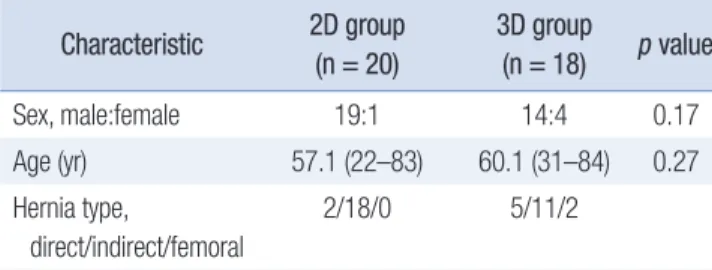
Patient characteristics and hernia types are shown in Table 1.
The male-to-female sex ratio in the 2D group was 19:1. In the 3D group, there were 14 males and four females. There was no Table 1.
Table 1. Patient characteristics Characteristic
Characteristic 2D group 2D group (n = 20) (n = 20)
3D group 3D group (n = 18)
(n = 18) pp value value
Sex, male:female 19:1 14:4 0.17
Age (yr) 57.1 (22–83) 60.1 (31–84) 0.27
Hernia type,
direct/indirect/femoral
2/18/0 5/11/2
Body mass index (kg/m2) 23.8 (16.0–28.6) 24.5 (17.6–30.4) 0.42 Values are presented as number only or mean (range).
2D, two-dimensional; 3D, three-dimensional.
significant difference in sex ratio between the two groups. The age distribution of the 2D group was 22 to 83 years, with a mean of 57.1 years. The age distribution of the 3D group was 31 to 84 years, with a mean of 60.1 years. There was no significant differ- ence in age distribution between the two groups. The 2D group included two patients with direct hernia and 18 patients with indirect hernia. In the 3D group, there were five patients with direct hernia, 11 with indirect hernia, and two with femoral her- nia. The BMI was 23.8 kg/m2 and 24.5 kg/m2 in the 2D and 3D groups, respectively.
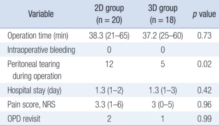
The results of these operations are summarized in Table 2.
The mean ± standard deviation of operation time was 38.2 ± 10.86 minutes in the 2D groups and 37.2 ± 13.74 minutes in the 3D groups, respectively. There was no intraoperative bleeding in either group. Intraoperative peritoneal tearing occurred in 12 patients in the 2D group compared to five patients in the 3D group (p = 0.02). The average length of hospital stay did not dif- fer between the two groups; however, a patient in the 3D group was hospitalized for 1 additional day due to difficulty in voiding.
The mean NRS scores were 3.3 and 3 in the 2D and 3D groups, respectively (p = 0.96). Patients visited the OPD approximately 1 week after discharge for their follow-up. There were no abnormal findings in the 3D group at the outpatient visit. However, in the
2D group, two patients revisited the OPD because of the postop- erative occurrence of seroma and varicocele.
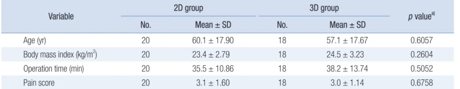
Tables 3 and 4 present the results of the normality test. Table 3 shows a comparison of the categorical data of 3D and 2D laparo- scopic surgeries. Sex, hernia type, number of hernias, and num- ber of OPD visits were analyzed using Fisher exact test. The ratio of sex, hernia type, hernia number, and OPD visits in the two Table 2.
Table 2. Operation results Variable
Variable 2D group 2D group (n = 20) (n = 20)
3D group 3D group (n = 18)
(n = 18) pp value value Operation time (min) 38.3 (21–65) 37.2 (25–60) 0.73
Intraoperative bleeding 0 0
Peritoneal tearing during operation
12 5 0.02
Hospital stay (day) 1.3 (1–2) 1.3 (1–3) 0.42
Pain score, NRS 3.3 (1–6) 3 (0–5) 0.96
OPD revisit 2 1 0.99
Values are presented as number only or mean (range).
2D, two-dimensional; 3D, three-dimensional; NRS, numeric rating scale;
OPD, outpatient department.
Table 3.
Table 3. Comparison of categorical data from the 3D and 2D laparoscopic surgeries Variable
Variable Total patients (n = 38)Total patients (n = 38) 2D group (n = 20)2D group (n = 20) 3D group (n = 18)3D group (n = 18) pp value value Sex (yr)
Female 5 1 (5.0) 4 (22.2) 0.3867b)
Male 33 19 (95.0) 14 (77.8)
Hernia type
Direct 7 2 (10.0) 5 (27.8) 0.0804b)
Indirect 29 16 (80.0) 11 (61.1)
Femoral 2 0 (0) 2 (11.1)
Pantaloon 2 2 (10.0) 0 (0)
No. of hernias
Unilateral 38 20 (100) 17 (94.4) 0.0735b)
Bilateral 1 0 (0) 1 (5.6)
Peritoneal tearing during operation
Yes 17 12 (60.0) 5 (27.8) 0.0105a)
No 21 8 (40.0) 13 (72.2)
No. of OPD visits
1 35 18 (90.0) 17 (94.4) >0.9999b)
2 3 2 (10.0) 1 (5.6)
Values are presented as number only or number (%).
2D, two-dimensional; 3D, three-dimensional; OPD, outpatient department.
Analyzed by a)chi-square test, b)Fisher exact test.
groups showed a difference, but not all variables were statistically significant. Only peritoneal tearing during the operation was analyzed using the chi-square test. The p value was 0.01, indicat- ing statistically significant results. Table 4 shows a comparison of 3D and 2D laparoscopic surgeries for continuous data. Age, BMI, operation time, and pain score were analyzed using an in- dependent t test. None of the variables in the 2D and 3D groups showed statistically significant differences.
DISCUSSION
Although there is little doubt that minimally invasive surgery, particularly laparoscopic surgery, benefits patients, surgeons require a lot of training to get used to a 2D laparoscope. Lapa- roscopic surgery is more challenging than open surgery partly because surgeons need to operate in a 3D space according to a 2D projection on a monitor, which results in the loss of depth per- ception [8]. The 3D laparoscopy has taken laparoscopic surgery to a new level, offering the surgeon stereoscopic vision, which is valuable for difficult activities such as suturing and knotting, which demand a high degree of spatial perception. Numerous recent studies have shown that 3D laparoscopic surgery can re- duce the operation time, intraoperative blood loss, and surgeon’s learning curve compared with 2D laparoscopic surgery [9–12].
Although 3D vision technology has substantially improved in recent years and various technical concepts have been imple- mented, it is still not the accepted standard for laparoscopic ab- dominal surgery. A key reason for this is that 3D laparoscopy is significantly more expensive than 2D laparoscopy [13]. Moreover, surgeons often complain about the poor quality of the 3D im- age and the discomfort of wearing 3D glasses [14–16]. In recent years, the development of 3D HD vision systems with better im- age quality and lower cost has made great progress in 3D vision technology [17]. Therefore, 3D laparoscopic surgery is currently performed more frequently than before, and research on its ef- fectiveness has also been conducted as the field of application has increased. Previously, comparative studies of 2D and 3D laparo- scopic surgeries were conducted in the field of gastrointestinal
surgery, obstetrics and gynecology, and urology; therefore, it is important to confirm the safety and effectiveness of the inguinal hernia repair in this study [18–20].
The learning curve of laparoscopic inguinal hernia repair is higher than that of other surgeries because of the ambiguity of the inguinal anatomy. Therefore, peritoneal tearing is frequent in laparoscopic inguinal hernia repair. The reported incidence of peritoneal tears is 10% to 64% during TEP [21]. Peritoneal tearing is the most common reason for the conversion of endoscopic ex- traperitoneal inguinal hernioplasty to TAPP or open approaches [22]. Peritoneal tearing not only leads to the loss of extraperito- neal space but also increases the risk of small bowel adhesions and internal herniation. Furthermore, as the mesh is no longer securely buttressed between the abdominal wall and peritoneum by intraabdominal pressure, it becomes susceptible to migration, particularly when a nonstapling technique is adopted [23]. It is important to note that 3D laparoscopic surgery can reduce the risk of peritoneal tear.
In conclusion, 3D laparoscopic surgery for inguinal hernia pa- tients was observed to be feasible and safe and showed less peri- toneal tearing than the 2D laparoscopic surgery group.
One of the limitations of our study was that it included surger- ies performed by a single experienced surgeon. The 3D laparos- copy is superior to 2D laparoscopy in terms of depth perception, so it can reduce the learning curve of beginners. In the case of a novice surgeon, the difference between the two groups could have been more dramatic. If further research compares the out- comes of 2D and 3D laparoscopic surgeries performed by novice surgeons, we believe the excellence of 3D laparoscopy can be bet- ter demonstrated.
Another limitation of this study is that it was conducted in a single institution, and the number of study subjects was small.
Hence, generalization and confirmation of these findings are limited. Therefore, multicenter studies involving a larger number of subjects are required to improve the reliability of our results.
Additionally, this was a retrospective study that was per- formed by reviewing the charts. Therefore, it was impossible to analyze the factors that might influence the outcome. In addi- Table 4.
Table 4. Comparison of continuous data from the 3D and 2D laparoscopic surgeries Variable
Variable 2D group2D group 3D group3D group
pp value valuea)a) No.
No. Mean ± SDMean ± SD No.No. Mean ± SDMean ± SD
Age (yr) 20 60.1 ± 17.90 18 57.1 ± 17.67 0.6057
Body mass index (kg/m2) 20 23.4 ± 2.79 18 24.5 ± 3.23 0.2604
Operation time (min) 20 35.5 ± 10.86 18 38.2 ± 13.74 0.5052
Pain score 20 3.1 ± 1.60 18 3.0 ± 1.14 0.6758
2D, two-dimensional; 3D, three-dimensional; SD, standard deviation.
a)Analyzed by independent t test.
tion, it was not possible to conduct preliminary investigations into the factors that could influence the results.
Although the 2D and 3D laparoscopic surgeries showed no statistical difference in operation time, intraoperative bleeding, hospital stay, pain score, and OPD revisit, the 3D laparoscopic surgery showed less peritoneal tearing during the TEP procedure for inguinal hernia repair. Therefore, we conclude that the 3D laparoscopic TEP procedure is feasible and safe for inguinal her- nia repair.
NOTES
Ethical statements
The study protocol was reviewed and approved by the Institu- tional Review Board of Kyung Hee University Hospital at Gang- dong (No. KHNMC 2021-02-049). The requirement for informed consent was waived because of the retrospective nature of the study.
Authors’ contributions
Conceptualization: All authors Data curation, Investigation: AYK Formal analysis: JHY
Funding acquisition: SIC
Methodology, Visualization: AYK, JHY Project administration: SIC, JHY Writing–original draft: AYK, SIC Writing–review & editing: All authors
All authors read and approved the final manuscript.
Conflict of interest
All authors have no conflicts of interest to declare.
ORCID
Ah Yoon Kim, https://orcid.org/0000-0003-4989-1636 Sung Il Choi, https://orcid.org/0000-0002-0662-0951 Jung Hyun Yeom, https://orcid.org/0000-0001-5368-0191
REFERENCES
1. Merola G, Cavallaro G, Iorio O, et al. Learning curve in open inguinal hernia repair: a quality improvement multicentre study about Lich- tenstein technique. Hernia 2020;24:651-659.
2. Arregui ME, Davis CJ, Yucel O, Nagan RF. Laparoscopic mesh repair of inguinal hernia using a preperitoneal approach: a preliminary re- port. Surg Laparosc Endosc 1992;2:53-58.
3. McKernan JB, Laws HL. Laparoscopic repair of inguinal hernias using a totally extraperitoneal prosthetic approach. Surg Endosc 1993;7:26-28.
4. Agrusa A, Di Buono G, Buscemi S, Cucinella G, Romano G, Gulotta G. 3D laparoscopic surgery: a prospective clinical trial. Oncotarget 2018;9:17325-17333.
5. Vărcuş F, Duţă C, Dobrescu A, Lazăr F, Papurica M, Tarta C. Laparo- scopic repair of inguinal hernia TEP versus TAPP. Chirurgia (Bucur) 2016;111:308-312.
6. Patrzyk M, Klee M, Stefaniak T, Heidecke CD, Beyer K. Randomized study of the influence of two-dimensional versus three-dimensional imaging using a novel 3D head-mounted display (HMS-3000MT) on performance of laparoscopic inguinal hernia repair. Surg Endosc 2018;32:4624-4631.
7. Usta TA, Ozkaynak A, Kovalak E, Ergul E, Naki MM, Kaya E. An assessment of the new generation three-dimensional high definition laparoscopic vision system on surgical skills: a randomized prospec- tive study. Surg Endosc 2015;29:2305-2313.
8. Nicolau S, Soler L, Mutter D, Marescaux J. Augmented reality in laparoscopic surgical oncology. Surg Oncol 2011;20:189-201.
9. Lu J, Zheng CH, Zheng HL, et al. Randomized, controlled trial com- paring clinical outcomes of 3D and 2D laparoscopic surgery for gas- tric cancer: an interim report. Surg Endosc 2017;31:2939-2945.
10. Aykan S, Singhal P, Nguyen DP, et al. Perioperative, pathologic, and early continence outcomes comparing three-dimensional and two- dimensional display systems for laparoscopic radical prostatectomy- -a retrospective, single-surgeon study. J Endourol 2014;28:539-543.
11. Blavier A, Gaudissart Q, Cadière GB, Nyssen AS. Comparison of learning curves and skill transfer between classical and robotic lapa- roscopy according to the viewing conditions: implications for train- ing. Am J Surg 2007;194:115-121.
12. Chiu CJ, Lobo Prabhu K, Tan-Tam CC, Panton ON, Meneghetti A. Using three-dimensional laparoscopy as a novel training tool for novice trainees compared with two-dimensional laparoscopy. Am J Surg 2015;209:824-827.
13. Hanani M, Cernat V, Beyer K, et al. Comparison of a 3D head- mounted display (HMS-3000MT) and 3D passive polarizing display with 2D technique for first laparoscopic inguinal hernia repair by novice surgeons. Hernia 2020;24:661-668.
14. Wang T, Zheng B. 3D presentation in surgery: a review of technology and adverse effects. J Robot Surg 2019;13:363-370.
15. Taffinder N, Smith SG, Huber J, Russell RC, Darzi A. The effect of a second- generation 3D endoscope on the laparoscopic precision of novices and experienced surgeons. Surg Endosc 1999;13:1087-1092.
16. Shah J, Buckley D, Frisby J, Darzi A. Depth cue reliance in surgeons and medical students. Surg Endosc 2003;17:1472-1474.
17. Mashiach R, Mezhybovsky V, Nevler A, Gutman M, Ziv A, Khaikin M. Three-dimensional imaging improves surgical skill performance in a laparoscopic test model for both experienced and novice laparo- scopic surgeons. Surg Endosc 2014;28:3489-3493.
18. Zhao B, Lv W, Mei D, et al. Comparison of short-term surgical out- come between 3D and 2D laparoscopy surgery for gastrointestinal cancer: a systematic review and meta-analysis. Langenbecks Arch Surg 2020;405:1-12.
19. Yazawa H, Takiguchi K, Imaizumi K, Wada M, Ito F. Surgical out- comes of total laparoscopic hysterectomy with 2-dimensional versus 3-dimensional laparoscopic surgical systems. Fukushima J Med Sci 2018;64:38-45.
20. Dirie NI, Wang Q, Wang S. Two-dimensional versus three-dimen- sional laparoscopic systems in urology: a systematic review and meta-
analysis. J Endourol 2018;32:781-790.
21. Mathew KG, Pokhrel G. Closing peritoneal tear during laparo- scopic inguinal hernia repair: simple and effective technique. Hernia 2020;24:1121-1124.
22. Felix EL, Harbertson N, Vartanian S. Laparoscopic hernioplasty: sig- nificant complications. Surg Endosc 1999;13:328-331.
23. Lau H, Patil NG, Yuen WK, Lee F. Management of peritoneal tear during endoscopic extraperitoneal inguinal hernioplasty. Surg En- dosc 2002;16:1474-1477.