학 술 논 문
189
특발정상압수두증에서 해마 및 외측 뇌실의 부피와 뇌척수액배액검사
강경훈
1· 한재환
2· 윤의철
3*
1경북대학교 의과대학 신경과학교실, 2경북대학교 대학원 의용생체공학과,
3대구가톨릭대학교 의공학과
Hippocampal and Ventricular Volumes of Idiopathic Normal-pressure Hydrocephalus and the Cerebrospinal Fluid Tap Test
Kyunghun Kang
1, Jaehwan Han
2and Uicheul Yoon
3*
1
Department of Neurology, School of Medicine, Kyungpook National University, Daegu, South Korea
2
Department of Medical and Biological Engineering, Graduate School, Kyungpook National University
3
Department of Biomedical Engineering, Daegu Catholic University
(Manuscript received 11 September 2019 ; revised 7 October 2019 ; accepted 11 October 2019)
Abstract: We investigated differences in ventricular and hippocampal volumes between CSF tap test (CSFTT) responders and non-responders in idiopathic normal-pressure hydrocephalus (INPH) patients and compared these parameters in INPH patients with that of age- and gender-matched healthy controls. We also evaluated relationships between ventricular and hippocampal volumes and clinical profiles in INPH patients. We enrolled 48 patients with INPH and 29 healthy controls. Ventricular and hippocampal volumes were measured on MRI, including 3-dimensional volumetric images. INPH patients, when compared to healthy controls, had significantly larger ventricular and smaller hip- pocampal volumes. No difference in ventricular and hippocampal volumes was found between CSFTT responders and non-responders in INPH patients. And hippocampal volumes showed significant negative correlations with Clin- ical Dementia Rating Scale scores, INPH grading scale cognitive scores, Timed Up and Go Test scores, and Unified Parkinson’s Disease Rating Scale motor scores in INPH patients. Volumetric assessment of ventricular and hippo- campal regions may have no predictive value in differentiating between CSFTT responders and non-responders in INPH patients. Our findings may help us understand the potential pathophysiology of unique symptoms associated with INPH.
Key words: Idiopathic normal pressure hydrocephalus, Cerebrospinal fluid tap test, Magnetic resonance imaging, Hippocampus
I. 서 론
특발정상압수두증(Idiopathic Normal-Pressure Hydrocephalus) 은 구조적인 뇌 영상에서 뇌척수액의 순환 경로에 막힘 없 이 뇌실 확장을 보이는 것이 특징이고, 주로 성인에서 발병
하며 보행장애와 배뇨장애, 그리고 인지기능장애를 보이는 신경퇴행질환이다[1]. 뇌신경영상에서 뇌실 확장을 확인하는 것이 특발정상압수두증을 진단할 때 가장 중요하다. 특발정 상압수두증의 진단에 가장 흔히 사용되는 Relkin 등의 진단 기 준과 Ishikawa 등의 진단 기준에서 Evans’ index가 0.3을 초과하는 것으로 뇌실 확장을 정의하며[1,2], Evans’ index는 좌우 외측 뇌실의 가장 긴 전각 사이의 거리를 두개골의 가장 긴 내경으로 나눈 값이다[3].
특발정상압수두증은 수술로 치료가 가능하며, 특발정상압 수두증이 의심되는 경우 뇌실복강션트(Ventriculoperitoneal Corresponding Author : Uicheul Yoon
D5 St. Dominic Hall, 204, Hayang-Ro 13-13, Hayang-Eup, Gyeongsan-si, Gyeongbuk, Rep.of Korea 38430
Tel : +82-53-850-2515 E-mail : [email protected]
이 결과물은 2017년도 대구가톨릭대학교 학술연구비 지원에 의한 것임.
190
shunt) 의 치료 예후를 예측하는 것이 매우 중요하다. 요추 천자로 뇌척수액을 다량 배액하는 것은 뇌실복강션트 치료 예후를 예측하는 데 가장 널리 사용되고 있다. 하지만, 요추 천자로 뇌척수액을 배액하는 것 역시 침습적인 방법이며, 비 교적 위험성이 낮지만 척수의 경막하 또는 거미막하의 출혈 등 심각한 부작용이 발생할 수 있다[4-6].
뇌 자기공명영상의 정량적인 분석은 비침습적인 방법으로 서 해부학적인 뇌의 구조적 변화를 연구하는데 유용하다[7].
퇴행성 뇌질환과 같이 미만성으로 서서히 진행되는 뇌질환 이나 조직학적 미세구조를 침범하는 질환에 있어서 이와 같은 분석 기법은 매우 중요하다[8-10]. 해마(Hippocampus)를 포함한 내측두엽(Temporal lobe)은 알츠하이머병 (Alzheimer’s disease) 초기부터 병리적 변성이 일어나는 것으로 잘 알려져 있다[8]. 이러한 신경병리학적 변화 양상은 알츠하이머병 환 자에서 질병 초기부터 해마를 포함한 내측두엽의 위축을 보 이는 뇌 자기공명영상 소견과도 일치한다[9]. 해마의 위축은 알츠하이머병 진행과정의 초기 특정 표지자로서 해마의 부 피는 알츠하이머병의 진단에도 도움이 된다. 알츠하이머병에서 치매로 진행하는 경도인지장애(Mild Cognitive Impairment, MCI) 환자가 해마의 부피 감소가 특히 심한 것으로 알려져 있다[10]. 특발정상압수두증 환자의 대부분이 알츠하이머 병 리를 가지는 것으로 보고되었다[11]. 또한, 알츠하이머 병리가 심한 특발정상압수두증 환자가 뇌실복강션트 치료에 대한 반응이 나쁜 것으로 알려져 있다[12]. 요추천자로 뇌척수액 배액 후 증상이 호전되는 기전에 대해서는 아직까지 정립된 이론은 없으나 동반된 알츠하이머 병리가 치료 결과에 영향을 미칠 수 있다[13].
본 연구에서는 외측 뇌실과 해마가 특발정상압수두증 환 자에서 요추천자로 뇌척수액배액 후에 증상이 호전되는 것 과 연관이 있는지를 알아보고자 한다. 이를 위해서 요추천 자를 통한 뇌척수액배액 치료 반응 집단과 반응이 없는 집 단 간에 외측 뇌실과 해마의 부피를 비교하였다. 추가적으 로 본 연구에서는 특발정상압수두증 환자들을 대상으로 외 측 뇌실 및 해마의 부피와 다양한 임상양상 간의 연관성을 분석하였다.
II. 대상 및 방법
1. 연구 대상
2013 년 6월부터 2016년 5월까지 칠곡경북대학교병원 뇌 신경센터에 내원한 환자 중에서 Relkin 등의 진단 기준에 따라 특발정상압수두증으로 진단받은 환자 48명을 대상으 로 하였다[1]. 특발정상압수두증의 진단은 1) 병력에서 점진 적 발병, 40세 이상의 증상 발병, 최소한 3-6개월의 기간, 진행 경과, 임상 증상이나 영상 소견을 설명할 만한 다른 질
환의 부재, 2) 뇌 영상에서 뇌실 확장의 소견, 3) 임상양상에서 보행장애가 반드시 존재하면서 인지기능장애 또는 배뇨장애의 존재, 4) 뇌척수액압의 증가 소견이 없음(70-245 mmH
2O) 의 진단 기준에 따라 이루어졌다[1]. 정상인군은 특발정상압 수두증 환자군의 나이 및 성별을 고려하여 동일하게 칠곡경 북대학교병원의 내원 대상자 29명으로 구성되었다. 본 연구는 칠곡경북대학교병원 임상연구윤리심의 위원회(Institutional Review Broad, IRB) 의 승인을 받아 진행되었다.
2. 특발정상압수두증 증상의 평가척도
보행기능 평가를 위해 ‘일어나 걸어가기’ 검사(Timed Up- and-Go test, TUG) 와 10미터 보행검사(10-meter walking test) 를 시행하였다. TUG는 의자에서 일어나기, 3미터 걸 어가기, 돌기, 걸어 돌아오기, 의자에 앉기의 순으로 진행하여 이 동작을 수행하는 동안에 소요된 시간을 기록한다[4]. 10-meter walking test 는 평소에 걷는 속도로 10미터를 걷게 하여 소 요된 시간을 기록한다[4]. TUG와 10-meter walking test는 연속적으로 4번하여 평균값을 기록하였다[4]. 인지기능 평 가를 위해 한국판 간이 정신상태 검사(Korean Mini-Mental State Examination, K-MMSE), 임상치매척도(Clinical Dementia Rating scale, CDR), 한국판 전두엽 기능평가 검사(Korean Version of Frontal Assessment Battery, K-FAB)를 시행하였다[4]. 보행상태척도(Gait Status Scale, GSS), 통합파킨슨병척도(Unified Parkinson’s Disease Rating Scale, UPDRS) 의 운동기능검사, INPHGS(idiopathic normal-pressure hydrocephalus grading scale)를 추가로 시행하였다[4].
3. 요추천자를 통한 뇌척수액배액검사
요추천자로 뇌척수액 30-50 mL를 배액한 후 증상의 호전 여 부를 관찰하였다[2]. TUG, K-MMSE, INPHGS를 요추천자 시행 전과 시행 후에 측정하여 비교하였다[2]. 보행장애는 1 일 뒤에 평가하였고, 인지기능장애 및 배뇨장애는 1주 뒤에 평 가하였다. 요추천자로 뇌척수액 배액 후 TUG의 시간이 10%를 초과하여 호전되거나, K-MMSE 점수가 3점을 초과하여 호 전된 경우, 또는 INPHGS 점수가 1점 이상 호전되면 치료 반응군으로 분류하였다[2].
4. 뇌 MRI 자료 획득
본 연구에 활용된 뇌 자기공명영상(Magnetic Resonance
Imaging, MRI) 은 3.0T 자기공명 영상기기(GE Discovery
MR 750, GE Healthcare) 를 사용하여 획득하였다. 구조적
뇌 MRI는 IR-FSPGR (Inversion-recovery fast spoiled
gradient echo) 프로토콜을 이용하여 T1 강조영상을 획득
하였다. 영상획득변수는 TR = 8.2 ms, TE = 3.2 ms, flip
191 angle = 12°, acquisition matrix = 256 × 256, field of view
= 240 mm, slice thickness = 1 mm 이다.
5. 뇌실과 해마 영역 분할: 템플릿 기반 그래프컷 알고리즘 대상자의 뇌실과 해마의 크기를 측정하기 위해 뇌 MRI 로부터 해당 영역을 정의하고 정확히 분할해야한다. 먼저, 뇌실과 해마 영역을 기준 템플릿 위에 정의하고, 정합 기술을 통해 각 대상자별로 그래프컷 알고리즘의 seeds 값으로 선정하 여 분할하는 방법으로 진행하였다. 기준 템플릿은 International Consortium for Brain Mapping (ICBM) 152 symmetric template 으로 신경과 전문의가 시상면을 기준으로 뇌실과 해마의 영역을 직접 정의하였다[14].
먼저, 각 대상자의 MRI 영상을 기준 템플릿에 선형으로 정합을 한 후, 영역 분할 성능 개선을 위해 영상의 밝기 불균 일성을 제거하였다[15]. 그리고, 계층적 다중 단계의 비선형 정합(Automatic Nonlinear Image Matching and Anatomical Labeling, ANIMAL)을 이용하여 기준 템플릿의 뇌실과 해 마 영역을 각 대상자의 MRI 영상에 정의하였다. 또한, 인 공신경망 기반의 분류기(Artificial neural network classifier) 를 적용하여 뇌의 주요 조직인 회백질, 백질, 뇌척수액을 분 할하였다[16]. 이를 통해 각 대상자의 MRI 영상에 정의된 뇌실 영역에 포함된 회백질 및 백질 성분을 제거하고, 뇌척 수액과 백질을 제외한 해마 영역을 정의함으로써 그래프컷 알고리즘의 foreground 및 background로 활용할 수 있는 seeds 영상을 생성했다[14,17,18].
가변형 모델(Deformable model) 방식의 그래프컷 알고 리즘은 영상 분할에 주로 활용되고 있으며 foreground와 background 에 해당하는 화소들을 seeds로 지정함으로써 객체와 배경에 대한 밝기 정보를 얻을 수 있어 매우 정확하고 안정적인 결과를 제시한다[19,20]. 본 연구에서는 템플릿을 통해 foreground와 background를 자동으로 지정함으로써 그래프컷 알고리즘의 결과로 뇌실과 해마영역을 높은 정확 도로 추출할 수 있다. 각 대상자의 MRI 영상에서 템플릿을 이용하여 추출한 뇌실 영역을 foreground로 백질과 회백질 영역을 background로 지정하여 경계 부분의 가중치를 최대 유 량-최소 컷(Maximum Flow Minimum Cut) 방법으로 분 할한다. 또한, 해마는 템플릿 기반의 분할 결과를 foreground 로, 뇌척수액과 백질 영역을 background로 지정하여 영역을 추 출한다[14,19,20]. 추가적인 형태학적 영상처리 기법(erosion과 dilation) 을 적용하여 분할 결과를 개선하였다[19]. 개인 간 대뇌 부피의 차이를 고려하여 선형 역변환을 통해 원 영상의 공간에서 분할된 뇌실과 해마 영역의 크기를 측정하였다.
6. 통계 분석
모든 통계 분석은 SPSS (IBM SPSS Statistics for Windows,
Version 25.0. Armonk, NY: IBM Corp.) 를 사용하였다.
요추천자를 통한 뇌척수액배액 치료 반응군과 비반응군 그 리고 정상대조군 간의 외측 뇌실과 해마의 부피의 비교는 일원배치분산분석(One-way ANOVA) 또는 Kruskal-Wallis test 로 분석하였다. 사후검정을 위해 Dunnett’s test, Bonferroni’s test, Mann-Whitney post-hoc test 를 이용하였다. 특발정 상압수두증 환자들을 대상으로 외측 뇌실 및 해마의 부피와 다양한 임상양상 간의 상관 관계 분석은 Pearson’s correlation 또는 Spearman’s correlation을 이용하였다. 통계학적 유 의성은 p값이 0.05 미만인 경우로 정의하였다.
III. 연구 결과
1. 연구대상자의 특성
본 연구에 참여한 대상자 수는 총 77명이었고, 특발정상 압수두증 환자들 중 뇌척수액배액 치료 반응군은 36명 그리고 비 반응군은 12명이었다. 각 집단의 평균 연령은 특발정상압수 두증 환자들 중 반응군 73.4±5.2세, 비반응군 71.1±5.3세 그리고 정상대조군 70.8±4.3세로 통계적으로 유의미한 차 이는 없었다. 세 집단에서 성별에 대해서도 통계적으로 유 의미한 차이는 없었다. 각 집단의 평균 학력은 특발정상압 수두증 환자들 중 반응군 9.3±4.5년, 비반응군 9.6±5.4년 그리고 정상대조군 11.6±4.9년이었고, 통계적으로 유의미한 차이는 없었다. 그러나, K-MMSE 점수는 세 집단 사이에서 유의 미한 차이를 보여 특발정상압수두증 환자들 중 반응군과 비 반응군에서 정상대조군 보다 K-MMSE 점수가 유의하게 낮 았다(Table 1).
특발정상압수두증 환자들 중 뇌척수액배액 치료 반응군과 비반응군 사이에 유병 기간, 임상증상, INPHGS 점수, TUG 시 간, 10-meter walking test 시간, GSS 점수, UPDRS 운동기 능검사 점수, K-MMSE 점수, CDR 점수, K-FAB 점수, Evans’
ratio 에 유의미한 차이는 없었다. 실비우스틈새(Sylvian fissure) 의 뇌척수액공간은 넓어져 있는 반면에 두정부대뇌볼록의 뇌척수액공간은 좁아져 있는 뇌 영상 소견을 보이는 환자의 비율도 두 집단에서 유의미한 차이는 없었다(Table 1).
2. 외측 뇌실과 해마의 부피 분석 결과
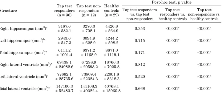
외측 뇌실의 부피는 세 집단 사이에서 유의한 차이를 보여 특발정상압수두증 환자들 중 뇌척수액배액 치료 반응군 (147.10±52.48 ( ml))과 비반응군(141.11±40.32)에서 정상 대조군(40.77±15.96)보다 외측 뇌실의 부피가 유의하게 컸 다(p<0.001). 특발정상압수두증 환자들 중 반응군과 비반응 군 사이에 외측 뇌실 부피에 유의한 차이는 없었다(Fig. 1, Table 2).
해마의 부피는 세 집단 사이에서 유의미한 차이를 보여
192
특발정상압수두증 환자들 중 뇌척수액배액 치료 반응군 (6.11±1.00) 과 비반응군(6.37±1.17)에서 정상대조군(8.67±1.12) 보다 해마의 부피가 유의미하게 작았다(p<0.001). 특발정상 압수두증 환자들 중 반응군과 비반응군 사이에 해마 부피에 유의미한 차이는 없었다(Fig. 1, Table 2).
3. 특발정상압수두증 환자에서 외측 뇌실 및 해마의 부피와 다양한 임상양상 간의 연관성
우측 해마의 부피는 CDR 점수(r=-0.407), INPHGS 배 뇨장애 점수(-0.312), UPDRS 운동기능검사 점수(-0.316)와 음 의 상관관계를 보였다(Table 3). 좌측 해마의 부피는 CDR 점
표 1. 특발정상압수두증 환자와 정상인의 주요 정보Table 1. Demographic data and clinical characteristics of idiopathic normal-pressure hydrocephalus patients and controls
Characteristics Tap test responders
(n = 36)
Tap test non-responders (n = 12)
Healthy controls (n = 29)
Gender, male 24 (66.7) 6 (50.0) 11 (37.9)
Age (year) 73.4 ± 5.2 71.1 ± 5.3 70.8 ± 4.3
Education (year) 9.3 ± 4.5 9.6 ± 5.4 11.6 ± 4.9
Duration of symptoms (year) 2.9 ± 2.6 2.6 ± 1.3
Initial symptoms
Gait disturbance 29 (80.6) 12 (100.0)
Cognitive impairment 7 (19.4) 0 (0.0)
Full-blown symptoms
Gait disturbance 36 (100.0) 12 (100.0)
Cognitive impairment 36 (100.0) 11 (91.7)
Urinary incontinence 22 (61.1) 6 (50.0)
Clinical triad 22 (61.1) 5 (41.7)
Drainage volume of CSF 38.2 ± 3.5 38.2 ± 4.6
CSF opening pressure (cmH2O) 9.0 ± 3.0 8.6 ± 3.3
Apolipoprotein E ε4+/ε4− 3/21 1/8
INPHGS
GS-Gait 1.7 ± 0.6 1.5 ± 0.5
GS-Cogn 2.7 ± 0.7 2.6 ± 0.5
GS-Urin 1.6 ± 1.2 1.6 ± 1.2
TUG 22.0 ± 15.0 15.3 ± 8.2
10-meter walking test 21.5 ± 21.3 15.7 ± 11.6
GSS 7.6 ± 2.7 6.9 ± 3.3
UPDRS motor score 22.1 ± 7.1 24.4 ± 9.6
K-MMSEa 20.1 ± 6.8b 20.1 ± 5.2b 27.3 ± 2.2
CDR (0:0.5:1:2:3) 0:19:9:5:3 0:8:3:1:0
FAB 9.7 ± 4.4 10.0 ± 3.5
Evans’ ratio 0.33 ± 0.01 0.32 ± 0.02
Narrowing of the CSF space at the high convexity 29 (82.9) 9 (75.0)
Values denote number (%) or mean ± standard deviation
a
Significant intergroup difference with Kruskal-Wallis analysis (p < 0.001)
b
Statistically significant versus healthy controls
INPH = idiopathic normal-pressure hydrocephalus; INPHGS = Idiopathic Normal-Pressure Hydrocephalus Grading Scale; GS-
Gait = INPHGS for gait; GS-Cogn = INPHGS for cognition; GS-Urin = INPHGS for urinary function; TUG = Timed Up-and-Go
test; GSS = Gait Status Scale; UPDRS = Unified Parkinson’s Disease Rating Scale; K-MMSE = Korean version of Mini-Mental
State Examination; CDR = Clinical Dementia Rating Scale; FAB = Frontal Assessment Battery
193
수(-0.331), INPHGS 인지기능장애 점수(-0.297), TUG 시 간(-0.399), GSS 점수(-0.304), UPDRS 운동기능검사 점수 (-0.345) 와 음의 상관관계를 보였다(Table 3). 외측 뇌실의 부피는 임상양상 간의 상관관계를 보이지 않았다.
IV. 고 찰
가변형 모델 방식의 그래프컷은 영역 분할에 좋은 성능을 보이지만, foreground와 background를 위한 사전 정보를 사용자가 직접 입력해야 하는 수동성이라는 단점을 지니고 있는 반면 본 연구에서 진행한 템플릿 기반의 그래프컷 알 고리즘은 seeds를 자동적으로 처리 할 수 있다. 또한, 뇌 MRI 영상 연구에서 뇌실과 해마 영역을 포함한 뇌 구조물의 분 할에 탁월함이 보고되고 있으나, 현재까지 특발정상압수두 증 환자 분석에 적용된 연구는 없었다[14,18,21-23]. 본 연 구는 템플릿 기반의 그래프컷 알고리즘을 특발정상압수두증 환자 분석에 적용하여 뇌실과 해마 영역을 추출하여 각 영 역의 부피를 구하고, 특발정상압수두증의 증상과 요추천자 반응 집단과 비반응 집단을 분류하여 집단간의 차이를 분석 하였다.
본 연구에서는 특발정상압수두증 환자들 중 뇌척수액배액 치료 반응군과 비반응군에서 정상대조군보다 외측 뇌실의 부피가 유의미하게 컸고, 해마의 부피가 유의미하게 작았다.
이는 이전 연구와 부합하는 결과이다[14,24]. 특발정상압수 두증은 뇌신경영상에서 뇌척수액의 순환 경로에 막힘 없이 뇌실 확장을 보이는 것이 특징인 것으로 알려져 있기 때문에 특발정상압수두증 진단을 위해 뇌신경영상에서 뇌실 확장을 확인하는 것은 아주 중요하다[14]. 특발정상압수두증 환자는 정상인에 비해 해마의 부피 감소를 보이는 것으로 보고되고 있다[24].
그림 1. 템플릿 기반 그래프컷 알고리즘 이용한 외측 뇌실과 해마 영 역 분할Fig. 1. Segmentation of lateral ventricle and hippocampus in MR images using template based graph cuts algorithm. The results of segmentation of lateral ventricle and hippocampus.
Cyan color represents hippocampus region. Yellow color indicates lateral ventricle region
표 2. 뇌 자기공명영상의 부피 분석 결과
Table 2. Volumetric measurements of brain MR Imaging
Structure
Tap test responders
(n = 36)
Tap test non- responders
(n = 12)
Healthy controls (n = 29)
Post-hoc test, p value Tap test responders
vs. tap test non-responders
Tap test responders vs.
healthy controls
Tap test non-responders vs.
healthy controls Right hippocampus (mm3)a 3167.6
± 582.1
3276.3
± 708.1
4426.8
± 564.9 0.353 <0.001c <0.001c
Left hippocampus (mm3)b 2943.6
± 547.3
3094.9
± 628.9
4244.2
± 598.2 0.715 <0.001c <0.001c
Total hippocampus (mm3)a 6111.2
± 1001.4
6371.2
± 1168.9
8671.0
± 1119.1 0.171 <0.001c <0.001c Right lateral ventricle (mm3)a 69438.1
± 24982.6
67298.9
± 20598.2
18766.3
± 7925.8 0.812 <0.001c <0.001c Left lateral ventricle (mm3)a 77662.1
± 28735.6
73809.4
± 22324.3
22001.8
± 8318.3 0.520 <0.001c <0.001c Total lateral ventricle (mm3)a 147100.3
± 52483.7
141108.3
± 40322.4
40768.1
± 15960.8 0.668 <0.001c <0.001c
Values denote mean ± standard deviation
a
Significant intergroup difference with Kruskal-Wallis analysis (p < 0.001).
b
Significant intergroup difference with analysis of variance (p < 0.001).
c
Significant intergroup difference with post-hoc comparisons
194 본 연구에서는 특발정상압수두증 환자들 중 뇌척수액배액 치료 반응군과 비반응군 사이에 외측 뇌실과 해마 부피에 유의미한 차이는 없었다. 요추천자를 통한 뇌척수액배액 후 증상이 호전되는 것은 특발정상압수두증의 중요한 특징이다 [14]. 요추천자를 통한 뇌척수액배액 후 치료 반응을 예측할 수 있는 뇌영상 바이오마커들에 대한 몇가지 연구들이 있었 다[13,24-27]. 요추천자로 뇌척수액배액 후에 증상이 호전 된 특발정상압수두증 환자의 대부분이 두정부대뇌볼록의 뇌 척수액공간이 좁아져 있는 뇌 영상 소견을 보였다[25]. 실비우 스틈새의 뇌척수액공간은 넓어져 있는 반면에 두정부대뇌볼 록의 뇌척수액공간은 좁아져 있는 뇌 영상 소견으로 특발정 상압수두증에서 요추천자로 뇌척수액배액 후에 증상이 호전 되는 것을 예측할 수 있었다[27]. 또한, 요추천자로 뇌척수 액배액 후에 증상이 호전된 특발정상압수두증 환자가 그렇지 않은 환자에 비해 상전두회(superior frontal gyrus)의 피질 두께가 두꺼웠다[26]. 그리고 확산텐서자기공명영상을 이용 하여 다양한 뇌 영역에서 분석된 분할비등방도(fractional anisotropy) 는 앞시상부챗살(anterior thalamic radiation) 등 의 영역에서 요추천자로 뇌척수액배액 후에 증상이 호전된 특발정상압수두증 환자가 그렇지 않은 환자에 비해 값이 유 의미하게 컸다[13]. 종합해 보면, 외측 뇌실 자체의 부피 변 화보다 두정부대뇌볼록의 뇌척수액공간이 좁아지는 현상, 대뇌 피질 두께 변화, 뇌백질 미세구조 변화가 요추천자를 통한
뇌척수액배액 후 치료 반응을 예측하는데 보다 더 중요한 요인으로 예상된다. 해마의 위축은 알츠하이머병 진행과정의 초 기부터 중요한 표지자이나 특발정상압수두증 환자들 중 대 뇌 피질에서 알츠하이머병리를 가지는 집단과 가지지 않는 집단 사이에 해마 부피에 유의미한 차이는 없었던 것으로 보고되었다[24]. 특발정상압수두증에서는 해마의 부피가 동 반된 알츠하이머병리를 예민하게 반영하지 못하는 것으로 추정되는데, 요추천자를 통한 뇌척수액배액 후의 경과에 대해 여러 가지 객관적인 평가방법들을 이용하여 변화를 관찰하 고, 정량적 뇌 자기공명영상 분석과 아밀로이드 양전자방출 단층촬영 분석 등 여러 가지 바이오마커를 동시에 이용하는 추가 연구들이 필요하다고 판단된다.
본 연구에서는 특발정상압수두증 환자에서 인지장애 및 보행장애의 정도는 해마의 위축과 연관이 있음을 보였다. 과 거 여러 연구에서 내측두엽 위축, 특히 해마의 위축은 기억 력과 일반적 인지 상태의 저하와 연관이 있다는 일관된 결과를 보여 왔다[28,29]. 그리고, 특발정상압수두증에서 보행장애의 기전에 대해서는 아직까지 정립된 이론은 없지만, 뇌실 주 위를 지나가는 전두교소뇌섬유(frontopontocerebellar fiber)가 확장된 뇌실로부터 손상되어 전두엽실조(frontal lobe ataxia) 증상을 보이는 것으로 추정된다[30]. 외측 뇌실 자체의 부 피변화보다는 뇌실 주위를 지나가는 신경섬유의 손상 정도가 중요한 요인으로 예상된다. 또한, 해마는 인지기능에 중요한
표 3. 특발정상압수두증 환자에서 뇌 자기공명영상의 부피 분석 결과와 임상 양상 간의 상관관계 분석 결과Table 3. Correlations between clinical measures and volumetric measurements of brain MR Imaging in idiopathic normal- pressure hydrocephalus patients
Tests
Correlation coefficients Right
hippocampus
Left hippocampus
Total hippocampus
Right lateral ventricle
Left lateral ventricle
Total lateral ventricle
K-MMSE 0.196 0.180 0.213 -0.043 0.001 -0.020
FAB 0.039 0.117 0.087 0.016 0.036 0.027
CDR -0.407a -0.331a -0.410a 0.244 0.148 0.216
INPHGS
GS-Gait -0.055 -0.103 -0.076 0.109 0.125 0.111
GS-Cogn -0.281 -0.297a -0.329a 0.192 0.266 0.257
GS-Urin -0.312a -0.027 -0.182 0.124 0.005 0.040
TUG -0.222 -0.399a -0.348a 0.066 0.134 0.105
10-meter walking test -0.083 -0.264 -0.192 0.059 0.142 0.106
GSS -0.146 -0.304a -0.252 0.228 0.198 0.218
UPDRS motor score -0.316a -0.345a -0.384a 0.063 -0.089 -0.018
a
Statistically significant relationships
K-MMSE = Korean version of Mini-Mental State Examination; FAB = Frontal Assessment Battery; CDR = Clinical Dementia Rating Scale; INPHGS = Idiopathic Normal-Pressure Hydrocephalus Grading Scale; GS-Gait = INPHGS for gait; GS-Cogn = INPHGS for cognition; GS-Urin = INPHGS for urinary function; TUG = Timed Up-and-Go test; GSS = Gait Status Scale;
UPDRS = Unified Parkinson’s Disease Rating Scale
195 역할을 할 뿐만 아니라, 이 부위의 손상 및 연결된 전전두
엽(prefrontal cortex)의 손상을 통한 인지기능 저하는 보 행장애를 유발할 수 있는 것으로도 알려져 있다[31]. 좌측 해마의 부피가 우측 해마의 부피에 비해 더 많은 증상 평가 항목에서 유의한 상관관계를 보였다. 신경퇴행질환에서는 좌 우 반구 비대칭성이 관찰될 수 있는 것으로 알려져 있다[32].
알츠하이머병에서 비대칭적 뇌 위축이 나타나는 것으로 보 고되었고, 이러한 비대칭성은 증상에도 영향을 미칠 수 있는 것 같다[32]. 알츠하이머병에서 좌반구에서 우반구보다 퇴행성 변화가 더 빨리 진행할 가능성이 있다[33]. 본 연구는 대상자 수가 제한적이므로, 이를 보완한 추가 연구가 필요할 것으로 생각된다.
V. 결 론
본 연구에서 특발정상압수두증 환자들 중 뇌척수액배액 치료 반응군과 비반응군에서 정상대조군보다 외측 뇌실의 부피가 컸고, 해마의 부피가 작았다. 하지만, 반응군과 비반 응군 사이에서는 유의미한 차이가 없었다. 요추천자를 통한 뇌척수액배액 후 치료 반응을 예측할 수 있는 뇌영상 바이 오마커를 찾기 위해서 정량적 뇌 자기공명영상 분석과 아밀 로이드 양전자방출단층촬영 분석 등 여러 가지 바이오마커 를 광범위하게 이용하는 추가 연구들이 필요하며, 특발정상 압수두증에서 해마는 인지기능과 보행 기능에 중요한 역할 을 하는 것으로 판단된다.
References
[1] Relkin N, Marmarou A, Klinge P, Bergsneider M, Black PM.
Diagnosing idiopathic normal-pressure hydrocephalus. Neu- rosurgery. 2005;57(3 Suppl):S4-16.
[2] Ishikawa M, Hashimoto M, Kuwana N, Mori E, Miyake H, Wachi A, et al. Guidelines for management of idiopathic normal pressure hydrocephalus. Neurologia medico-chirurgica. 2008;48 Suppl:S1-23.
[3] Toma AK, Holl E, Kitchen ND, Watkins LD. Evans' index revis- ited: the need for an alternative in normal pressure hydrocepha- lus. Neurosurgery. 2011;68(4):939-44.
[4] Kubo Y, Kazui H, Yoshida T, Kito Y, Kimura N, Tokunaga H, et al. Validation of grading scale for evaluating symptoms of idiopathic normal-pressure hydrocephalus. Dementia and geriatric cognitive disorders. 2008;25(1):37-45.
[5] Kilic K, Czorny A, Auque J, Berkman Z. Predicting the outcome of shunt surgery in normal pressure hydrocephalus. Journal of clinical neuroscience : official journal of the Neurosurgi- cal Society of Australasia. 2007;14(8):729-36.
[6] Ravdin LD, Katzen HL, Jackson AE, Tsakanikas D, Assuras S, Relkin NR. Features of gait most responsive to tap test in normal pressure hydrocephalus. Clinical neurology and neu- rosurgery. 2008;110(5):455-61.
[7] Johnston B, Atkins MS, Mackiewich B, Anderson M. Segmen-
tation of multiple sclerosis lesions in intensity corrected mul- tispectral MRI. IEEE transactions on medical imaging. 1996;
15(2):154-69.
[8] Braak H, Braak E. Neuropathological stageing of Alzheimer- related changes. Acta neuropathologica. 1991;82(4):239-59.
[9] Fox NC, Schott JM. Imaging cerebral atrophy: normal age- ing to Alzheimer’s disease. Lancet. 2004;363(9406):392-4.
[10] Chetelat G, Landeau B, Eustache F, Mezenge F, Viader F, de la Sayette V, et al. Using voxel-based morphometry to map the structural changes associated with rapid conversion in MCI:
a longitudinal MRI study. NeuroImage. 2005;27(4):934-46.
[11] Cabral D, Beach TG, Vedders L, Sue LI, Jacobson S, Myers K, et al. Frequency of Alzheimer’s disease pathology at autopsy in patients with clinical normal pressure hydrocephalus. Alz- heimer’s & dementia : the journal of the Alzheimer’s Associa- tion. 2011;7(5):509-13.
[12] Hamilton R, Patel S, Lee EB, Jackson EM, Lopinto J, Arnold SE, et al. Lack of shunt response in suspected idiopathic nor- mal pressure hydrocephalus with Alzheimer disease pathol- ogy. Annals of neurology. 2010;68(4):535-40.
[13] Kang K, Yoon U, Choi W, Lee HW. Diffusion tensor imaging of idiopathic normal-pressure hydrocephalus and the cerebrospinal fluid tap test. Journal of the neurological sciences. 2016;364:90-6.
[14] Park S, Yoon U. Automated Segmentation of the Lateral Ventricle Based on Graph Cuts Algorithm and Morphologi- cal Operations. Journal of Biomedical Engineering Research.
2017;38(2):82-8.
[15] Sled JG, Zijdenbos AP, Evans AC. A nonparametric method for automatic correction of intensity nonuniformity in MRI data.
IEEE transactions on medical imaging. 1998;17(1):87-97.
[16] Zijdenbos AP, Forghani R, Evans AC. Automatic “pipeline”
analysis of 3-D MRI data for clinical trials: application to multiple sclerosis. IEEE transactions on medical imaging.
2002;21(10):1280-91.
[17] Collins DL, Neelin P, Peters TM, Evans AC. Automatic 3D intersubject registration of MR volumetric data in standard- ized Talairach space. Journal of computer assisted tomogra- phy. 1994;18(2):192-205.
[18] Kwak K, Yoon U, Lee DK, Kim GH, Seo SW, Na DL, et al.
Fully-automated approach to hippocampus segmentation using a graph-cuts algorithm combined with atlas-based segmenta- tion and morphological opening. Magnetic resonance imag- ing. 2013;31(7):1190-6.
[19] Firbank MJ, Barber R, Burton EJ, O'Brien JT. Validation of a fully automated hippocampal segmentation method on patients with dementia. Human brain mapping. 2008;29(12):1442-9.
[20] Boykov Y, Kolmogorov V. An experimental comparison of min-cut/max-flow algorithms for energy minimization in vision.
IEEE transactions on pattern analysis and machine intelli- gence. 2004;26(9):1124-37.
[21] Lotjonen J, Wolz R, Koikkalainen J, Julkunen V, Thurfjell L, Lundqvist R, et al. Fast and robust extraction of hippocampus from MR images for diagnostics of Alzheimer’s disease.
NeuroImage. 2011;56(1):185-96.
[22] van der Lijn F, den Heijer T, Breteler MM, Niessen WJ. Hip- pocampus segmentation in MR images using atlas registration, voxel classification, and graph cuts. NeuroImage. 2008;
43(4):708-20.
[23] Wolz R, Heckemann RA, Aljabar P, Hajnal JV, Hammers A, Lotjonen J, et al. Measurement of hippocampal atrophy using
196
4D graph-cut segmentation: application to ADNI. NeuroImage.
2010;52(1):109-18.
[24] Savolainen S, Laakso MP, Paljarvi L, Alafuzoff I, Hurska- inen H, Partanen K, et al. MR imaging of the hippocampus in normal pressure hydrocephalus: correlations with cortical Alzheimer’s disease confirmed by pathologic analysis. AJNR American journal of neuroradiology. 2000;21(2):409-14.
[25] Ishikawa M, Oowaki H, Matsumoto A, Suzuki T, Furuse M, Nishida N. Clinical significance of cerebrospinal fluid tap test and magnetic resonance imaging/computed tomography find- ings of tight high convexity in patients with possible idio- pathic normal pressure hydrocephalus. Neurologia medico- chirurgica. 2010;50(2):119-23; disucussion 23.
[26] Kang K, Yoon U, Lee JM, Lee HW. Idiopathic normal-pressure hydrocephalus, cortical thinning, and the cerebrospinal fluid tap test. Journal of the neurological sciences. 2013;334(1-2):55- 62.
[27] Lee WJ, Wang SJ, Hsu LC, Lirng JF, Wu CH, Fuh JL. Brain MRI as a predictor of CSF tap test response in patients with idiopathic normal pressure hydrocephalus. Journal of neurol- ogy. 2010;257(10):1675-81.
[28] Kohler S. Quantitative characterization of verbal learning
deficits in patients with Alzheimer’s disease. Journal of clin- ical and experimental neuropsychology. 1994;16(5):749-53.
[29] Laakso MP, Soininen H, Partanen K, Helkala EL, Hartika- inen P, Vainio P, et al. Volumes of hippocampus, amygdala and frontal lobes in the MRI-based diagnosis of early Alzhei- mer’s disease: correlation with memory functions. Journal of neu- ral transmission Parkinson’s disease and dementia section.
1995;9(1):73-86.
[30] Thompson PD, Nutt JG. Higher level gait disorders. Journal of neural transmission. 2007;114(10):1305-7.
[31] Malouin F, Richards CL, Jackson PL, Dumas F, Doyon J. Brain activations during motor imagery of locomotor-related tasks:
a PET study. Human brain mapping. 2003;19(1):47-62.
[32] Derflinger S, Sorg C, Gaser C, Myers N, Arsic M, Kurz A, et al.
Grey-matter atrophy in Alzheimer’s disease is asymmetric but not lateralized. Journal of Alzheimer’s disease : JAD. 2011;
25(2):347-57.
[33] Thompson. PM, Hayashi. KM, Zubicaray. Gd, Jank. AL, Rose.
SE, Semple. J, et al. Dynamics of Gray Matter Loss in Alz- heimer’s Disease. The Journal of Neuroscience. 2003;23(3):
994-1005.