319 http://dx.doi.org/10.4196/kjpp.2011.15.6.319
ABBREVIATIONS: ROS, reactive oxygen species; IL, interleukin;
NADPH, nicotinamide adenine dinucleotide phosphate (reduced form); MAPK, mitogen-activated protein kinase; ERK, extracellular signal-regulated kinases; PI3K, phosphatidyl inositol 3-kinase; TNF, tumor necrosis factor; HPLC, high performance liquid chromato- graphy; DMSO, dimethyl sulfoxide; MTT, 3-(4, 5-dimethylthiozol-2- yl)-2,5-diphenyltetrazolium bromide; GAPDH, Glyceraldehyde-3-pho- sphate dehydrogenase.
Received September 30, 2011, Revised October 24, 2011, Accepted October 30, 2011
Corresponding to: Uy Dong Sohn, Department of Pharmacology, College of Pharmacy, Chung-Ang University, 221, Heukseok-dong, Dongjak-gu, Seoul 156-756, Korea. (Tel) 82-2-820-5614, (Fax) 82-2-826-8752, (E-mail) [email protected]
*These authors are equally contributed to this study.
CCThis is an Open Access article distributed under the terms of the Creative Commons Attribution Non-Commercial License (http://creativecommons.org/licenses/
by-nc/3.0) which permits unrestricted non-commercial use, distribution, and reproduction in any medium, provided the original work is properly cited.
The Protective Effect of Quercetin-3-O-β-D-Glucuronopyranoside on Ethanol-induced Damage in Cultured Feline Esophageal Epithelial Cells
Jung Hyun Cho1,*, Sun Young Park1,*, Ho Sung Lee1, Wan Kyunn Whang2, and Uy Dong Sohn1 Departments of 1Pharmacology, 2Pharmacognosy, College of Pharmacy, Chung-Ang University, Seoul 156-756, Korea
Quercetin-3-O-β -D-glucuronopyranoside (QGC) is a flavonoid glucoside extracted from Rumex Aquaticus Herba. W e aimed to explore its protective effect against ethanol-induced cell damage and the mechanism involved in the effect in feline esophageal epithelial cells (EEC). Cell viability was tested and 2',7'-dichlorofluorescin diacetate assay was used to detect intracellular H2O2 production.
W estern blotting analysis was performed to investigate MAPK activation and interleukin 6 (IL-6) expression. Exposure of cells to 10% ethanol time-dependently decreased cell viability. Notably, exposure to ethanol for 30 min decreased cell viability to 43.4%. When cells were incubated with 50μM QGC for 12 h prior to and during ethanol treatment, cell viability was increased to 65%. QGC also inhibited the H2O2 production and activation of ERK 1/2 induced by ethanol. Pretreatment of cells with the NADPH oxidase inhibitor, diphenylene iodonium, also inhibited the ethanol-induced ERK 1/2 activation. Treatment of cells with ethanol for 30 or 60 min in the absence or presence of QGC exhibited no changes in the IL-6 expression or release compared to control. Taken together, the data indicate that the cytoprotective effect of QGC against ethanol-induced cell damage may involve inhibition of ROS generation and downstream activation of the ERK 1/2 in feline EEC.
Key Words: Flavonoid, Hydrogen peroxide, ERK, Esophageal epithelial cell, Ethanol
INTRODUCTION
Ethanol is a commonly used and abused substance.
Notably, 10∼13% ethanol is used in beverage such as wines [1]. Alcohol has diverse effects on human health and organ systems. Notably, ethanol intake injures the functional and structural integrity of the intestinal mucosa [2]. Clinical ex- perience suggests that the frequency of chronic esophagitis is increased in patients who abuse alcohol [3]. Both the in- duction of gastro-esophageal reflux and disordered motility of the esophagus caused by acute ethanol ingestion may promote the development of mucosal lesions [3-5].
Reactive oxygen species (ROS) participate and regulate diverse downstream signaling pathways leading to specific cellular functions [6,7] such as growth, metabolic rate, cell division, necrosis, apoptosis and aging processes [8,9].
Oxygen-derived free radicals have been known to play a
key role in the generation of gastrointestinal diseases, in- cluding the acid-related peptic diseases and inflammatory disorders [10,11]. It was demonstrated that cell damage caused by free radicals in gastric or esophageal mucosa can be prevented by the administration of free radical scav- engers [12-14].
Many cellular responses to ethanol are mediated by the modulation of mitogen activated protein kinases (MAPK) signaling [15,16]. MAPK signaling cascades regulate im- portant cellular processes including gene expression, cell proliferation, cell survival and death, and cell motility [17]
and the induction of most cytokine genes requires activa- tion of the ERK and p38 MAPK in response to a variety of extracellular stimuli [18,19]. ERK has been classically associated with growth and differentiation inducing sig- nals, whereas p38 MAPK is involved in inflammatory cyto- kines and environmental stress inducers [20]. Several stud- ies in human and animal models have shown that pro- longed alcohol consumption is associated with elevated se- rum levels of not only tumor necrosis factor (TNF), but also interleukins such as IL-1, IL-6, and IL-8 [21-25]. Enhanced
production of IL-1β and IL-6 has been documented in esophageal tissue of cats with experimental esophagitis [26,27], and both cytokines contribute to reduce esophageal circular muscle contractility [28].
Flavonoids are a large heterogeneous group of benzo-γ- pyron derivatives which are present in fruits, vegetables and medicinal herbs. Flavonoids have received a great deal of attention over the last several decades, and several bio- logical activities including antioxidant, apoptosis-induction and anti-inflammatory effects have been identified [29,30].
Plant-originated flavonoids are highly gastroprotective against gastric mucosal lesions induced by ethanol in rats in vivo [31]. Among them, quercetin (3,5,7,3’4’-pentahy- droxy flavon) has been known to possess a broad range of pharmacological properties, including anti-inflammatory [32], antioxidative [33] and anti-proliferative activities [34].
It has been reported that quercetin exerts potential gastro- protective effect against ethanol-induced cellular damages [35] and prevents the HCl- as well as ethanol-induced gas- tric mucosal injuries [36]. Quercetin-3-O-β-D-glucurono- pyranoside (QGC) is a flavonoid glucoside extracted from Rumex Aquaticus Herba. In our previous study, QGC was more potent than quercetin on inhibition of experimental reflux esophagitis and indomethacin-induced gastritis in rats [37]. In another previous study, QGC enhanced anti- oxidant enzyme defense systems via heme oxygenase-1 (HO-1) expression and NF-E2-related factor 2 (Nrf2) trans- location involving both ERK and PI3K-Akt pathways as well as PKC pathways in esophageal epithelial cell (EEC) [38]. In this study, we aimed to investigate the mechanism of the cytoprotective effect of QGC against cell damage in- duced by 10% ethanol in cultured feline EECs. As cell dam- age factors induced by 10% ethanol, we investigated intra- cellular ROS production, MAPK activation and IL-6 pro- duction and expression in this study.
METHODS Materials
QGC (molecular weight: 477), which isolated from the herba of Rumex Aquaticus and its purity was 96∼97%, was provided by Pharmacal Botany Resources Laboratory (Dr.
Whang, Chung-Ang Univ., Seoul, Korea) and dissolved in serum free DMEM containing 0.01% dimethy sulfoxide (DMSO). Fetal bovine serum (FBS), antibiotic-antimycotic (penicillin, streptomycin, amphotericin B), trypsin-EDTA were purchased from Invitrogen (Grand Island, NY, USA);
Dulbecco’s modified Eagle’s medium (DMEM), 0.1 N hydro- chloric acid and phosphate-buffered saline (PBS) were from Welgene Inc. (Daegu, South Korea); absolute ethanol and DMSO were from Thermo Fisher Scientific (Waltham, MA, USA); actin and IL-6 antibodies were from Santa Cruz Biotechnology (Santa Cruz, CA, USA); goat anti-rabbit IgG-HRP and goat anti-mouse IgG-HRP were from Zymed Laboratories, Inc. (Eccles Avenue, CA, USA); phospho-spe- cific ERK 1/2, phospho-JNKs and phospho-p38 MAPK were from Cell Signaling (Beverly, MA); Rainbow molecular weight marker were from Amersham (Arlington Heights, IL,USA); enhanced chemiluminescence (ECL) agents were from PerkinElmer Life Sciences (Boston, MA, USA); sodium dodecyl sulfate (SDS) sample buffer were from Owl Scientific, Inc. (Woburn, MA, USA); nitrocellulose mem- brane, Tris/Glycine/SDS buffer and Tris/Glycine buffer
were from BioRad (Richmond, CA, USA); RestoreTM Wes- tern Blot Stripping Buffer was from Pierce (Rockford, IL, USA); Hank’s Balanced Salt Solution-Modified (HBSS), am- monium persulfate, ponceau S, bovine serum albumin (BSA), leupeptin, aprotinin, β-mercaptoethanol, N,N,N’,N’- tetramethylethylene diamine (TEMED), ethylene glycol- bis-(β-aminoethylether)-N,N,N’,N’-tetraacetic acid (EGTA), ethylenediamine tetraacetic acid (EDTA), phenylmethyl- sulfonylfluoride (PMSF), 2’,7’-Dichloro-fluorescein diacetate (DCF-DA) and other reagents were purchased from Sigma Chemical Co. (St. Louis, MO, USA);human IL-6 enzyme im- munometric assay kit was from assay designs (Ann arbor, MI,USA).
Extraction and isolation of QGC from Rumex aqua- ticus herba
Fresh folium (600 g) of Rumex aquaticus (Korean name:
Todaehwang) was extracted with ethanol under sonicator.
After filtration, the ethanol solution was evaporated under a vacuum to yield an ethanol extract (72 g). The extract was partitioned between chlorform and water to give a chloroform-soluble fraction (16 g) and a water-soluble frac- tion (54 g). Based on target-guided fractionation, the water- soluble fraction was chromatographed on Sephadex LH 20 by elution with 50% methanol to give sub-fraction 1, 2, 3, 4. A portion of sub-fraction 2 was chromatographed with an ODS column using 30% methanol as eluent to give QGC (1.2 g) at a purity of 96∼97% by HPLC.
QGC is yellow amorphous powder: IR (KBr) cm−1: 3385 (OH), 1657 (C=O), 1651, 1602, 1502 (aromatic ring), 1052 (glucuronide −CO); FAB-MS (neg.) m/z: 477[M-H]−, 301 [M-Glu]−; 1H-NMR and 13C-NMR data were consistent with literature values [39].
Preparation of cat esophageal epithelial tissue squares All animal experiments were approved by the Institutio- nal Animal Care and Use Committee of Chung-Ang University, in accordance with the guide for the Care and Use of Laboratory Animals in Seoul, Republic of Korea.
Adult cats of either sex weighing between 2.5 and 4.0 kg were anesthetized with Zoletil 50 (12.5 mg/0.25 ml/kg), which composed of tiletamine and zolazepam, and euthan- ized with an overdose of 25% urethane (Aldrich, St. Louis, MO, USA). Then, the abdomen was opened with a midline incision and the esophagus was excised, cleaned and main- tained in Krebs buffer with the following composition: 118 mM NaCl, 24 mM NaHCO3, 1.2 mM KH2PO4, 4.8 mM KCl, 2.5 mM CaCl2, 11 mM glucose and 1.2 mM MgSO4. The esophagus was opened along the lesser curvature. The loca- tion of the squamocolumnar junction was identified and the mucosa was peeled off. The submucosal connective tissue was then removed by micro spring scissors. The mucosa from the esophagus was sliced off into 0.5 mm thick sec- tions with a Stadie Riggs tissue slicer (Thomas Scientific Apparatus, Philadelphia, PA, USA). The last slices were cut into 2×2 mm tissue squares with scissors.
Primary culture and identification of EECs
EECs were successfully isolated from mucosa and sub- cultured as previously described [40]. Briefly, the sliced tis- sue was then placed into DMEM supplemented with 10%
FBS containing 100 U/ml penicillin, 0.1 mg/ml streptomy-
cin and 0.25μg/ml amphotericin B, then incubated in a hu- midified atmosphere of 5% CO2 and 95% air at 37oC. After 10 days, the medium was exchanged with fresh DMEM con- taining 10% FBS. After reaching confluence, cells were de- tached with 1% Trypsin-EDTA in HBSS with bicarbonate.
Cells were then counted, seeded at 2×10⁵ cells/ml on 100-mm culture dishes and maintained in DMEM contain- ing 10% FBS. The medium was changed every 48 h until the cells reached confluence. Experiments were performed on cells at passage 3 or 4.
In order to confirm typical epithelial morphology of the primary-cultured EECs, cells were captured and confirmed using a phase contrast microscope (model ULWCD 0.30 Olympus, Tokyo, Japan) and a digital closed-circuit video camera (CCD color camera, Toshiba, Tokyo, Japan) con- nected to a Macintosh computer (Apple, Cupertino, CA) with NIH Image 1.57 software (National Institutes of Health, Bethesda, MD). In order to characterize epithelial cells and to exclude contamination by smooth muscle cells and fibroblasts, cells were fixed with 10% formalin contain- ing 0.1% Triton X-100 at passage 1 and 2, and identified by an indirect immunofluorescent staining method using a cytokeratin monoclonal antibody from Dako and captured and confirmed by a Olympus BX51 microscope.
Measurement of cell viability
Cell viability was determined by the conventional MTT reduction assay as previously described [40]. In this assay, viable cells convert MTT to insoluble blue formazan crystals by the mitochondrial respiratory chain enzyme succinate dehydrogenase. Cells were seeded at a density of 2×10⁴/ 6-well plates and maintained in DMEM containing 10%
FBS. Then, cells were made quiescent at confluence by in- cubation in serum-free DMEM for 24 h, followed by treat- ment with indicated concentrations of each compound for the desired time. After the incubation, cells were washed with PBS three times and treated with MTT solution (final concentration, 5 mg/ml) for 4 h at 37oC. Then, the super- natant was removed and the formazan crystals were dis- solved with 500μl DMSO. Absorbance at 570 nm was measured with a microplate reader (Molecular Devices, Sunnyvale, CA).
Measurement of ROS production
2’,7’-Dichlorofluorescein diacetate (DCF-DA) was used to measure the level of intracellular H2O2 production induced by ethanol in EECs [40]. Cells were seeded and grown on 6-well plates for 2 day and then serum-starved in DMEM for 24 h. The cells were treated with 50μM QGC for 3 h and then incubated with H2O2-sensitive fluorophore DCF- DA (20μM) for 1 h at 37oC in the dark. The cells were then washed with Krebs solution and exposed with ethanol for indicated times at 37oC in the dark. The cells were har- vested and centrifuged for 5 min at 10,000 rpm at 4oC to remove the supernatants. After the pellets were resus- pended with Krebs buffer, DCF fluorescence was measured using a fluorospectrophotometer using excitation and emis- sion wavelengths of 485 and 535 nm, respectively (Tecan, GENios Pro).
Measurement of IL-6 production
Cells were cultured in 24-well plates until confluent and
then treated with 50μM QGC for 12 h before incubation with 10% ethanol for indicated times. After the incubation, supernatants were collected at various time points, and cy- tokine content was measured by an IL-6 enzyme immuno- metric assay kit. Assays were performed according to the manufacturer’s instruction.
Preparation of cell extracts and protein determination When the cells reached confluence, they were serum starved by incubation in serum-free DMEM for 24 h. The cells were then stimulated with each compound for in- dicated time periods or at indicated concentrations. After incubation, the cells were rapidly washed twice with ice- cold PBS and lysed with ice-cold lysis buffer for 5 min con- taining: 20 mM Tris- HCl (pH 7.4), 0.5 mM EDTA, 0.5 mM EGTA, 1% (w/v) Triton X-100, 0.01% (w/v) SDS, 10μg/ml leupeptin, 10μg/ml aprotinin, 1 mM phenylmethylsulfonyl fluoride, and 0.7μg/ml β-mercaptoethanol. The lysates were scraped with a cell scraper and collected in eppendorf tubes. They were then sonicated and centrifuged for 10 min at 13,000 rpm at 4oC to remove cellular debris and the su- pernatants were collected.
Equal amounts of the protein from each sample were re- solved on a SDS-polyacrylamide gel by electrophoresis; the protein concentrations of supernatants were determined with Bradford reagent according to the instructions of the manufacturer (Bio-Rad Chemical Division, Richmond, CA).
Absorption was monitored at 595 nm.
Western blotting analysis
Equal amounts of protein from each sample were sub- jected to electrophoresis on a 10% SDS-polyacrylamide gel and transferred to a nitrocellulose (NC) membrane, using a Power Pac 1000 power supply (Bio-Rad, Melville, NY).
To block nonspecific binding, the NC membrane was in- cubated in 5% nonfat dry milk in PBS for 60 min followed by three rinses in milk-free PBS. The membranes were in- cubated for 1 h with shaking with primary antibodies raised against each phospho-specific ERK 1/2, phos- pho-JNKs, phospho-p38 MAPK and IL-6 protein followed by three washes with PBS containing 0.05% Tween 20. This was followed by 60 min incubation in horseradish perox- idase-conjugated secondary antibody. Detection was per- formed with an enhanced chemiluminescence agent. Mole- cular masses were estimated by comparison with a pre- stained molecular mass marker. To confirm uniformity of protein loading, the same blots were subsequently stripped with western blot stripping buffer and reprobed with ERK 1/2, p38 MAPK, JNK, and GAPDH antibody. Developed films were scanned and analyzed densitometrically using Scion Image. Percent of MAPKs activation and IL-6 ex- pression were calculated as the ratio of phosphorylated MAPK to total MAPK and IL-6 to actin, respectively.
Statistical analysis
Data are expressed as means±SEM of n separate experi- ments and differences between means were analyzed by a Student’s t test (two-tailed), with p<0.05 considered as significant.
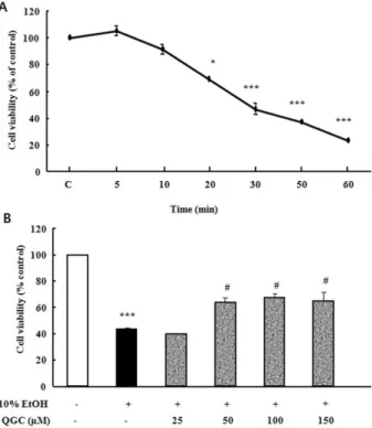
Fig. 1. Time course analysis for the effect of 10% ethanol in the absence or presence of QGC on the viability of cultured EECs. (A) Serum-starved EECs were stimulated with ethanol for the indicated times and their survival was estimated using the MTT assay. (B) Serum-starved EECs were pre-incubated in the presence of QGC for 12 h at indicated concentrations. EECs were then stimulated with 10% ethanol for 30 min and their survival was estimated using MTT assay. Data are expressed as means±SEM of three experiments. Student’s t-test; *p<0.05, ***p<0.001 vs.
control, #p<0.05 vs. cells in 10% ethanol alone.
Fig. 2. Effect of QGC on intracellular H2O2 levels induced by ethanol in EECs. Serum-starved EECs were pre-incubated in the presence of 50μM QGC for 4 h. EECs were then stimulated with ethanol for 10 min and ROS production was estimated using DCF-DA assay. Data are expressed as means±SEM of three experiments. Student’s t-test; *p<0.05 vs. control, ##p<0.001 vs.
cells in 10% ethanol alone.
RESULTS
Time-course analysis for the effect of 10% ethanol on cell viability
To assess whether 10% ethanol causes cell death in cul- tured EECs, serum starved cells were exposed to 10% etha- nol for the indicated times and then cell viability was meas- ured using MTT assay (Fig. 1A). There was no significant decrease in cell viability of cultured EECs incubated in ethanol for 10 min compared to control. After exposure to ethanol for 10 min, however, cell viability was decreased to about 70%. In addition, exposure of cells to ethanol for 30 min led to less than 50% cell viability.
Concentration-related protective effect of QGC against the ethanol-induced cytotoxicity
In a previous study, we have reported the chemical struc- ture of QGC compared to quercetin [38]. QGC alone did not exhibit any significant cytotoxicity on EECs at various concentration of 25∼200μM when incubated for 24 h (data not shown). In this time, to assess the cytoprotective effect of QGC against 10% ethanol-induced cell damage, serum starved cells were pre-incubated with 25∼150μM QGC for 12 h and then exposed to 10% ethanol for 30 min, followed
by measurement of cell viability (Fig. 1B). Ethanol treat- ment alone for 30 min caused a significant decrease to about 45% in cell viability. When cells were pretreated with 50∼150μM QGC for 12 h, the viability of cells exposed to ethanol significantly increased to 68%.
Effect of QGC on intracellular H2O2 levels
Serum starved EECs were pre-incubated with the pres- ence of 50μM QGC for 4 h. Cells were then stimulated with the 10% ethanol for 10 min and intracellular ROS pro- duction was estimated using DCF-DA (Fig. 2). 10% ethanol alone induced significant increase in intracellular ROS pro- duction by 135.3% vs. control. However, when cells were pretreated with 50μM QGC for 4 h, the ROS levels were significantly decreased below about 45%.
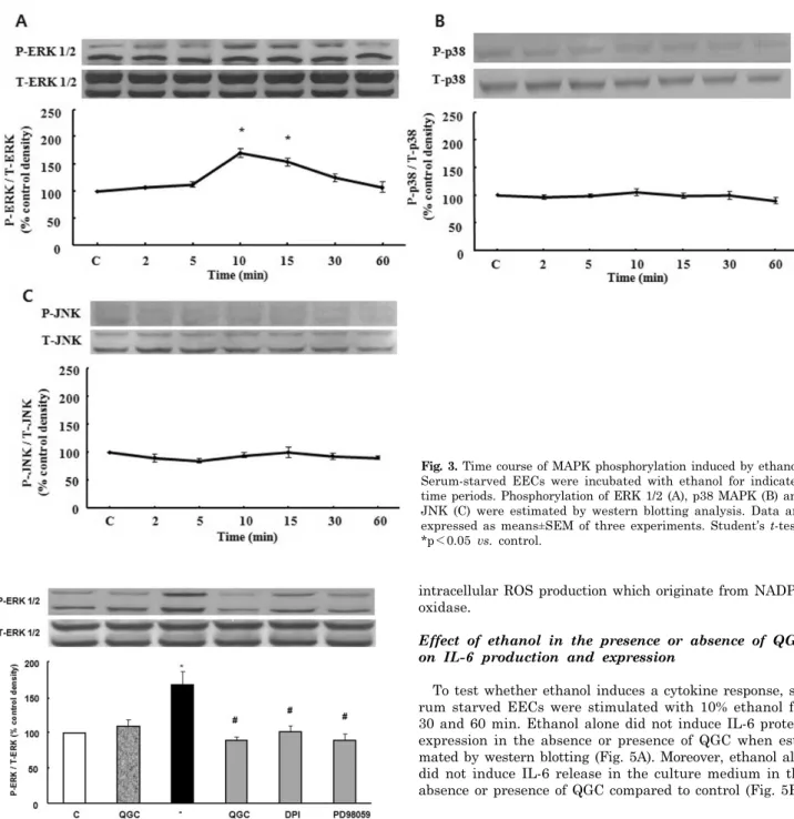
Ethanol-induced MAPK activation
To test whether ethanol induces activation of MAPK, se- rum starved cells were treated with 10% ethanol at the in- dicated time periods. Ethanol induced the activation of ERK 1/2, which reached maximally at 10 min (Fig. 3A).
Longer stimulation with 10% ethanol, only slightly reduced ERK 1/2 phosphorylation. As shown in Fig. 3B, phosphor- ylation of p38 MAPK did not cause significant changes after treatment of ethanol. Similarly, JNK was also not changed (Fig. 3C).
Effect of QGC and diphenylene iodonium (DPI) on ethanol-induced ERK 1/ 2 activation
Next, serum starved EECs were pre-incubated in the presence of 50μM QGC for 12 h and then stimulated with 10% ethanol for 10 min. Pretreatment with QGC inhibited the expression of 10% ethanol-induced ERK 1/2 phos- phorylation. To further clarify whether intracellular ROS production is involved in 10% ethanol-induced ERK 1/2 ac- tivation, DPI was utilized. Serum starved cells were pre- treated with 10μM DPI for 30 min and then exposed to
Fig. 3. Time course of MAPK phosphorylation induced by ethanol.
Serum-starved EECs were incubated with ethanol for indicated time periods. Phosphorylation of ERK 1/2 (A), p38 MAPK (B) and JNK (C) were estimated by western blotting analysis. Data are expressed as means±SEM of three experiments. Student’s t-test;
*p<0.05 vs. control.
Fig. 4. Effects of QGC and DPI on ethanol-induced ERK 1/2 phos- phorylation. Serum-starved EECs were incubated with QGC (50μM, 12 h), DPI (a NADPH oxidase inhibitor, 10μM, 0.5 h), PD98059 (a MEK inhibitor, 30μM, 1 h) prior to ethanol treatment for 10 min. Phosphorylation of ERK 1/2 was estimated by western blot analysis. Data are expressed as means±SEM of three experiments.
Student’s t-test; *p<0.05 vs. control, #p<0.05 vs. cells in 10%
ethanol alone.
10% ethanol for 10 min. DPI blocked ethanol-induced acti- vation of ERK 1/2 (Fig. 4). These results indicated that the inhibitory effect of QGC on ERK 1/2 activation by ethanol treatment may be mediated by reducing ethanol-induced
intracellular ROS production which originate from NADPH oxidase.
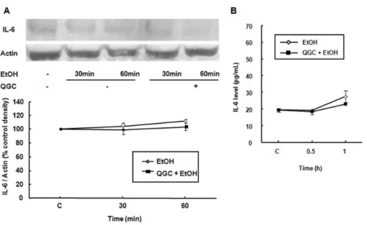
Effect of ethanol in the presence or absence of QGC on IL-6 production and expression
To test whether ethanol induces a cytokine response, se- rum starved EECs were stimulated with 10% ethanol for 30 and 60 min. Ethanol alone did not induce IL-6 protein expression in the absence or presence of QGC when esti- mated by western blotting (Fig. 5A). Moreover, ethanol also did not induce IL-6 release in the culture medium in the absence or presence of QGC compared to control (Fig. 5B).
DISCUSSION
Alcohol has many effects on the esophagus and stomach, and changes in these two organs can significantly increase morbidity due to alcohol consumption [3]. Ethanol is related to inflammation of the esophagus and stomach [41]. Both acute and chronic alcohol consumption have severe effects on the structure and function of the entire gastrointestinal tract which results in a vicious cycle [42]. Ethanol intake injures the functional and structural integrity of the in- testinal mucosa [2]. Previous study showed that ethanol-in- duced cell injury was dependent on both the concentration of ethanol applied and the duration of exposure in rat gas- tric epithelial cells. 6∼10% of ethanol was used as a cyto- toxic agent [43]. In the present study, we also confirmed
Fig. 5. Effect of ethanol in the absence or presence of QGC on IL-6 protein expression or release in EECs. Serum-starved EECs were stimulated with ethanol for 30 and 60 min in the absence or presence of 50μM QGC for 12 h. (A) IL-6 ex- pression was estimated by western blot analysis. (B) IL-6 production in culture medium was estimated by an EIA kit. Data are expressed as means±SEM of three experiments.
that 10% ethanol induces cytotoxicity to cat EECs.
It is well known that flavonoids, which are natural prod- uct of plants, have antioxidative and the antiinflammatory effects [44]. Quercetin has already been reported that anti- oxidative and antiinflammatory flavonoid [45,46]. In our previous study, QGC, a flavonoid glycoside extracted from Rumex Aquaticus, also acts as a non-stressful and non-cyto- toxic antioxidant and antiinflammatory flavonoid in rat model [37]. Flavonoids are known as free radical scavengers and cytoprotective compounds [43], and exhibited pro- tection against H2O2-mediated cytotoxicity. It was known that antioxidants protect cells against ethanol-induced cy- totoxicity and apoptosis [47]. In our study, QGC also ex- hibited a protective effect against 10% ethanol which caused EEC death. We have already confirmed that QGC enhances antioxidant enzyme defense systems via HO-1 ex- pression and Nrf2 translocation involving both the ERK and PI3K-Akt pathways as well as partial involvement of PKC pathways in EEC [38]. Mice lacking functional HO-1 exhibit chronic inflammation and increased mortality after lipopolysaccharide challenge [48,49]. Overexpression of HO-1 in cells resulted in a marked reduction in injury and cytotoxicity induced by oxidative stress [50,51]. An HO-1 inhibitor, ZnPP significantly attenuated the cytoprotective effect of eupatilin, one of the pharmacologically active fla- vones derived from Artemisia plants, in indomethacin- in- duced cytotoxicity [52]. We presumed that the antioxidative effect mediated by QGC-induced HO-1 expression may con- tribute to cytoprotection of the ethanol-induced cytotoxicity.
In addition, we measured directly the intracellular ROS production induced by ethanol in the absence or presence of QGC in order to confirm the antioxidative effect of QGC.
Exposure to oxidant molecules from the environment, food or during pathologies can generate ROS. Overproduction of ROS can cause oxidative stress and disease [53]. Several studies have suggested that the generation of ROS may be involved in the pathogenesis of ethanol-induced gastric mu- cosal injury in vivo, or resulted in cell death of human colon cancer cell and neuronal cell [54-57]. Natural and synthetic antioxidants or induction of cellular antioxidant systems can modulate the adverse effects of oxidative stress [53].
Several previous studies have indicated that flavonoids ex- hibit potent down regulation of ROS generation [58,59]. In the present study, our results suggest that QGC acts as a scavenger of intracellular ROS generation induced by 10%
ethanol in EECs.
Ethanol exposure causes the depletion of glutathione (GSH) and the formation of ROS [60,61]. It has been shown that ROS causes deleterious effects by oxidizing important structures in the cells [62] or acting as a second messenger stimulating intracellular signaling pathways including MAPK [63]. Modulation of MAPK signaling pathway by ethanol is distinctive, depending on the cell type, ethanol concentration and duration of exposure [15]. Acute ex- posure to ethanol results in activation of ERK in astrocytes.
However, chronic ethanol treatment causes activation of ERK and p38 MAPK leading to increased synthesis of TNF [64]. Ethanol treatment of stromal osteoblasts increases the ROS associated with induction of NADPH oxidase (NOX) and downstream signaling cascades involving sustained ac- tivation of ERK [65]. Also, ROS produced by ethanol in liver Kupffer cells and stellate cells as well as in the lung occur, in part, through activation of NADPH oxidase [66-68].
Concordantly, our data also showed that ethanol induced ERK 1/2 activation via NADPH oxidase-derived ROS pro- duction and reduction of the ROS generation by QGC may contribute to inhibition of ethanol-induced ERK 1/2 activa- tion in EECs.
Alcohol consumption is associated with elevated serum levels of interleukins such as IL-1, IL-6, and IL-8 [25,69].
Enhanced production of IL-1β and IL-6 has been docu- mented in esophageal tissue of cats with experimental esophagitis. Mucosa from esophagitis patients has the high- est concentrations of IL-1β and IL-6, cytokines known to reduce esophageal muscle contractility [28,70]. However, in our study, 10% ethanol in the absence or presence of QGC did not elicit any changes in the level of IL-6 protein ex- pression or release. Although the ethanol did not induce any change of IL-6 level in our experimental condition, ethanol reduced cell viability (45% compared to control) and induced ERK 1/2 activation via intracellular H2O2 pro- duction. Conversely, co-treatment with QGC significantly
reversed the reduction of ethanol-induced cell viability from 45% (ethanol alone) to 68% (ethanol with QGC). Moreover, QGC exhibited inhibitory effect on the ROS-dependent ERK 1/2 activation induced by ethanol. Although the mechanism underlying the effect of QGC was not detail determined within this study, these phenomena indicate that QGC has cytoprotective effect against ethanol-induced cytotoxicity.
Just, the relationship between the cytoprotective effect by QGC and QGC-induced inhibitory effect on the ROS-de- pendent ERK 1/2 activation induced by ethanol should be explored via further study.
In conclusion, QGC reduce the 10% ethanol-induced cyto- toxicity, and inhibits the production of intracellular ROS and ERK 1/2 activation induced by ethanol. Whether or not QGC-induced inhibitory effect on the ROS-dependent ERK 1/2 activation induced by ethanol is involved in the cytopro- tective effect of QGC should be determined by further study.
ACKNOWLEDGEMENTS
This study was supported by a grant of the Traditional Korean Medicine R&D Project, Ministry for Health &
Welfare, Republic of Korea (B090062).
REFERENCES
1. Soleas GJ, Diamandis EP, Goldberg DM. Wine as a biological fluid: history, production, and role in disease prevention. J Clin Lab Anal. 1997;11:287-313.
2. Bjorkman DJ, Jessop LD. Effects of acute and chronic ethanol exposure on intestinal microvillus membrane lipid composition and fluidity. Alcohol Clin Exp Res. 1994;18:560-565.
3. Teyssen S, Singer MV. Alcohol-related diseases of the oesophagus and stomach. Best Pract Res Clin Gastroenterol.
2003;17:557-573.
4. Hogan WJ, Viegas de Andrade SR, Winship DH. Ethanol- induced acute esophageal motor dysfunction. J Appl Physiol.
1972;32:755-760.
5. Silver LS, Worner TM, Korsten MA. Esophageal function in chronic alcoholics. Am J Gastroenterol. 1986;81:423-427.
6. Halliwell B. Free radicals, antioxidants, and human disease:
curiosity, cause, or consequence? Lancet. 1994;344:721-724.
7. Aviram M. Review of human studies on oxidative damage and antioxidant protection related to cardiovascular diseases. Free Radic Res. 2000;33 Suppl:S85-S97.
8. de Magalhães JP, Church GM. Cells discover fire: employing reactive oxygen species in development and consequences for aging. Exp Gerontol. 2006;41:1-10.
9. Menon SG, Goswami PC. A redox cycle within the cell cycle:
ring in the old with the new. Oncogene. 2007;26:1101-1109.
10. Olyaee M, Sontag S, Salman W, Schnell T, Mobarhan S, Eiznhamer D, Keshavarzian A. Mucosal reactive oxygen species production in oesophagitis and Barrett's oesophagus. Gut.
1995;37:168-173.
11. Yamaguchi T, Yoshida N, Tomatsuri N, Takayama R, Katada K, Takagi T, Ichikawa H, Naito Y, Okanoue T, Yoshikawa T.
Cytokine-induced neutrophil accumulation in the pathogenesis of acute reflux esophagitis in rats. Int J Mol Med. 2005;16:
71-77.
12. Odeleye OE, Eskelson CD, Mufti SI, Watson RR. Vitamin E inhibition of lipid peroxidation and ethanol-mediated promotion of esophageal tumorigenesis. Nutr Cancer. 1992;17:223-234.
13. Stein HJ, Esplugues J, Whittle BJ, Bauerfeind P, Hinder RA, Blum AL. Direct cytotoxic effect of oxygen radicals on the gastric mucosa. Surgery. 1989;106:318-323.
14. Stein HJ, Hinder RA, Oosthuizen MM. Gastric mucosal injury
caused by hemorrhagic shock and reperfusion: protective role of the antioxidant glutathione. Surgery. 1990;108:467-473.
15. Aroor AR, Shukla SD. MAP kinase signaling in diverse effects of ethanol. Life Sci. 2004;74:2339-2364.
16. Chen J, Ishac EJ, Dent P, Kunos G, Gao B. Effects of ethanol on mitogen-activated protein kinase and stress-activated protein kinase cascades in normal and regenerating liver.
Biochem J. 1998;334:669-676.
17. Chang L, Karin M. Mammalian MAP kinase signalling cas- cades. Nature. 2001;410:37-40.
18. Shapiro L, Dinarello CA. Hyperosmotic stress as a stimulant for proinflammatory cytokine production. Exp Cell Res. 1997;
231:354-362.
19. Zu YL, Qi J, Gilchrist A, Fernandez GA, Vazquez-Abad D, Kreutzer DL, Huang CK, Sha'afi RI. p38 mitogen-activated protein kinase activation is required for human neutrophil function triggered by TNF-alpha or FMLP stimulation. J Immunol. 1998;160:1982-1989.
20. Ludwig S, Hoffmeyer A, Goebeler M, Kilian K, Häfner H, Neufeld B, Han J, Rapp UR. The stress inducer arsenite activates mitogen-activated protein kinases extracellular signal-regulated kinases 1 and 2 via a MAPK kinase 6/p38- dependent pathway. J Biol Chem. 1998;273:1917-1922.
21. Hanck C, Rossol S, Böcker U, Tokus M, Singer MV. Presence of plasma endotoxin is correlated with tumour necrosis factor receptor levels and disease activity in alcoholic cirrhosis.
Alcohol Alcohol. 1998;33:606-608.
22. Tilg H, Wilmer A, Vogel W, Herold M, Nölchen B, Judmaier G, Huber C. Serum levels of cytokines in chronic liver diseases.
Gastroenterology. 1992;103:264-274.
23. Tilg H, Vogel W, Wiedermann CJ, Shapiro L, Herold M, Judmaier G, Dinarello CA. Circulating interleukin-1 and tumor necrosis factor antagonists in liver disease. Hepatology. 1993;
18:1132-1138.
24. Yin M, Wheeler MD, Kono H, Bradford BU, Gallucci RM, Luster MI, Thurman RG. Essential role of tumor necrosis factor alpha in alcohol-induced liver injury in mice. Gastroenterology.
1999;117:942-952.
25. Das SK, Vasudevan DM. Alcohol-induced oxidative stress. Life Sci. 2007;81:177-187.
26. Cheng L, Cao W, Behar J, Biancani P, Harnett KM. Inflam- mation induced changes in arachidonic acid metabolism in cat LES circular muscle. Am J Physiol Gastrointest Liver Physiol.
2005;288:G787-G797.
27. Cheng L, Cao W, Fiocchi C, Behar J, Biancani P, Harnett KM.
Platelet-activating factor and prostaglandin E2 impair eso- phageal ACh release in experimental esophagitis. Am J Physiol Gastrointest Liver Physiol. 2005;289:G418-G428.
28. Cao W, Cheng L, Behar J, Fiocchi C, Biancani P, Harnett KM.
Proinflammatory cytokines alter/reduce esophageal circular muscle contraction in experimental cat esophagitis. Am J Physiol Gastrointest Liver Physiol. 2004;287:G1131-G1139.
29. Shen SC, Ko CH, Tseng SW, Tsai SH, Chen YC. Structurally related antitumor effects of flavanones in vitro and in vivo:
involvement of caspase 3 activation, p21 gene expression, and reactive oxygen species production. Toxicol Appl Pharmacol.
2004;197:84-95.
30. Ko CH, Shen SC, Lee TJ, Chen YC. Myricetin inhibits matrix metalloproteinase 2 protein expression and enzyme activity in colorectal carcinoma cells. Mol Cancer Ther. 2005;4:281-290.
31. Zayachkivska OS, Konturek SJ, Drozdowicz D, Konturek PC, Brzozowski T, Ghegotsky MR. Gastroprotective effects of flavonoids in plant extracts. J Physiol Pharmacol. 2005;56 Suppl 1:219-231.
32. Morikawa K, Nonaka M, Narahara M, Torii I, Kawaguchi K, Yoshikawa T, Kumazawa Y, Morikawa S. Inhibitory effect of quercetin on carrageenan-induced inflammation in rats. Life Sci. 2003;74:709-721.
33. Reiterer G, Toborek M, Hennig B. Quercetin protects against linoleic acid-induced porcine endothelial cell dysfunction. J Nutr. 2004;134:771-775.
34. Borska S, Gebarowska E, Wysocka T, Drag-Zalesińska M, Zabel
M. The effects of quercetin vs cisplatin on proliferation and the apoptotic process in A549 and SW1271 cell lines in in vitro conditions. Folia Morphol (Warsz). 2004;63:103-105.
35. Kahraman A, Erkasap N, Köken T, Serteser M, Aktepe F, Erkasap S. The antioxidative and antihistaminic properties of quercetin in ethanol-induced gastric lesions. Toxicology. 2003;
183:133-142.
36. Suzuki Y, Ishihara M, Segami T, Ito M. Anti-ulcer effects of antioxidants, quercetin, alpha-tocopherol, nifedipine and tetra- cycline in rats. Jpn J Pharmacol. 1998;78:435-441.
37. Min YS, Lee SE, Hong ST, Kim HS, Choi BC, Sim SS, Whang WK, Sohn UD. The Inhibitory Effect of Quercetin-3-O-beta- D-Glucuronopyranoside on Gastritis and Reflux Esophagitis in Rats. Korean J Physiol Pharmacol. 2009;13:295-300.
38. Kim JS, Song HJ, Ko SK, Whang WK, Sohn UD. Quercetin-3- O-beta-d-glucuronopyranoside (QGC)-induced HO-1 expression through ERK and PI3K activation in cultured feline esophageal epithelial cells. Fitoterapia. 2010;81:85-92.
39. Jürgenliemk G, Nahrstedt A. Phenolic compounds from Hyperi- cum perforatum. Planta Med. 2002;68:88-91.
40. Park SY, Sohn UD. Inhibitory effect of rosiglitazone on the acid-induced intracellular generation of hydrogen peroxide in cultured feline esophageal epithelial cells. Naunyn Schmiede- bergs Arch Pharmacol. 2011;383:191-201.
41. Franke A, Teyssen S, Singer MV. Alcohol-related diseases of the esophagus and stomach. Dig Dis. 2005;23:204-213.
42. Rajendram R, Preedy VR. Effect of alcohol consumption on the gut. Dig Dis. 2005;23:214-221.
43. Zhang J, Stanley RA, Adaim A, Melton LD, Skinner MA. Free radical scavenging and cytoprotective activities of phenolic antioxidants. Mol Nutr Food Res. 2006;50:996-1005.
44. Nijveldt RJ, van Nood E, van Hoorn DE, Boelens PG, van Norren K, van Leeuwen PA. Flavonoids: a review of probable mechanisms of action and potential applications. Am J Clin Nutr. 2001;74:418-425.
45. Chow JM, Shen SC, Huan SK, Lin HY, Chen YC. Quercetin, but not rutin and quercitrin, prevention of H2O2-induced apoptosis via anti-oxidant activity and heme oxygenase 1 gene expression in macrophages. Biochem Pharmacol. 2005;69:1839- 1851.
46. Wang L, Tu YC, Lian TW, Hung JT, Yen JH, Wu MJ. Dis- tinctive antioxidant and antiinflammatory effects of flavonols.
J Agric Food Chem. 2006;54:9798-9804.
47. Kaviarasan S, Ramamurthy N, Gunasekaran P, Varalakshmi E, Anuradha CV. Epigallocatechin-3-gallate(-)protects Chang liver cells against ethanol-induced cytotoxicity and apoptosis.
Basic Clin Pharmacol Toxicol. 2007;100:151-156.
48. Takahashi T, Morita K, Akagi R, Sassa S. Heme oxygenase-1:
a novel therapeutic target in oxidative tissue injuries. Curr Med Chem. 2004;11:1545-1561.
49. Zhou H, Lu F, Latham C, Zander DS, Visner GA. Heme oxygenase-1 expression in human lungs with cystic fibrosis and cytoprotective effects against Pseudomonas aeruginosa in vitro.
Am J Respir Crit Care Med. 2004;170:633-640.
50. Soares MP, Lin Y, Anrather J, Csizmadia E, Takigami K, Sato K, Grey ST, Colvin RB, Choi AM, Poss KD, Bach FH. Ex- pression of heme oxygenase-1 can determine cardiac xenograft survival. Nat Med. 1998;4:1073-1077.
51. Taillé C, El-Benna J, Lanone S, Dang MC, Ogier-Denis E, Aubier M, Boczkowski J. Induction of heme oxygenase-1 inhibits NAD(P)H oxidase activity by down-regulating cyto- chrome b558 expression via the reduction of heme availability.
J Biol Chem. 2004;279:28681-28688.
52. Song HJ, Shin CY, Oh TY, Sohn UD. The protective effect of eupatilin on indomethacin-induced cell damage in cultured feline ileal smooth muscle cells: involvement of HO-1 and ERK.
J Ethnopharmacol. 2008;118:94-101.
53. Gaté L, Paul J, Ba GN, Tew KD, Tapiero H. Oxidative stress induced in pathologies: the role of antioxidants. Biomed Phar-
macother. 1999;53:169-180.
54. Mizui T, Sato H, Hirose F, Doteuchi M. Effect of anti- peroxidative drugs on gastric damage induced by ethanol in rats. Life Sci. 1987;41:755-763.
55. Szelenyi I, Brune K. Possible role of oxygen free radicals in ethanol-induced gastric mucosal damage in rats. Dig Dis Sci.
1988;33:865-871.
56. Lee HO, Byun YJ, Cho KO, Kim SY, Lee SB, Kim HS, Kwon OJ, Jeong SW. GS28 protects neuronal cell death induced by hydrogen peroxide under glutathione-depleted condition. Korean J Physiol Pharmacol. 2011;15:149-156.
57. Lee YJ, Kim NY, Suh YA, Lee C. Involvement of ROS in curcumin-induced autophagic cell death. Korean J Physiol Pharmacol. 2011;15:1-7.
58. Kandaswami C, Middleton E Jr. Free radical scavenging and antioxidant activity of plant flavonoids. Adv Exp Med Biol.
1994;366:351-376.
59. Pietta PG. Flavonoids as antioxidants. J Nat Prod. 2000;63:
1035-1042.
60. Koch OR, Pani G, Borrello S, Colavitti R, Cravero A, Farrè S, Galeotti T. Oxidative stress and antioxidant defenses in ethanol-induced cell injury. Mol Aspects Med. 2004;25:191-198.
61. Li Y, Walker DW, King MA. Peroxide mediates ethanol-induced cytotoxicity in PC12 cells. Free Radic Biol Med. 2001;30:389- 392.
62. Adachi M, Ishii H. Role of mitochondria in alcoholic liver injury.
Free Radic Biol Med. 2002;32:487-491.
63. Li SY, Li Q, Shen JJ, Dong F, Sigmon VK, Liu Y, Ren J.
Attenuation of acetaldehyde-induced cell injury by overex- pression of aldehyde dehydrogenase-2 (ALDH2) transgene in human cardiac myocytes: role of MAP kinase signaling. J Mol Cell Cardiol. 2006;40:283-294.
64. Zima T, Kalousová M. Oxidative stress and signal transduction pathways in alcoholic liver disease. Alcohol Clin Exp Res.
2005;29(11 Suppl):110S-115S.
65. Chen JR, Shankar K, Nagarajan S, Badger TM, Ronis MJ.
Protective effects of estradiol on ethanol-induced bone loss involve inhibition of reactive oxygen species generation in osteoblasts and downstream activation of the extracellular signal-regulated kinase/signal transducer and activator of transcription 3/receptor activator of nuclear factor-kappaB ligand signaling cascade. J Pharmacol Exp Ther. 2008;324:
50-59.
66. Novitskiy G, Traore K, Wang L, Trush MA, Mezey E. Effects of ethanol and acetaldehyde on reactive oxygen species pro- duction in rat hepatic stellate cells. Alcohol Clin Exp Res.
2006;30:1429-1435.
67. Polikandriotis JA, Rupnow HL, Elms SC, Clempus RE, Campbell DJ, Sutliff RL, Brown LA, Guidot DM, Hart CM.
Chronic ethanol ingestion increases superoxide production and NADPH oxidase expression in the lung. Am J Respir Cell Mol Biol. 2006;34:314-319.
68. Thakur V, Pritchard MT, McMullen MR, Wang Q, Nagy LE.
Chronic ethanol feeding increases activation of NADPH oxidase by lipopolysaccharide in rat Kupffer cells: role of increased reactive oxygen in LPS-stimulated ERK1/2 activation and TNF-alpha production. J Leukoc Biol. 2006;79:1348-1356.
69. Jeong HJ, Hong SH, Park RK, An NH, Kim HM. Ethanol induces the production of cytokines via the Ca2+, MAP kinase, HIF-1alpha, and NF-kappaB pathway. Life Sci. 2005;77:2179- 2192.
70. Rieder F, Cheng L, Harnett KM, Chak A, Cooper GS, Isenberg G, Ray M, Katz JA, Catanzaro A, O'Shea R, Post AB, Wong R, Sivak MV, McCormick T, Phillips M, West GA, Willis JE, Biancani P, Fiocchi C. Gastroesophageal reflux disease-asso- ciated esophagitis induces endogenous cytokine production leading to motor abnormalities. Gastroenterology. 2007;132:
154-165.