• 검색 결과가 없습니다.

Morphological and Electrical Characteristics in Patient with Hypertrophic Cardiomyopathy: Quantitative Analysis of 864 Korean Cohort

N/A
N/A
Protected

Academic year: 2022

Share "Morphological and Electrical Characteristics in Patient with Hypertrophic Cardiomyopathy: Quantitative Analysis of 864 Korean Cohort"

Copied!
7
0
0

로드 중.... (전체 텍스트 보기)

전체 글

(1)

INTRODUCTION

The evaluation for hypertrophic cardiomyopathy (HCM) re- lies principally on echocardiography and electrocardiogra-

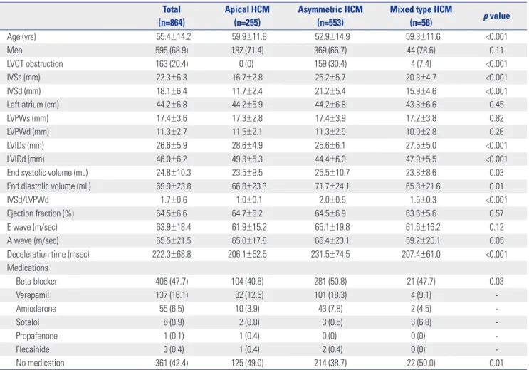
phy. Historically, the evolution of echocardiography has dem- onstrated the important morphological characteristics such as asymmetric pattern, systolic anterior motion of the mitral valve, obstructive form, and apical involvement.

1-4

Therefore, echocardiography is considered as the gold standard for iden- tifying the clinical phenotype of HCM.
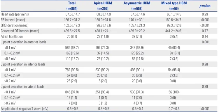
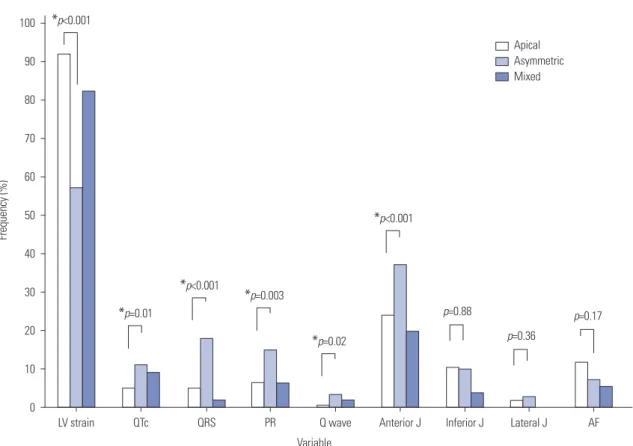
5

The electrocardiogra- phy also provides substantial information of HCM including mimic myocardial ischemia or infarction, and thereby creates diagnostic confusion.

6-9

However, most data were produced a long time ago and on the basis of relatively small population before the establishment of diagnostic consensus for HCM.

10,11

In addition, most data is about asymmetric HCM, while there are limited data on the apical HCM which was introduced lat- er and was a relatively rare form in Western country.

5,6,12,13

We, therefore, investigated the morphological and electrical char-

Morphological and Electrical Characteristics in Patient with Hypertrophic Cardiomyopathy:

Quantitative Analysis of 864 Korean Cohort

Sung-Hwan Kim

1

, Yong-Seog Oh

1

, Gi-Byoung Nam

2

, Kee-Joon Choi

2

, Dae Hee Kim

2

, Jong-Min Song

2

, Duk-Hyun Kang

2

, Jae-Kwan Song

2

, and You-Ho Kim

2

1

Division of Cardiology, Department of Internal Medicine, Seoul St. Mary’s Hospital, College of Medicine, The Catholic University of Korea, Seoul;

2

Department of Cardiology, University of Ulsan College of Medicine, Asan Medical Center, Seoul, Korea.

Purpose: Although several studies have reported the morphological and electrical characteristics in patients with hypertrophic cardiomyopathy (HCM), comparison between asymmetric and apical HCM has not been investigated in a reasonably sized cohort.

Materials and Methods: Echocardiography and electrocardiography were quantitatively analyzed in patients with HCM in a Ko- rean tertiary referral center.

Results: Of 864 patients (mean age 55.4±14.2 years, 68.9% men), 255 (29.5%) patients had apical HCM, 553 (64.0%) patients asym- metric HCM, and 56 (6.4%) patients mixed type HCM. In echocardiographic evaluations, about three quarters of patients (75.8%) had left atrial enlargement. Left ventricular (LV) dilatations and systolic dysfunction were observed in 6.1% and 2.4%, respective- ly. QRS widening, PR prolongation, and pathologic Q wave are frequent in patients with asymmetric HCM, while LV strain is fre- quent in patient with apical HCM. The prevalence of J-point elevations (9.4% in inferior, 2.2% in lateral leads) were substantially higher than that in general population. Giant negative T wave was observed in 15.0% of total patients (32.2% in apical, 6.2% in asym- metric, 25% in mixed type). There was no significant correlation between the thickness of the apical wall and the amplitude of T wave inversion (r=-0.005, p=0.71).

Conclusion: In a large cohort of HCM including apical type, repolarization abnormalities, including early repolarization and QT prolongation as well as LV strain, were significantly observed. T wave inversion was not appropriate for screening of HCM and not correlated with apical wall thickness.

Key Words: Hypertrophic cardiomyopathy, electrocardiography, echocardiography Yonsei Med J 2015 Nov;56(6):1515-1521

http://dx.doi.org/10.3349/ymj.2015.56.6.1515 pISSN: 0513-5796 · eISSN: 1976-2437

Received: July 10, 2014 Revised: December 4, 2014 Accepted: January 3, 2015

Corresponding author: Dr. Yong-Seog Oh, Division of Cardiology, Department of Internal Medicine, The Catholic University of Korea, Seoul St. Mary’s Hospital, 222 Banpo-daero, Seocho-gu, Seoul 06591, Korea.

Tel: 82-2-3779-1325, Fax: 82-2-3779-1374, E-mail: [email protected]

•The authors have no financial conflicts of interest.

© Copyright: Yonsei University College of Medicine 2015

This is an Open Access article distributed under the terms of the Creative Com- mons Attribution Non-Commercial License (http://creativecommons.org/ licenses/

by-nc/3.0) which permits unrestricted non-commercial use, distribution, and repro-

duction in any medium, provided the original work is properly cited.

(2)

acteristics in a large Korean cohort, and they were compared among three HCM groups (asymmetric, apical, and mixed). It should be noted that we had already reported long-term sur- vival between the two types of HCM.

14

MATERIALS AND METHODS

Our study enrolled patients with HCM who underwent echo- cardiography at a large tertiary referral center in Korea, be- tween March 1996 and November 2005. We excluded 93 pa- tients that had no electrocardiogram or otherwise unreadable traces. Thus, a total of 864 patients were enrolled. The diagno- sis of HCM was based on the presence of a hypertrophied left ventricle in the absence of other diseases capable of produc- ing the degree of observed hypertrophy. The patients were di- vided into three types (apical, asymmetric, and mixed) as an involved myocardium. The diagnostic criteria for apical HCM included demonstration of asymmetric left ventricular (LV) hypertrophy, confined predominantly to the LV apex with an apical wall thickness ≥15 mm and a ratio of maximal apical to posterior wall thickness ≥1.5, based on two-dimensional echo- cardiography.

15

Asymmetric HCM was diagnosed in case of a septal-to-posterior (or posterior-to septal) wall thickness ratio of equal to or greater than 1.3.

2,16,17

Mixed type HCM was de- fined when a patient had a coexistent apical and asymmetric HCM. The 12-lead electrocardiograms for each of the eligible Fig. 1. Proportion of hypertrophic cardiomyopathy type relative to pa-

tient’s age. In contrast to asymmetric hypertrophy, the proportion of pa- tients with apical hypertrophy increased with age.

30

20

10

0

Age (years)

–39 40–49 50–59 60–69 70–

13

20

26 27

15 Apical Asymmetric Mixed

Frequency (%)

Table 1. Demographic and Echocardiographic Characteristics Total (n=864)

Apical HCM (n=255)

Asymmetric HCM (n=553)

Mixed type HCM

(n=56) p value

Age (yrs) 55.4±14.2 59.9±11.8 52.9±14.9 59.3±11.6 <0.001

Men 595 (68.9) 182 (71.4) 369 (66.7) 44 (78.6) 0.11

LVOT obstruction 163 (20.4) 0 (0) 159 (30.4) 4 (7.4) <0.001

IVSs (mm) 22.3±6.3 16.7±2.8 25.2±5.7 20.3±4.7 <0.001

IVSd (mm) 18.1±6.4 11.7±2.4 21.2±5.4 15.9±4.6 <0.001

Left atrium (cm) 44.2±6.8 44.2±6.9 44.2±6.8 43.3±6.6 0.45

LVPWs (mm) 17.4±3.6 17.3±2.8 17.4±3.9 17.2±3.8 0.82

LVPWd (mm) 11.3±2.7 11.5±2.1 11.3±2.9 10.9±2.8 0.26

LVIDs (mm) 26.6±5.9 28.6±4.9 25.6±6.1 27.5±5.0 <0.001

LVIDd (mm) 46.0±6.2 49.3±5.3 44.4±6.0 47.9±5.5 <0.001

End systolic volume (mL) 24.8±10.3 23.5±9.5 25.5±10.7 23.8±8.6 0.03

End diastolic volume (mL) 69.9±23.8 66.8±23.3 71.7±24.1 65.8±21.6 0.01

IVSd/LVPWd 1.7±0.6 1.0±0.1 2.0±0.5 1.5±0.3 <0.001

Ejection fraction (%) 64.5±6.6 64.7±6.2 64.5±6.9 63.6±5.6 0.57

E wave (m/sec) 63.9±18.4 61.9±15.2 65.1±19.8 61.6±16.2 0.12

A wave (m/sec) 65.5±21.5 65.0±17.8 66.4±23.1 59.2±20.1 0.05

Deceleration time (msec) 222.3±68.8 206.1±52.5 231.5±74.5 207.4±61.0 <0.001

Medications

Beta blocker 406 (47.7) 104 (40.8) 281 (50.8) 21 (47.7) 0.03

Verapamil 137 (16.1) 32 (12.5) 101 (18.3) 4 (9.1) -

Amiodarone 55 (6.5) 10 (3.9) 43 (7.8) 2 (4.5) -

Sotalol 8 (0.9) 2 (0.8) 3 (0.5) 3 (6.8) -

Propafenone 1 (0.1) 1 (0.4) 0 (0) 0 (0) -

Flecainide 3 (0.4) 1 (0.4) 2 (0.4) 0 (0) -

No medication 361 (42.4) 125 (49.0) 214 (38.7) 22 (50.0) 0.01

HCM, hypertrophic cardiomyopathy; LVOT, left ventricular outflow tract; IVSd, interventricular septum end diastole; IVSs, interventricular septum end systole;

LVPWs, left ventricular posterior wall end systole; LVPWd, left ventricular posterior wall end diastole; LVIDs, left ventricular internal diameter end systole; LVIDd,

left ventricular internal diameter end diastole.

(3)

patients were obtained at the time of the initial evaluation for HCM. All electrocardiograms were retrospectively analyzed.

The electrocardiographic parameters included heart rate, PR duration, corrected QRS duration, the presence of atrial fibril- lation, the presence of pathologic Q wave, the presence of LV strain, the amplitude of T wave inversion in V4–6, and the am- plitude of J-point elevation. LV strain was defined as ST-T seg- ment vector shifted in direction opposite to the mean QRS complex.

18,19

Early repolarization patterns were stratified ac- cording to the degree of J-point elevation (<0.1 mV, 0.1–0.2 mV, or >0.2 mV) that was either notched or slurred in at least two consecutive anterior (V1–3), lateral (V4–6, I, aVL), or inferior (II, III, aVF) leads. A prolonged corrected QT interval was de- fined as at least 440 msec for men and at least 460 msec for women. A pathologic Q wave was defined as at least 25% or more of the height of the partner R wave and greater than 40 msec in width in at least two consecutive leads. The presence of atrial fibrillation was decided with the electrocardiography at the time of diagnosis of HCM. The electrocardiogram find- ings were compared among the three different HCM groups.

Categorical variables are presented as raw numbers and percentages, and were compared with the chi-square test or Fisher’s exact test. Continuous variables are presented as mean±standard deviation and were compared by Kruskal Wallis test. A probability value <0.05 was considered statisti- cally significant. SPSS for Windows (12.0, SPSS Inc., Chicago, IL, USA) was used for all statistical analyses. This study was approved by the Institutional Review Board at Asan Medical Center, Seoul, Korea.

RESULTS

Demographic findings

Baseline demographic and echocardiographic findings are summarized in Table 1, according to the type of HCM. Of 864 patients, 255 patients had apical HCM, 553 patients had asym- metric HCM, and 56 patients had mixed type HCM. The mean age was 55.4±14.2 years (from 12 to 92), and the patient with apical HCM was likely to be young compared to those with asymmetric HCM. The majority of patients were men. The proportion of patients with apical HCM increased in elder pa- tients compared with stationary proportion of the patient with asymmetric and mixed type HCM (Fig. 1). Beta blocker was prescribed in about a half of patients, while 42.4% of patients had no medication. The patients with verapamil were 16.1%.

Comparison of morphologic differences

30.4% of patients with asymmetric HCM had resting LV out- flow tract obstruction. About three quarters of patients (75.8%) had left atrial enlargement, defined as equal to or more than 40 mm of anterior-posterior diameter. LV dilatations, consid- ered as more than 55 mm of LV internal diameter in diastole, were observed in 6.1% of total patients. It was more frequent in patients with apical HCM (12.2% in apical HCM, 3.1% in asymmetric HCM, and 8.9% in mixed type HCM, p<0.001, by Kruskal Wallis test). The prevalence of LV dilatation was in- creased with age (6.1% in total). Mean ejection fraction was within normal range, and LV systolic dysfunction, defined as equal to or less than 50%, was observed in 2.4% of patients.

Table 2. Electrocardiographic Findings

Total (n=864)

Apical HCM (n=255)

Asymmetric HCM (n=553)

Mixed type HCM

(n=56) p value

Heart rate (per mins) 67.5±14.7 68.0±14.9 67.5±14.6 65.1±14.5 0.29

PR interval (msec) 166.7±31.2 160.0±31.6 170.4±30.1 160.6±34.7 <0.001

QRS duration (msec) 102.5±19.3 96.8±13.6 105.4±21.3 99.3±12.8 <0.001

Corrected QT interval (msec) 439.5±27.5 438.1±24.1 439.9±29.2 441.2±24.6 0.77

Atrial fibrillation 70 (8.1) 28 (11.0) 39 (7.1) 3 (5.4) 0.14

J point elevation in anterior leads 0.001

<0.1 mV 585 (67.7) 192 (75.3) 348 (62.9) 45 (80.4)

0.1–0.2 mV 169 (19.6) 37 (14.5) 123 (22.2) 9 (16.1)

>0.2 mV 110 (12.7) 26 (10.2) 82 (14.8) 2 (3.6)

J point elevation in inferior leads 0.28

<0.1 mV 782 (90.5) 230 (90.2) 498 (90.1) 54 (96.4)

0.1–0.2 mV 57 (6.6) 20 (7.8) 35 (6.3) 2 (3.6)

>0.2 mV 25 (2.9) 5 (2.0) 20 (3.6) 0 (0)

J point elevation in lateral leads 0.29

<0.1 mV 845 (97.8) 251 (98.4) 538 (97.3) 56 (100)

0.1–0.2 mV 12 (1.4) 1 (0.4) 11 (2.0) 0 (0)

>0.2 mV 7 (0.8) 3 (1.2) 4 (0.7) 0 (0)

Amplitude of negative T wave (mV) 0.6±0.5 0.8±0.5 0.5±0.4 0.7±0.5 <0.001

HCM, hypertrophic cardiomyopathy.

(4)

There were no difference of incidence according to the type of HCM. Deceleration time in patient with asymmetric HCM was longer than those with apical HCM. Prolonged decelera- tion time, considered as more than 220 msec, were prevalent in patient with asymmetric HCM (41.5% in total, 29.5% in api- cal HCM, 48.0% in asymmetric HCM, and 33.3% in mixed type HCM, p<0.001, by Kruskal Wallis test).

Comparisons of ECG findings

Electrocardiographic findings are summarized in Table 2. Pa- tients with asymmetric HCM were likely to have long PR in- terval and long QRS duration. Wide QRS (17.7% vs. 4.7%, p<0.001) and PR prolongation (14.8% vs. 6.8%, p=0.003) were more prevalent in patients with asymmetric HCM compared to those with apical HCM. The prevalence of atrial fibrillation at the diagnosis was 8.1% (11.0% in apical HCM, 7.1% in asym- metric HCM, p=0.17), and it increased in elder patients.

Early repolarization, considered as more than 0.1 mV of J- point elevation, was found in 32.3% of anterior leads, 9.5% of inferior leads, and 2.2% of lateral leads. Significant J-point ele- vations, defined as more than 0.2 mV, were found in 12.7% (an- terior leads), 2.9% (inferior leads), and 0.8% (lateral leads) of patients.

Repolarization abnormalities

Patients with apical HCM were likely to have LV strain and deep negative T wave (Figs. 2 and 3). Giant negative T wave, de- fined as a negative T wave with depth ≥1 mV, was observed in 15.0% of total patients (32.2% in apical HCM, 6.2% in asym- metric HCM, 25% in mixed type HCM). In patients with apical HCM, there was no significant correlation between the thick- ness of the apical wall and the amplitude of T wave inversion (r=-0.005, p=0.71) (Fig. 4). Abnormal findings of electrocardi- Fig. 2. Electrocardiographic abnormalities according to the type of hypertrophy. *Advanced electrical abnormalities such as QT prolongation, QRS widening, PR prolongation, and pathologic Q wave were more prevalent in patients with asymmetric hypertrophy. LV, left ventricle; QTc, corrected QT duration ≥480 msec; QRS, QRS duration ≥120 msec; PR, PR interval ≥200 msec; Q wave, pathologic Q wave; anterior J, J point in anterior leads ≥0.1 mV; inferior J, J point in inferior leads ≥0.1 mV; lateral J, J point in lateral leads ≥0.1 mV; AF, atrial fibrillation.

Apical Asymmetric Mixed 100

90 80 70 60 50 40 30 20 10

0

Variable

LV strain QTc QRS PR Q wave Anterior J Inferior J Lateral J AF

Frequency (%)

* p<0.001

* p=0.01

* p<0.001

* p=0.003

* p=0.02

* p<0.001

p=0.88

p=0.36

p=0.17

Fig. 3. The amplitude T wave inversion according to the type of hyper- trophic cardiomyopathy. HCM, hypertrophic cardiomyopathy.

100 80 60 40 20 0

Type of HCM

Apical Asymmetric Mixed

<0.1 mV

≥0.1 mV, <0.5 mV

≥0.5 mV, <1 mV

≥1 mV

Frequency (%)

(5)

ography were included the pattern of LV strain, longer than 480 msec of corrected QT interval, longer than 120 msec of PR interval, the presence of atrial fibrillation, the presence of LV strain, and more than 0.2 mV of the elevation of J-point. Their frequencies of abnormal findings according to the type of HCM are shown in Fig. 2. LV strain and QT prolongation were frequently observed in patients with HCM (68.9% and 52.5%, respectively).

DISCUSSION

Major findings

In 864 Korean cohort of patients with HCM, significant elec- trical as well as morphological abnormalities were found. The major findings on electrical abnormalities were as follows; 1) more than half of patients had a repolarization abnormality presenting LV strain and QT prolongation, 2) giant negative T wave were observed in 15.0% of total patients, mostly patients with apical HCM, 3) the prevalence of wide QRS and PR pro- longation were not infrequent, especially in patients with asymmetric HCM.

Echocardiographic evaluation revealed that the prevalence of apical HCM was increased with age until seventh decade, and substantial portion of patients had left atrial enlargement (75.8%) and deceleration time prolongation (41.5%), suggest- ing diastolic dysfunction.

Comparison with previous studies

HCM is a complex and relatively common genetic cardiac disorder.

20

Although the majority of patients are asymptomat-

ic, that is allowed to delayed diagnosis, some present with se- vere symptoms and some may even die suddenly because of fatal ventricular arrhythmia. LV obstruction by asymmetrical septal hypertrophy is the first discovered and a very specific finding in HCM.

21,22

Therefore, most morphological and elec- trical observations for HCM have been based on the popula- tion with these findings.

6,7,9,11,23,24

However, the evolution of echocardiography demonstrated heterogeneous clinical phe- notypes such as non-obstruction form and apical hypertro- phy,

1,4

and established the diagnosis of HCM.

5,11,25

However, there is a little data about morphological and electrical char- acteristics since the consensus for diagnosis and manage- ment of HCM were established. Especially, the differences of asymmetric HCM and apical HCM are hardly known.

In the demographic results, the patients with apical HCM were likely to be old compared to those with asymmetric HCM, and their prevalence was increased with age (Fig. 1). It is quite possible that almost all patients with apical HCM had no ob- struction of the LV outflow tract and relatively benign progno- sis. Although it was possible to be under-diagnosed in women of such a tertiary referral population, the majority of patients were men, in consistance with previous reports, especially in patients with apical HCM.

15,26

In the echocardiographic re- sults, substantial portion of patients were likely to have dia- stolic dysfunction, and it was consistent with previous data.

27,28

In other word, systolic dysfunction suggesting burned out phase was rare at the time of diagnosis.

17,29

With age, the prev- alence of LV dilatation was increased, while those of LV ob- struction was decreased. Although these findings were de- rived from cross-sectional observation, it was supported by the previous reports for progressive LV remodeling in patients with HCM.

29-31

There are some uncertainties over medical treatment of the patients with HCM. While 40% of patients had no medication and near half of patients were prescribed beta blocker in our routine practice, it was highly dependent on individual situa- tions such as symptom, compliance, and physician’s discretion, which are not described in the present study. It is one of the limitations of the present study. However, the initiation and maintenance of pharmacologic treatment are largely subject- ed to patient’s compliance and physician’s discretion. Symp- tom and response for medication are highly variable in terms of magnitude and duration of benefit. Therefore, it is difficult to objectively categorize patient’s symptom and medication.

Repolarization abnormalities in HCM

Various electrocardiographic abnormalities were found in our study. They included LV strain, QT prolongation, QRS prolon- gation, PR prolongation, atrial fibrillation, and J point elevation more than 0.2 mV in anterior leads, and pathologic Q wave.

More than half of patients showed repolarization abnor- mality like LV strain (68.9%) and QT prolongation (52.5%). Re- versal of the sequence of depolarization secondary to severe Fig. 4. Correlation between apical wall thickness and the amplitude of T

wave inversion in patients with apical hypertrophy. No significant cor- relation was observed.

3

2

1

0

r=-0.005, p=0.71

0 15 20 25 30

The amplitude of T wave inversion (mV)

The thickness of apical wall (mm)

(6)

and localized LV hypertrophy was considered as mechanism of LV strain.

32

LV strain was defined as ST-T segment vector shifted in direction opposite to the mean QRS complex in the present study, and it is similar to T wave inversion in other studies. It was reported that T wave inversion in the lateral leads is seen commonly in HCM. In the asymptomatic young athletes with HCM, 62% exhibited T wave inversion.

33

Similar- ly, in HCM patients with a positive genetic test, 54% demon- strate T wave inversion.

34

Our study confirmed that T wave in- version was common in patients with HCM and demonstrated that its prevalence was different as the type of HCM. Giant negative T wave was considered as classic electrocardiograph- ic finding of HCM. However, it seemed not to be sensitive pa- rameter of HCM, since only 14.8% of our patients showed gi- ant negative T wave. No significant relationship between apical wall thickness and the degree of T wave inversion was found in the present study, in agreement with previous re- port.

35

Other ECG abnormalities in patients with HCM Recently, early repolarization has received attention for asso- ciation with sudden cardiac death.

36

Quantitative analysis showed that significant J wave elevation, more than 2 mV, was more frequently observed in patients with HCM (2.9% in infe- rior leads, 0.8% in lateral leads) than that previously reported in general population (0.3% in inferior leads, 0.3% in lateral leads).

36

Clinical significance of repolarization abnormality in patients with HCM should be clarified in further longitudinal study. The prevalence of atrial fibrillation in our population was 8.1% (11.0% in apical, 7.1% in asymmetric, 5.4% in mixed type, mean age 55.4 years), much higher than that in Eastern general populations including Korean (0.7%, mean age 54.6 years),

37

Japanese (1.6%, mean age 72.1 years),

38

Hong Kong (1.3%, mean age 71 years)

39

as well as in Western general popu- lation (0.95%, mean age 71 years).

40

And it is comparable with that in Western HCM population (5% at the time of diagnosis, mean age 55 years).

41

Atrial fibrillation in patients with HCM was challenging and associated with substantial risk for mor- tality and morbidity.

41

In our results, only persistent atrial fi- brillation might be detected, indicating an another limitation of our study: the prevalence of atrial fibrillation could be high- er, about three times in a previous study, if paroxysmal atrial fibrillation was included.

41

Study limitation

Our results had several limitations. First, we could not evalu- ate genetic analysis or magnetic resonance imaging for diag- nostic test. Second, this study was based on the database of a tertiary referral center, and a community-based study is war- ranted to avoid referral bias.

Conclusion

This large Korean cohort of HCM including apical type dem-

onstrated that repolarization abnormalities including early repolarization and QT prolongation as well as LV strain were significantly observed. T wave inversion was not appropriate for screening of HCM and not correlated with apical wall thickness.

ACKNOWLEDGEMENTS

This study was supported by a grant of the Korea Healthcare technology R&D Project, Ministry for Health, Welfare & Fami- ly Affairs, Republic of Korea (A100909).

REFERENCES

1. Abbasi AS, MacAlpin RN, Eber LM, Pearce ML. Echocardiograph- ic diagnosis of idiopathic hypertrophic cardiomyopathy without outflow obstruction. Circulation 1972;46:897-904.

2. Henry WL, Clark CE, Epstein SE. Asymmetric septal hypertrophy.

Echocardiographic identification of the pathognomonic anatom- ic abnormality of IHSS. Circulation 1973;47:225-33.

3. Pridie RB, Oakley C. Mitral valve movement in hypertrophic ob- structive cardiomyopathy. Br Heart J 1969;31:390.

4. Sakamoto T, Tei C, Murayama M, Ichiyasu H, Hada Y. Giant T wave inversion as a manifestation of asymmetrical apical hyper- trophy (AAH) of the left ventricle. Echocardiographic and ultraso- no-cardiotomographic study. Jpn Heart J 1976;17:611-29.

5. Klues HG, Schiffers A, Maron BJ. Phenotypic spectrum and pat- terns of left ventricular hypertrophy in hypertrophic cardiomyop- athy: morphologic observations and significance as assessed by two-dimensional echocardiography in 600 patients. J Am Coll Cardiol 1995;26:1699-708.

6. Maron BJ, Wolfson JK, Ciró E, Spirito P. Relation of electrocardio- graphic abnormalities and patterns of left ventricular hypertro- phy identified by 2-dimensional echocardiography in patients with hypertrophic cardiomyopathy. Am J Cardiol 1983;51:189-94.

7. Savage DD, Seides SF, Clark CE, Henry WL, Maron BJ, Robinson FC, et al. Electrocardiographic findings in patients with obstruc- tive and nonobstructive hypertrophic cardiomyopathy. Circula- tion 1978;58(3 Pt 1):402-8.

8. Prescott R, Quinn JS, Littmann D. Electrocardiographic changes in hypertrophic subaortic stenosis which simulate myocardial in- farction. Am Heart J 1963;66:42-8.

9. Cosio FG, Moro C, Alonso M, de la Calzada CS, Liovet A. The Q waves hypertrophic cardiomyopathy: an electrophysiologic study.

N Engl J Med 1980;302:96-9.

10. Maron BJ, Maron MS, Wigle ED, Braunwald E. The 50-year histo- ry, controversy, and clinical implications of left ventricular outflow tract obstruction in hypertrophic cardiomyopathy from idiopath- ic hypertrophic subaortic stenosis to hypertrophic cardiomyopa- thy: from idiopathic hypertrophic subaortic stenosis to hypertro- phic cardiomyopathy. J Am Coll Cardiol 2009;54:191-200.

11. Maron BJ, McKenna WJ, Danielson GK, Kappenberger LJ, Kuhn HJ, Seidman CE, et al. American College of Cardiology/European Society of Cardiology clinical expert consensus document on hy- pertrophic cardiomyopathy. A report of the American College of Cardiology Foundation Task Force on Clinical Expert Consensus Documents and the European Society of Cardiology Committee for Practice Guidelines. J Am Coll Cardiol 2003;42:1687-713.

12. Yamaguchi H, Ishimura T, Nishiyama S, Nagasaki F, Nakanishi S,

Takatsu F, et al. Hypertrophic nonobstructive cardiomyopathy

(7)

with giant negative T waves (apical hypertrophy): ventriculograph- ic and echocardiographic features in 30 patients. Am J Cardiol 1979;44:401-12.

13. Chikamori T, Doi YL, Akizawa M, Yonezawa Y, Ozawa T, McKenna WJ. Comparison of clinical, morphological, and prognostic fea- tures in hypertrophic cardiomyopathy between Japanese and western patients. Clin Cardiol 1992;15:833-7.

14. Kim SH, Kim SO, Han S, Hwang KW, Lee CW, Nam GB, et al. Long- term comparison of apical versus asymmetric hypertrophic car- diomyopathy. Int Heart J 2013;54:207-11.

15. Eriksson MJ, Sonnenberg B, Woo A, Rakowski P, Parker TG, Wigle ED, et al. Long-term outcome in patients with apical hypertrophic cardiomyopathy. J Am Coll Cardiol 2002;39:638-45.

16. Otto CM. The practice of clinical echocardiography. 2nd ed. Phil- adelphia: W.B. Saunders; 2002. p.589.

17. Feigenbaum H, Armstrong WF, Ryan T. Feigenbaum’s echocar- diography. 6th ed. Philadelphia: Lippincott Williams & Wilkins;

2005. p.545.

18. Romhilt DW, Bove KE, Norris RJ, Conyers E, Conradi S, Rowlands DT, et al. A critical appraisal of the electrocardiographic criteria for the diagnosis of left ventricular hypertrophy. Circulation 1969;40:185-95.

19. Wagner GS, Marriott HJL. Marriott’s practical electrocardiography.

10th ed. Philadelphia: Lippincott Williams & Wilkins; 2001. p.90-3.

20. Maron BJ, Gardin JM, Flack JM, Gidding SS, Kurosaki TT, Bild DE.

Prevalence of hypertrophic cardiomyopathy in a general popula- tion of young adults. Echocardiographic analysis of 4111 subjects in the CARDIA Study. Coronary Artery Risk Development in (Young) Adults. Circulation 1995;92:785-9.

21. Brock R. Functional obstruction of the left ventricle; acquired aor- tic subvalvar stenosis. Guys Hosp Rep 1957;106:221-38.

22. Teare D. Asymmetrical hypertrophy of the heart in young adults.

Br Heart J 1958;20:1-8.

23. Braunmald E, Lambrew CT, Rockoff SD, Ross J Jr, Morrow AG. Id- iopathic hypertrophic subaortic stenosis. I. A description of the disease based upon an analysis of 64 patients. Circulation 1964;

30(Suppl 4):3-119.

24. Maron BJ, Bonow RO, Cannon RO 3rd, Leon MB, Epstein SE. Hy- pertrophic cardiomyopathy. Interrelations of clinical manifesta- tions, pathophysiology, and therapy (2). N Engl J Med 1987;316:

844-52.

25. Maron BJ. Hypertrophic cardiomyopathy: a systematic review.

JAMA 2002;287:1308-20.

26. Louie EK, Maron BJ. Apical hypertrophic cardiomyopathy: clini- cal and two-dimensional echocardiographic assessment. Ann In- tern Med 1987;106:663-70.

27. Ho CY, Sweitzer NK, McDonough B, Maron BJ, Casey SA, Seid- man JG, et al. Assessment of diastolic function with Doppler tissue imaging to predict genotype in preclinical hypertrophic cardio- myopathy. Circulation 2002;105:2992-7.

28. Maron BJ, Spirito P, Green KJ, Wesley YE, Bonow RO, Arce J. Non- invasive assessment of left ventricular diastolic function by pulsed Doppler echocardiography in patients with hypertrophic cardio- myopathy. J Am Coll Cardiol 1987;10:733-42.

29. Maron BJ, Spirito P. Implications of left ventricular remodeling in hypertrophic cardiomyopathy. Am J Cardiol 1998;81:1339-44.

30. Spirito P, Maron BJ, Bonow RO, Epstein SE. Occurrence and sig- nificance of progressive left ventricular wall thinning and relative cavity dilatation in hypertrophic cardiomyopathy. Am J Cardiol 1987;60:123-9.

31. Hina K, Kusachi S, Iwasaki K, Nogami K, Moritani H, Kita T, et al.

Progression of left ventricular enlargement in patients with hy- pertrophic cardiomyopathy: incidence and prognostic value. Clin Cardiol 1993;16:403-7.

32. Tsunakawa H, Wei D, Mashima S, Harumi K. Study on the genesis of giant negative T wave in apical hypertrophic cardiomyopathy using a three-dimensional computer model. Jpn Heart J 1991;32:

799-809.

33. Rowin EJ, Maron BJ, Appelbaum E, Link MS, Gibson CM, Lesser JR, et al. Significance of false negative electrocardiograms in pre- participation screening of athletes for hypertrophic cardiomyopa- thy. Am J Cardiol 2012;110:1027-32.

34. Lakdawala NK, Thune JJ, Maron BJ, Cirino AL, Havndrup O, Bund- gaard H, et al. Electrocardiographic features of sarcomere muta- tion carriers with and without clinically overt hypertrophic cardio- myopathy. Am J Cardiol 2011;108:1606-13.

35. Park SY, Park TH, Kim JH, Baek HK, Seo JM, Kim WJ, et al. Relation- ship between giant negative T-wave and severity of apical hypertro- phy in patients with apical hypertrophic cardiomyopathy. Echocar- diography 2010;27:770-6.

36. Tikkanen JT, Anttonen O, Junttila MJ, Aro AL, Kerola T, Rissanen HA, et al. Long-term outcome associated with early repolarization on electrocardiography. N Engl J Med 2009;361:2529-37.

37. Jeong JH. Prevalence of and risk factors for atrial fibrillation in Ko- rean adults older than 40 years. J Korean Med Sci 2005;20:26-30.

38. Iguchi Y, Kimura K, Aoki J, Kobayashi K, Terasawa Y, Sakai K, et al.

Prevalence of atrial fibrillation in community-dwelling Japanese aged 40 years or older in Japan: analysis of 41,436 non-employee residents in Kurashiki-city. Circ J 2008;72:909-13.

39. Lok NS, Lau CP. Prevalence of palpitations, cardiac arrhythmias and their associated risk factors in ambulant elderly. Int J Cardiol 1996;54:231-6.

40. Go AS, Hylek EM, Phillips KA, Chang Y, Henault LE, Selby JV, et al.

Prevalence of diagnosed atrial fibrillation in adults: national impli- cations for rhythm management and stroke prevention: the AnTi- coagulation and Risk Factors in Atrial Fibrillation (ATRIA) Study.

JAMA 2001;285:2370-5.

41. Olivotto I, Cecchi F, Casey SA, Dolara A, Traverse JH, Maron BJ.

Impact of atrial fibrillation on the clinical course of hypertrophic

cardiomyopathy. Circulation 2001;104:2517-24.

수치

Table 1. Demographic and Echocardiographic Characteristics Total  (n=864) Apical HCM(n=255) Asymmetric HCM(n=553) Mixed type HCM(n=56) p value Age (yrs) 55.4±14.2 59.9±11.8 52.9±14.9 59.3±11.6 &lt;0.001 Men 595 (68.9) 182 (71.4) 369 (66.7) 44 (78.6) 0.11 L
Table 2. Electrocardiographic Findings
Fig. 3. The amplitude T wave inversion according to the type of hyper- hyper-trophic cardiomyopathy

참조

관련 문서