Blockade of VEGFR-1 and VEGFR-2 Enhances Paclitaxel Sensitivity in Gastric Cancer Cells
Jun-Eul Hwang, Ji-Hee Lee, Mi-Ra Park, Dae-Eun Kim, Woo-Kyun Bae, Hyun-Jeong Shim, Sang-Hee Cho, and Ik-Joo Chung
Division of Hematology-Oncology, Department of Internal Medicine, Chonnam National University Hwasun Hospital, Hwasun, Korea.
Received: February 24, 2012 Revised: April 10, 2012 Accepted: April 23, 2012
Corresponding author: Dr. Ik-Joo Chung, Division of Hematology-Oncology, Department of Internal Medicine, Chonnam National University Hwasun Hospital, 322 Seoyang-ro, Hwasun-eup, Hwasun 519-763, Korea.
Tel: 82-61-379-7632, Fax: 82-61-379-7628 E-mail: [email protected]
∙ The authors have no financial conflicts of interest.
© Copyright:
Yonsei University College of Medicine 2013 This is an Open Access article distributed under the terms of the Creative Commons Attribution Non- Commercial License (http://creativecommons.org/
licenses/by-nc/3.0) which permits unrestricted non- commercial use, distribution, and reproduction in any medium, provided the original work is properly cited.
Purpose: Hypoxia-inducible factor-1α (HIF-1α) increases transcription of the vascular endothelial growth factor (VEGF) gene. Inhibition of VEGF abolishes VEGF mediated induction of HIF-1α. Recent reports suggested that HIF-1α also mediated the induction of class III β-tubulin (TUBB3) in hypoxia. TUBB3 con- fers resistance to taxanes. Inhibition of VEGF may decrease the expression of HIF-1α and TUBB3. This study was undertaken to investigate the roles of vascu- lar endothelial growth factor receptor (VEGFR) in gastric cancer cell behavior and to identify methods to overcome paclitaxel resistance in vitro. Materials and Methods: The protein expression levels of HIF-1α and TUBB3 were measured in human gastric cancer cell lines (AGS) under normoxic and hypoxic conditions.
The relationship between TUBB3 and paclitaxel resistance was assessed with small interfering TUBB3 RNA. AGS cells were treated with anti-VEGFR-1, anti- VEGFR-2, placental growth factor (PlGF), bevacizuamb, and paclitaxel. Results:
Hypoxia induced paclitaxel resistance was decreased by knockdown of TUBB3.
Induction of HIF-1α and TUBB3 in AGS is VEGFR-1 mediated and PlGF de- pendent. Hypoxia-dependent upregulation of HIF-1α and TUBB3 was reduced in response to paclitaxel treatment. Expressions of HIF-1α and TUBB3 were most decreased when AGS cells were treated with a combination of paclitaxel and anti- VEGFR-1. AGS cell cytotoxicity was most increased in response to paclitaxel, anti-VEGFR-1, and anti-VEGFR-2. Conclusion: We suggest that blockade of VEGFR-1 and VEGFR-2 enhances paclitaxel sensitivity in TUBB3-expressing gastric cancer cells.
Key Words: Vascular endothelial growth factor, Hypoxia-inducible factor 1 alpha, class III beta-tubulin, paclitaxel
INTRODUCTION
Although the incidence and mortality of gastric cancer have decreased in the West- ern world, gastric cancer persists as a common malignancy and leading cause of cancer-related death in Asian contries.1,2 Surgery is the only curative treatment for gastric cancer. Chemotherapy achieves favorable results for unresectable advanced
vival and overall response rate, but not associated with significant increases in overall survival.25 To date, we had insufficient evidences for the benefit of adding bevacizum- ab to chemotherapy in the treatment of gastric cancer.
Placental growth factor (PlGF) was discovered shortly after VEGF. Whereas VEGF binds both VEGFR-1 and VEGFR-2, PlGF binds to VEGFR-1 but not VEGFR-2. The key role of VEGF and its receptor VEGFR-2 in tumor an- giogenesis is firmly established, but the contribution of VEGFR-1 remains poorly defined.26 In the present study, we investigated the relationships between VEGF, HIF-1α, and TUBB3 in gastric cancer cells (AGS). We report that blockade of VEGFR-1 and VEGFR-2 with concomitant pa- clitaxel treatment increase the cell cytotoxicity of TUBB3- expressing gastric cancer cells.
MATERIALS AND METHODS
Drugs and reagents
Paclitaxel (BMS Pharmaceuticals, New York, NY, USA) and bevacizumab (Roche Pharmaceuticals, Basel, Switzer- land) were diluted in absolute DMSO. This solution was diluted to 0.1% DMSO on each day of experimentation.
The PlGF and VEGFR-1/2 neutralizing monoclonal anti- bodies were purchased from research and development systems.
Cell culture and hypoxic treatment
The human gastric cancer cell lines, AGS, SNU, and TMK1 were used as in vitro model systems. Cells were cultured in RPMI 1640 with 10% fetal bovine serum. For normoxia, cultures were incubated at 37°C in a humidified chamber of 5% CO2. For hypoxia, cells were treated with CoCl2. Confir- matory experiments under hypoxic conditions (1% O2/5%
CO2/94% N2) were performed using a Modular Incubator Chamber-101. Each hypoxia experiment was conducted under CoCl2-treated and true hypoxic conditions, and the results were equivalent.
siRNA
The TUBB3 siRNA duplex targeted a sequence 531 bases downstream of the TUBB3 start codon (siTUBB3: 5’-CAC GGTGGTGGAGCCCTACAA-3’). siRNA duplex oligonu- cleotides were purchased from QIAGEN (Gaithersburg, MD, USA) and transfected at 100 nM using Lipofectamine RNAiMAX Reagent (Invitrogen, Carlsbad, CA, USA).
and recurrent gastric cancers; however, the prognosis for these cancers is very poor.
Taxanes such as paclitaxel and docetaxel are a class of an- ticancer agents that bind to the β tubulin subunit of polymer- ized microtubules and induce hyperstabilization, which causes cell cycle arrest and apoptosis.3 Taxanes are used most commonly for the treatment of breast, lung, ovary, and gastric cancer. The taxanes paclitaxel and docetaxel exhibit similar efficacies in gastric cancer treatment.4-11
There are at least eight mammalian β tubulin isotypes, and the isotype composition in humans varies across tis- sues.12 Overexpression of class III β tubulin (TUBB3) has been observed in several human cancer cell lines such as prostate, ovarian, breast, and non small cell lung cancer.
Cells expressing TUBB3 show resistance to docetaxel and paclitaxel.12-15 A small cohort study of advanced gastric can- cer patients who were receiving preoperative docetaxel- based chemotherapy detected a correlation between expres- sion of TUBB3 and poor response to chemotherapy.16
Hypoxia in solid tumors is associated with resistance to chemotherapy, induction of angiogenesis, and poor patient prognosis, and angiogenesis is a hallmark of human malig- nancies. The induction of vascular endothelial growth factor (VEGF), mediated by interacting genetic and environmental signals, is an essential component of tumor angiogenesis.17 The transcription factor hypoxia-inducible factor-1α (HIF-1α) is a primary regulator of VEGF during hypoxic conditions, and Calvani, et al.18 reported that inhibition of VEGF or the VEGF receptor-2 (VEGFR-2), abolishes VEGF-mediated induction of HIF-1α. That is, HIF-1α in- creases VEGF induction, and VEGF also induces HIF-1α expression. A recent report suggested that HIF-1α also me- diates TUBB3 induction in hypoxia.19-21 Through these re- sults, we postulated that blockade of VEGF in gastric can- cer cells would be associated with a decrease in HIF-1α and TUBB3 expression and a concomitant increase in sensitivi- ty to paclitaxel.
The response of gastric cancer cells to anti-VEGF anti- bodies is of interest because bevacizumab, a humanized monoclonal antibody against VEGF, is used currently as an anti-angiogenic treatment for metastatic colorectal can- cer, non-squamous non-small cell lung cancer, and meta- static breast cancer.22-24 Recent the Avastin in Gastric Can- cer trial evaluated the efficacy of adding bevacizumab to chemotherapy in the first-line treatment of advanced gas- tric cancer. Adding bevacizumab to chemotherapy was as- sociated with significant increases in progression free sur-
containing assembled tubulin. Each fraction was separated by SDS-PAGE. Immunoblots then were performed using anti-pan-α-tubulin rabbit polyclonal antibody (Santa Cruz Biotechonology, Santa Cruz, CA, USA).
Immunoblotting
Cells were lysed in Mammalian Cell Lysis Reagent (Fer- mentas, Waltham, MA, USA), and approximately 30 μg protein was separated in 8-10% polyacrylamide gels and transferred to polyvinylidene difluoride membrane (Milli- pore, Billerica, MA, USA). The following antibodies were used: anti-HIF-1α and anti-TUBB3 (Abcam PLC, Cam- bridge, UK), anti-tubulin (Invitrogen, Carlsbad, CA, USA), β-actin (Santa Cruz Biotechnology, Santa Cruz, CA, USA).
Statistical analysis
The Student’s t-test was used to detect significant differenc- es among treatment groups. Statistical significance was as- signed at p<0.05.
RESULTS
Induction of HIF-1α, TUBB3 and VEGF in hypoxia The expression levels of TUBB3 and VEGF were measured during the induction of HIF-1α in hypoxic AGS. AGS exhib- ited well-defined hypoxic induction of HIF-1α, VEGF, and TUBB3 compared with other cell lines (SNU638, TMK1) (Fig. 1).
TUBB3 confers gastric cancer cells with resistance to paclitaxel
To assess whether TUBB3 expressions is associated with chemotherapeutic drug resistance in AGS, TUBB3 was knocked down with small interfering RNA (siTUBB3), and the effects of paclitaxel on AGS growth under normoxic and hypoxic conditions were evaluated over a 24-48 h period.
AGS was strongly resistant to paclitaxel during hypoxia.
Knockdown of TUBB3 decreased the resistance to paclitax- el during hypoxia, and cytotoxicity was increased (Fig. 2).
VEGFR-1 signals increase HIF-1α and TUBB3 expressions in AGS cells
After treatment with paclitaxel, cytoskeletal component were isolated from AGS cells, and tubulin polymerization was monitored. Western blots were performed using a pan α-tubulin polyclonal antibody to probe lysates from cells that had been treated with paclitaxel or paclitaxel and beva- Cell proliferation assay
To estimate viability, 105 cells were seeded in 24-well plates, subjected to normoxia or hypoxia, and exposed to neutral- izing antibodies in the culture media for 4 h. Cells then were treated with bevacizuamb and/or paclitaxel for 24 h. Cells were incubated for 24, 48, or 72 h, and EZ-CyTox (Daeil Lab Service, Seoul, Korea) was added to each well. After 3 h, absorbances at 420 nm were measured using a micro- plate reader (EL800, BioTek, Winooski, VT, USA).
Tubulin polymerization assay
Cells were treated with various concentrations of drugs for 24 h, washed twice with phosphate buffered saline, and lysed for 5 min in 100 μL hypotonic buffer [1 mM MgCl2, 2 mM EGTA, 0.5% NP40, 20 mM Tris-HCL (pH 6.8), prote- ase and phosphatase inhibitors]. Samples then were centri- fuged at 16000×g for 10 min at room temperature. Superna- tants, containing soluble tubulin, were separated from pellets,
Fig. 1. Induction of HIF-1α and its target genes, VEGF and TUBB3, in human gastric cancer cell lines under hypoxia. HIF-1α, VEGF and TUBB3 expres- sion levels were increased during hypoxia, compared to normoxia. Actin was used as a loading control. Band intensities were quantified by densi- tometry. HIF-1α, hypoxia inducible factor-1α; VEGF, vascular endothelial growth factor; TUBB3, class III β-tubulin.
Hypoxia 0
SNU638 AGS TMK1
4
8 0
4 0 8 4 8 h
HIF-1α
VEGF
TUBB3 β-actin
HIF-1α 23.14 63.81 135.84 25.11 84.3 120.39 39.48 133.25 143.84 VEGF 59.73 60.34 93.28 61.16 73.01 79.64 61.74 55.08 47.52 TUBB3 59.87 113.88 107.66 87.06 119.72 102.22 83.28 75.52 114.74
0 20 40 60 80 100 120 140 160
Intensity of each protein
HIF-1α VEGF TUBB3
SNU638 AGS TMK1
cizumab under normoxic conditions or after 24 h of hypox- ia. Paclitaxel activity was inhibited in hypoxia. Tubulin po- lymerization was decreased in hypoxia compared with normoxia and it was statistically significant (p<0.01). Hy- poxic decrease of polymerization was lessened after treated with bevacizumab (Fig. 3A).
As previously mentioned, we postulated that blockade of VEGF in AGS would be associated with a decrease in HIF- 1α and TUBB3 expression and a concomitant increase in sensitivity to paclitaxel. We treated cells with bevacizumab, anti-VEGFR-1 and anti-VEGFR-2 neutralizing antibodies to test this hypothesis. AGS cells were cultured under normoxic and hypoxic conditions for 24 h without exogenous growth factors, in the absence or presence of anti-VEGFR-1, anti- VEGFR-2, or bevacizumab with simultaneous paclitaxel treatment. HIF-1α and TUBB3 were increased in a hypoxic condition. Paclitaxel treatment decreased the hypoxia-depen- dent increased expression of HIF-1α and TUBB3. When pa- clitaxel was combined with bevacizumab or anti-VEGFR-2, expression levels of HIF-1α and TUBB3 were further de- creased. The greatest decrease in HIF-1α and TUBB3 levels occurred in response to paclitaxel and anti-VEGFR-1 treat- ment in AGS cells (Fig. 3B). Our results using AGS cells
Fig. 2. TUBB3 knockdown sensitizes gastric cancer cells to paclitaxel. (A) Western blot analysis of TUBB3 in AGS cells without treatment or in the presence of si-scramble control or si-TUBB3 RNAs. Actin was used as a loading control. (B) AGS cells were treated with si-scramble or si-TUBB.
Cells were maintained in normoxia or hypoxia for 24 h with paclitaxel. Cell proliferation was normalized to the normoxic control. Values are expressed as means±SEM (n=3). *p<0.05. NS, not significant. TUBB3, class III β-tubulin;
SEM, standard error of the mean.
Fig. 3. VEGFR-1/2 contributes to tubulin polymerization. (A) Western blots using a pan α-tubulin polyclonal antibody to probe cell lysates treated with pacli- taxel with or without bevacizumab for 24 h during normoxia or hypoxia. Paclitaxel activity was inhibited in hypoxia. Tubulin polymerization was decreased in hypoxia compared with normoxia and it was statistically significant. Cells were treated with paclitaxel with/without bevacizumab at 100 nM and 100 ug/mL, respectively. Lane 1: no drug, 2: paclitaxel, 3: paclitaxel with bevacizumab. Actin was used as a loading control. Band intensities were quantified by densi- tometry. (B) Hypoxia-dependent upregulation of HIF-1α and TUBB3 was most reversed in the presence of paclitaxel and anti-VEGFR-1 for 24 h. HIF-1α was measured from nuclear fractions, and TUBB3 was measured from total cell lysates. Band intensities were quantified by densitometry. *p<0.01. HIF-1α, hy- poxia inducible factor-1α; TUBB3, class III β-tubulin; VEGFR, vascular endothelial growth factor receptor.
Wild type si-scramble si-TUBB3-
24 hr si-TUBB3- 48 hr TUBB3
β-actin
0 0.2 0.4 0.6 0.8 1.0 1.2
Relative cell proliferation
No drug Paclitaxel Normoxia
No drug Paclitaxel Hypoxia
si-scramble si-TUBB3 NS
*
0 0 0
50 40
100 12080
150 160
200 200
20 40 60 80 100 120 140
Intensity of pellet α-tubulin Intensity of each protein
1 HIF-1α TUBB3 HIF-1α TUBB3
1
2 2
3 3
1 1
2 2
3 3
Normoxia AGS cells HCT116 cells
Normoxia Normoxia
Paclitaxel (100 nM) + + + + Bevacizumab (100 ug/mL) +
anti-VEGFR-2 (50 ng/mL) + anti-VEGFR-1 (50 ng/mL) +
Hypoxia
Hypoxia Hypoxia
* * *
Pellet α-tubulin
Soluble α-tubulin
HIF-1α
TUBB3 β-actin
β-actin
AGS cells
AGS cells HCT116 cells
HCT116 cells
A B
A
B
without anti-VEGFR-1, anti-VEGFR-2, and bevacizumab with simultaneous paclitaxel exposure. The cell viability as- say demonstrated that AGS cell cytotoxicity was increased in response to combined treatment with paclitaxel and bev- acizumab compared with paclitaxel treatment alone. Cyto- toxicity was increased synergistically in response to simul- taneous treatment with paclitaxel, anti-VEGFR-1, and anti- VEGFR-2 (p<0.05) (Fig. 5).
DISCUSSION
Bevacizumab is accepted as the standard treatment for ad- vanced metastatic colorectal cancer, but to date is not con- sidered as the standard treatment of gastric cancer.24 VEGF can bind with the receptor tyrosine kinases, VEGFR-1 and VEGFR-2. The key roles of VEGF and its receptor VEG- FR-2 in tumor angiogenesis have been established.26,27 Acti- vation of VEGFR-2 leads to auto-phosphorylation and the activation of downstream signaling pathways, such as Raf/
MEK/ERK and PI3K/Akt kinase cascades.25 In contrast, the function of VEGFR-1 remains poorly defined. VEG- FR-1 has been associated with tumor growth, tumor cell ac- contrast with results using HCT116 cells, in which hypoxia-
induced HIF-1α was inhibited via VEGFR-2.18
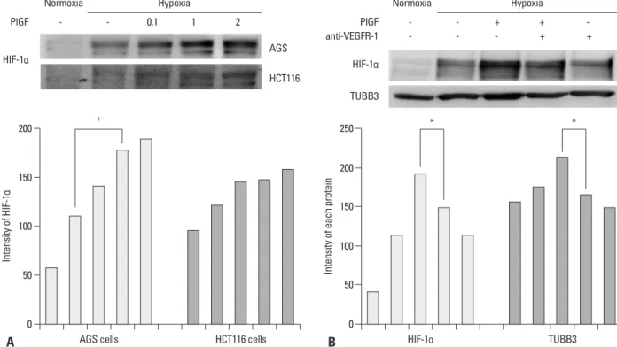
To examine VEGFR-1 function, we investigated whether the VEGFR-1 ligand, PlGF, influences the expression of HIF-1α in AGS and HCT116 cells. Cells were cultured un- der normoxic or hypoxic conditions in the presence of PlGF (range, 0.1-2.0 ng/mL). HIF-1α expression was in- creased in hypoxia and was dose dependently more in- creased according to the concentration of PlGF (Fig. 4A).
In contrast with AGS cells, PlGF rarely influenced HIF-1α expression in HCT116 cells. AGS cells were cultured under normoxic and hypoxic conditions in the absence or pres- ence of PlGF and anti-VEGFR-1. Expression levels of HIF-1α and TUBB3 were increased in hypoxia and were more increased in response to PlGF treatment. The in- creased expressions of HIF-1α and TUBB3 were suppressed when anti-VEGFR-1 antibodies were combined with PlGF treatment (Fig. 4B). We concluded that induction of HIF-1α in AGS is VEGFR-1 mediated and PlGF dependent.
Blockade of VEGFR-1/VEGFR-2 increases sensitivity to paclitaxel in AGS cells
AGS cells were cultured under hypoxic conditions with or
Fig. 4. PlGF induces HIF-1α. (A) Nuclear HIF-1α protein expression was increased in hypoxia and was dose dependently increased according to the in- creased concentration of PlGF (range 0.1-2 ng/mL) in AGS. PlGF rarely influenced HIF-1α expression in HCT116 cells. Band intensities were quantified by densitometry. (B) Expression levels of HIF-1α and TUBB3 were increased in hypoxia and were more increased with PlGF treatment (1 ng/mL) in AGS cells.
HIF-1α is measured from nuclear fractions, and TUBB3 is measured from total cell lysates. The observed increases in HIF-1α and TUBB3 expression levels were decreased when anti-VEGFR-1 was combined with PlGF treatment. Band intensities were quantified by densitometry. *p<0.05, †p<0.01. PlGF, placental growth factor; HIF-1α, hypoxia inducible factor-1α; TUBB3, class III β-tubulin; VEGFR, vascular endothelial growth factor receptor.
Normoxia Hypoxia Normoxia Hypoxia
HIF-1α HIF-1α
TUBB3
PlGF PlGF
anti-VEGFR-1
- - 0.1 1 2 - - + + - - - - + + AGS
HCT116
0 0
50 50
100
100
150 200
150
200 250
Intensity of HIF-1α Intensity of each protein
AGS cells HCT116 cells HIF-1α TUBB3
† * *
A B
sults, we suggest that AGS cells express functional VEGFR-1 and VEGFR-2.
TUBB3 confers chemoresistance to taxanes. Our experi- ment results of paclitaxel resistance in AGS cells are con- sistent with other reports suggesting that TUBB3 affects chemoresistance to taxanes in diverse cancer cells.12-16 Ras- paglio, et al.21 also reported that TUBB3 is not only a pa- rameter related to the chemoresponsiveness, but also could be a pure prognostic marker. TUBB3 expression levels dif- fer by cell and tissue types. In some tissues, TUBB3 is con- stitutively expressed and is not inducible upon hypoxia.21 In AGS cells, we observed TUBB3 was constitutively ex- pressed in normoxia and became more induced in hypoxia.
The present study demonstrated that blockade of both VEGFR-1 and VEGFR-2 in conjunction with paclitaxel synergistically depresses induction of HIF-1α and TUBB3 expression, and more effectively increases cytotoxicity in gastric cancer cells.
ACKNOWLEDGEMENTS
This study was supported by a grant (CRI11073-1) Chon- nam national university hospital research institute of clini- cal medicine.
REFERENCES
1. Alberts SR, Cervantes A, van de Velde CJ. Gastric cancer: epide- miology, pathology and treatment. Ann Oncol 2003;14 Suppl 2:ii31-6.
2. Won YJ, Sung J, Jung KW, Kong HJ, Park S, Shin HR, et al. Na- tionwide cancer incidence in Korea, 2003-2005. Cancer Res Treat 2009;41:122-31.
3. Hruban RH, Yardley JH, Donehower RC, Boitnott JK. Taxol tox- icity. Epithelial necrosis in the gastrointestinal tract associated with polymerized microtubule accumulation and mitotic arrest.
Cancer 1989;63:1944-50.
4. Ajani JA, Fodor MB, Tjulandin SA, Moiseyenko VM, Chao Y, Cabral Filho S, et al. Phase II multi-institutional randomized trial of docetaxel plus cisplatin with or without fluorouracil in patients with untreated, advanced gastric, or gastroesophageal adenocarci- noma. J Clin Oncol 2005;23:5660-7.
5. Constenla M, Garcia-Arroyo R, Lorenzo I, Carrete N, Campos B, Palacios P. Docetaxel, 5-fluorouracil, and leucovorin as treatment for advanced gastric cancer: results of a phase II study. Gastric Cancer 2002;5:142-7.
6. Kim YH, Shin SW, Kim BS, Kim JH, Kim JG, Mok YJ, et al. Pa- clitaxel, 5-fluorouracil, and cisplatin combination chemotherapy for the treatment of advanced gastric carcinoma. Cancer 1999;85:
295-301.
tivation, and metastasis. Treatment with VEGFR-1 inhibi- tors, such as anti-VEGFR-1 antibody, suppresses tumor growth and metastasis in various models.26 However, the degree of tumor growth inhibition varies. Some studies have reported that VEGFR-1 inhibition is not sufficient to block tumor growth without combined inhibition of VEGFR-2.28,29 In addition, it is unclear if VEGFR-1 inhibitors, by prevent- ing the binding of VEGF to VEGFR-1, increase the con- centration of free VEGF that can subsequently activate VEGFR-2 and stimulate angiogenesis by an alternate path- way. The precise mechanisms of VEGFR-1 inhibitor func- tioning warrant further exploration.26
We demonstrated that HIF-1α expressions increase in as- sociation with elevated PlGF. HIF-1α and TUBB3 were up- regulated in hypoxia and became more induced in response to PlGF treatment. We also demonstrated that inhibition of VEGFR-1 is associated with decreases in HIF-1α and TUBB3 expression. Calvani, et al.18 reported that VEGFR-2, rather than VEGFR-1, mediates VEGF-dependent induction of HIF-1α in hypoxic HCT116 colon cancer cells, suggesting that functional VEGFR-2, but not functional VEGFR-1, ex- ists in this colon cancer cell line. Cell viability assay results demonstrated that AGS cell cytotoxicity was most pro- nounced when cells were treated simultaneously with pacli- taxel, anti-VEGFR-1, and anti-VEGFR-2. Through these re-
Fig. 5. Blockade of VEGFR-1 and VEGFR-2 decreases resistance to pacli- taxel in AGS cells. The cell viability assay demonstrated that AGS cell cyto- toxicity was increased following combined treatment with paclitaxel and bevacizumab compared with treatment with paclitaxel alone. The increase of cytotoxicity was most pronounced when paclitaxel, anti-VEGFR-1, and anti-VEGFR-2 were applied simultaneously. Cells were maintained in nor- moxia or hypoxia for 24 h with paclitaxel following a 4-h pre-incubation with neutralizing antibody. Cell proliferation was normalized on the nor- moxic control. Values are expressed as means±SEM (n=3). *p<0.05.
VEGFR, vascular endothelial growth factor receptor; SEM, standard error of the mean.
0 0.2 0.4 0.6 0.8 1 1.2
Relative cell proliferation
Normoxia
+ +
+ +
++ +
+ +
+ ++ + Hypoxia
*
Paclitaxel (100 nM) anti-VEGFR-2 (50 ng/mL) anti-VEGFR-1 (50 ng/mL) Bevacizumab (100 ug/mL)
93-102.
18. Calvani M, Trisciuoglio D, Bergamaschi C, Shoemaker RH, Mel- illo G. Differential involvement of vascular endothelial growth factor in the survival of hypoxic colon cancer cells. Cancer Res 2008;68:285-91.
19. Kuwai T, Kitadai Y, Tanaka S, Onogawa S, Matsutani N, Kaio E, et al. Expression of hypoxia-inducible factor-1alpha is associated with tumor vascularization in human colorectal carcinoma. Int J Cancer 2003;105:176-81.
20. Pugh CW, Ratcliffe PJ. Regulation of angiogenesis by hypoxia:
role of the HIF system. Nat Med 2003;9:677-84.
21. Raspaglio G, Filippetti F, Prislei S, Penci R, De Maria I, Cicchi- llitti L, et al. Hypoxia induces class III beta-tubulin gene expres- sion by HIF-1alpha binding to its 3’ flanking region. Gene 2008;
409:100-8.
22. Hurwitz H, Fehrenbacher L, Novotny W, Cartwright T, Hain- sworth J, Heim W, et al. Bevacizumab plus irinotecan, fluoroura- cil, and leucovorin for metastatic colorectal cancer. N Engl J Med 2004;350:2335-42.
23. Miller K, Wang M, Gralow J, Dickler M, Cobleigh M, Perez EA, et al. Paclitaxel plus bevacizumab versus paclitaxel alone for met- astatic breast cancer. N Engl J Med 2007;357:2666-76.
24. Sandler A, Gray R, Perry MC, Brahmer J, Schiller JH, Dowlati A, et al. Paclitaxel-carboplatin alone or with bevacizumab for non- small-cell lung cancer. N Engl J Med 2006;355:2542-50.
25. Ohtsu A, Shah MA, Van Cutsem E, Rha SY, Sawaki A, Park SR, et al. Bevacizumab in combination with chemotherapy as first-line therapy in advanced gastric cancer: a randomized, double-blind, placebo-controlled phase III study. J Clin Oncol 2011;29:3968-76.
26. Fischer C, Mazzone M, Jonckx B, Carmeliet P. FLT1 and its li- gands VEGFB and PlGF: drug targets for anti-angiogenic thera- py? Nat Rev Cancer 2008;8:942-56.
27. Gerber HP, Ferrara N. Pharmacology and pharmacodynamics of bevacizumab as monotherapy or in combination with cytotoxic therapy in preclinical studies. Cancer Res 2005;65:671-80.
28. Gille J, Heidenreich R, Pinter A, Schmitz J, Boehme B, Hicklin DJ, et al. Simultaneous blockade of VEGFR-1 and VEGFR-2 ac- tivation is necessary to efficiently inhibit experimental melanoma growth and metastasis formation. Int J Cancer 2007;120:1899- 908.
29. Lyden D, Hattori K, Dias S, Costa C, Blaikie P, Butros L, et al.
Impaired recruitment of bone-marrow-derived endothelial and he- matopoietic precursor cells blocks tumor angiogenesis and growth. Nat Med 2001;7:1194-201.
7. Murad AM, Petroianu A, Guimaraes RC, Aragao BC, Cabral LO, Scalabrini-Neto AO. Phase II trial of the combination of paclitaxel and 5-fluorouracil in the treatment of advanced gastric cancer: a novel, safe, and effective regimen. Am J Clin Oncol 1999;22:580-6.
8. Park SH, Lee WK, Chung M, Lee Y, Han SH, Bang SM, et al. Pa- clitaxel versus docetaxel for advanced gastric cancer: a random- ized phase II trial in combination with infusional 5-fluorouracil.
Anticancer Drugs 2006;17:225-9.
9. Roth AD, Maibach R, Martinelli G, Fazio N, Aapro MS, Pagani O, et al. Docetaxel (Taxotere)-cisplatin (TC): an effective drug com- bination in gastric carcinoma. Swiss Group for Clinical Cancer Research (SAKK), and the European Institute of Oncology (EIO).
Ann Oncol 2000;11:301-6.
10. Roth AD, Fazio N, Stupp R, Falk S, Bernhard J, Saletti P, et al.
Docetaxel, cisplatin, and fluorouracil; docetaxel and cisplatin;
and epirubicin, cisplatin, and fluorouracil as systemic treatment for advanced gastric carcinoma: a randomized phase II trial of the Swiss Group for Clinical Cancer Research. J Clin Oncol 2007;25:
3217-23.
11. Van Cutsem E, Moiseyenko VM, Tjulandin S, Majlis A, Consten- la M, Boni C, et al. Phase III study of docetaxel and cisplatin plus fluorouracil compared with cisplatin and fluorouracil as first-line therapy for advanced gastric cancer: a report of the V325 Study Group. J Clin Oncol 2006;24:4991-7.
12. Banerjee A. Increased levels of tyrosinated alpha-, beta(III)-, and beta(IV)-tubulin isotypes in paclitaxel-resistant MCF-7 breast cancer cells. Biochem Biophys Res Commun 2002;293:598-601.
13. Kavallaris M, Kuo DY, Burkhart CA, Regl DL, Norris MD, Haber M, et al. Taxol-resistant epithelial ovarian tumors are associated with altered expression of specific beta-tubulin isotypes. J Clin In- vest 1997;100:1282-93.
14. Kavallaris M, Burkhart CA, Horwitz SB. Antisense oligonucle- otides to class III beta-tubulin sensitize drug-resistant cells to Tax- ol. Br J Cancer 1999;80:1020-5.
15. Ranganathan S, Benetatos CA, Colarusso PJ, Dexter DW, Hudes GR. Altered beta-tubulin isotype expression in paclitaxel-resistant human prostate carcinoma cells. Br J Cancer 1998;77:562-6.
16. Urano N, Fujiwara Y, Doki Y, Kim SJ, Miyoshi Y, Noguchi S, et al. Clinical significance of class III beta-tubulin expression and its predictive value for resistance to docetaxel-based chemotherapy in gastric cancer. Int J Oncol 2006;28:375-81.
17. Otrock ZK, Hatoum HA, Awada AH, Ishak RS, Shamseddine AI.
Hypoxia-inducible factor in cancer angiogenesis: structure, regu- lation and clinical perspectives. Crit Rev Oncol Hematol 2009;70: