61
o 서론
뇌졸중은 다양한 증상을 발생시키지만, 편마비 또 는 편측 부전마비와 같은 운동기능 장애가 가장 많 다. 이 때문에 일상생활을 독립적으로 수행하기 어 렵게 되고, 삶의 질이 떨어지게 된다2). 환자가 독립 적인 일상생활로 복귀하기 위해서는 자가 보행 가능
여부가 가장 중요하다3).
뇌졸중 후 편마비 환자는 반신마비로 인해 정상 인들과는 다른 보행양상을 보인다4). 특히, 발목 관 절 강직으로 인해 족하수가 발생하고, 체중전달기 (loading response) 단계에서 발뒤꿈치가 아닌 발의 앞부분이 먼저 지면에 닿게 되는 경우가 많다5). 이 에 따라 보행 양상도 변화게 되는데, 입각기(stance phase)와 유각기(swing phase)의 좌우측 균형이 깨지고6), 족하수(foot drop)로 인해 초기 접지기(ini-
Sanho Choi, Ilsuk Lee, Haejin Hong
1, Jaegun Oh, Kang-keyng Sung, Sangkwan Lee
*Department of internal medicine and neuroscience, College of Korean medicine, Wonkwang University
1School of Korean Medicine, Pusan National University
n Objectives
The goal of this pilot study is to observe the change of gait pattern in a patient after peroneal nerve electrical stimulation(PNST).n Methods
We analyzed the gait pattern of stroke patient using treadmill gait analysis system before and after PNST for seven weeks. The PNST was carried out for 20minutes every day except Sunday. In addition, the measurement was carried out every Saturday. At the fifth week, the PNST was not carried out to confirm whether the effect of PNST was disappeared immediately when PNST was not applied.n Results
After PNST, while heel contact time and heel max force increased and forefoot and midfoot max force decreased, the gait parameters such as cadence, velocity, swing phase, stance phase, total double support, step length, stride length, step time, stride time and forefoot contact time, were not changed.n Conclusion
Gait of a patient with cerebral stroke was changed positively after PNST.n Key words
Stroke, Peroneal nerve stimulation, Gait analysis 최산호, 이일석, 홍해진1, 오재건, 성강경, 이상관*원광대학교 한의과대학 광주한방병원 심계내과학교실 ,
1부산대학교 한의학전문대학원
뇌졸중 환자의 비골신경 자극에 따른 보행 양상의 변화
Change of gait pattern of a patient with cerebral stroke by peroneal nerve stimulation therapy
* 교신저자: 이상관
전화: 062 670 6407, E-mail: [email protected]
force)과 관련된 변수를 위주로 측정하였다.
o 재료 및 방법
1. 연구대상
보행분석 반복측정 대상은 광주 원광대학교 한의 과대학 부속한방병원에 우측마비를 주소증으로 입 원 치료 중인 54세의 남자 환자 1명으로, 기저질환 으로는 고혈압이 있다. 2012년 10월 10일에 의식 저하가 발생하여 광주 00병원에서 뇌출혈로 진단 받았고, 두개골 절제술을 2차례 받았다(Fig. 1). 이 후 8개월간 두 곳의 다른 병원에서 재활치료를 받 고, 2013년 6월 20일 두개골복원술이 시행되었으며, 2013년 7월 16일부터 본원에서 입원치료 중이다.
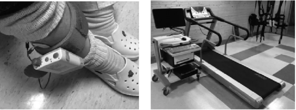
2. 비골신경 전기자극 및 보행 분석 측정 방법 사람이 보행할 때 유각기에서 심부 비골신경의 자 극에 의해 전경골근이 수축하면서 족배굴이 일어 난다14). 비골신경 전기자극기는 족하수가 있는 환 자의 환측 심부 비골신경이 지나는 부위에 두 개의 전극을 부착하여 전기자극을 주는 FES(Functional electrical stimulation)장비로 보행 중 전유각기(pre- swing phase)에 족배굴을 유도하게 된다. 마비된 우 측하지에 대한 비골신경 전기자극 치료는 일주일을 제외하고 매일 오전 11시부터 20분간 시행하였다.
비골신경 전기자극치료는 처음부터 4주동안 시행되 었는데, 비골신경 전기자극치료 후 변하는 보행양상 을 파악하기 위해 치료이전과 4주 동안 보행 분석하 였다. 4주 이후 1주일간 비골신경 전기자극 치료를 중지한 이유는 치료가 중지되면 변화된 보행양상이 원래의 상태로 회귀되는지 파악하기 위해서이다.
tial contact)에 편평족(flat foot)으로 착지하여 균형 잡기가 불안정하고, 유각기에 발이 지면에서 제대로 떨어지지 않게 된다7).
이렇게 변화된 보행 양상은 뇌졸중 환자가 보행 하는데 많은 어려움을 야기하므로, 이를 치료하기 위해서 AFO착용8), 비골신경 전기 자극9, 10), 보톡 스주사11) 등의 다양한 방법들이 빈번하게 사용된 다. 최근에는 비골신경 전기 자극치료를 하면, 뇌졸 중 후 편마비 환자들의 다양한 보행 변수와 보행 속 도가 긍정적인 방향으로 개선된다고 보고되었고12,
13), 뇌졸중 후 편마비 환자에게 비골신경 전기 치료 를 통해 족배굴근(Dorsi-flexor)과 족저굴근(Plan- tar-flexor)을 자극하는 것은 유각기에서 치고나가기 (push off)와 무릎관절이 굽혀지는 각도 등을 증가 시킨다는 보고가 있다14).
이와 같은 다양한 치료 방법의 효과를 측정하고, 더욱 적절한 재활 치료를 시행하기 위해서는 객관적 이고 정량적인 측정과 평가가 필요하다. 그러나 한 방 임상에서는 뇌졸중 환자의 비율이 높고, 뇌졸중 환자의 재활 치료에 대한 수요가 많음에도 불구하 고, 체계적인 보행분석 결과를 바탕으로 보행장애를 치료하는 방법이 개발되지 않고 있다.
본 연구는 뇌졸중 후 강직으로 인하여 족하수 증 상이 있는 뇌졸중 환자에게 비골신경 전기자극을 가 했을 때, 보행 양상이 즉각적으로 변화하는 것과, 4 주 동안 비골신경 전기자극 치료를 시행 한 후 변화 하는 것을 측정하였다. 단, 한방임상에서 적용하기 쉽게 측정이 쉽고 측정시간이 짧은 트레드밀 보행분 석 장비로 보행의 간격과 시간 변수(spatiotemporal gait parameter)와 지면반발력(Ground reaction
Fig. 1. Brain CT images of hemorrhage(left), after operation(middle) and after cranioplasty surgery(right).
o 결과
1. 마비된 측 발의 지면 접촉시간 비율(con- tact time rate, %)의 변화
보행 중에 우측 발을 forefoot, midfoot, heel로 나 누어 접촉시간을 비율로 환산하여 비교하였다(Fig.
3). Forefoot의 지면접촉시간비율(%)은 첫번째부터 일곱번째까지 순서대로 94.79±0.36, 94.43±0.32, 94.55±0.47, 94.75±0.33, 94.66±0.27 95.20±
0.30, 95.15±0.29로 측정되었다. Midfoot의 지 면접촉시간비율(%)은 첫번째부터 일곱번째까지 76.12±8.53, 85.52±3.89, 78.74±3.47, 81.94±
5.20, 79.82±3.81, 82.17±3.52, 85.93±4.16으 로 측정되었다. Heel의 지면접촉시간비율(%)은 첫 번째부터 일곱번째까지 순서대로 47.72±11.36, 57.84±11.76, 62.12±11.67, 72.48±3.28, 72.03±
3.82, 72.14±7.10, 73.98±3.58로 측정되었다.
보행분석은 트레드밀 보행 분석 시스템을 사 용하여 매주 시행하여 총 7회 시행하였다. 보행 분석을 하기 직전 환자의 MI(motricity index)와 MMT(manual muscle test)를 먼저 측정하였다(Ta- ble 1).
3. 비골신경 전기자극기 및 보행분석 장비 비골신경 전기 자극은 walkaide 제품을 사용하 였으며, 트레드밀 보행분석 장비는 Zebris Co.Ltd FDM-T를 사용하였다(Fig. 2). 보행분석 중에 운동 역학적 변수로 지면반발력의 최대값과 지면접지율 을 측정하였고, 시간과 공간에 대한 변수로 1분당 보행수(cadence), 보행속도(velocity), 입각기(stance phase), 유각기(swing phase), 양하지 지지기(dou- ble support), 한 발짝 시간(step time), 한 걸음 시간 (stride time), 한 발짝 길이(step length), 한 걸음 길 이(stride length)를 측정하였다.
Fig. 2. Peroneal nerve stimulation equipment(left) and treadmill gait analysis system(right).
Table 1. Change of Manual muscle test(MMT) and Motricity Index(MI) for Seven Weeks.
1
stmeasure
2
ndmeasure
3
rdmeasure
4
thmeasure
5
thmeasure
6
thmeasure
7
thmeasure
치료전 1주째
치료후
2주째 치료후
3주째 치료후
4주째 치료후
5주째 치료중지후
6주째 치료후
MMT 3 , 4 3 , 4 3 , 4 3 , 4 3 , 4 3 , 4 3 , 4
MI hip 25 25 25 25 25 25 25
knee 19 19 19 19 19 19 19
ankle 0 0 0 0 0 0 0
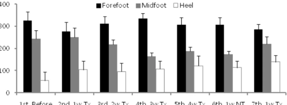
42.80, 218.10±20.95, 164.20±16.82, 187.46±
19.50, 173.50±13.74, 221.04±32.26로 측정되 었다. Heel의 지면반발력 최대값(N)은 첫번째부 터 일곱번째까지 순서대로 55.13±38.06, 104.46±
38.74, 95.23±38.17, 107.55±36.12, 121.84±
44.87, 113.32±28.71, 139.01±30.53으로 측정되 었다. Heel의 지면반발력 최대값이 현저하게 변화하 는 양상을 보였는데, 특징적인 것은 4주 치료후부터 는 치료를 중지하거나 치료를 재시행하여도 비슷한 상태를 유지하였다.
3. 한 발짝 길이(step length, cm)와 한 걸음 길이(stride length, cm)의 변화
보행 중에 양측 발의 한 발짝 길이와 한 걸음 길 이를 비교하였다(Fig. 5). 마비된 측 다리의 한 발 Heel의 지면접촉시간비율이 현저하게 변화하는 양
상을 보였는데, 특징적인 것은 4주 치료후부터는 치 료를 중지하거나 치료를 재시행하여도 비슷한 상태 를 유지하였다.
2. 마비된 측 발의 지면반발력 최대값(max force, N)의 변화
보행 중에 우측 발을 forefoot, midfoot, heel로 나 누어 지면반발력의 최대값을 비교하였다(Fig. 4).
Forefoot의 지면반발력 최대값(N)은 첫번째부터 일곱번째까지 순서대로 325.74±39.86, 276.05±
42.30, 311.56±32.12, 333.92±24.83, 307.22±
31.34, 306.84±31.83, 285.72±24.30로 측정되었 다. Midfoot의 지면반발력 최대값(N)은 첫번째부터 일곱번째까지 순서대로 243.32±38.89, 251.10±
Fig. 3. Contact time rate(%) of three different portions in right foot during walking. Before, before peroneal nerve stimulation treatment(Tx); 1w Tx, after Tx for 1week; 2w Tx, after Tx for 2weeks; 3w Tx, after Tx for 3weeks; 4w Tx, after Tx for 4weeks; 1w NT, after pause of Tx for 1week; 1w RTx, after Re-Tx for 1week.
Fig. 4. Max force(N) of three different portions in right foot during walking. Before, before peroneal nerve stimulation treatment(Tx); 1w Tx, after Tx for 1week; 2w Tx, after Tx for 2weeks; 3w Tx, after Tx for 3weeks;
4w Tx, after Tx for 4weeks; 1w NT, after pause of Tx for 1week; 1w RTx, after Re-Tx for 1week.
4. 한 발짝 시간(step time, second)과 한 걸 음 시간(stride time, second)의 변화 보행 중에 양측 발의 한 발짝 시간과 한 걸음 시 간을 비교하였다(Fig. 6). 마비된 측 다리의 한 발짝 시간(second)은 첫번째부터 일곱번째까지 순서대로 1.02±0.06, 1.00±0.04, 1.02±0.05, 1.02±0.06, 1.01±0.04, 1.06±0.06, 0.99±0.04로 측정되었다.
비마비된 측 다리의 한 발짝 시간(second)은 0.67±
0.05, 0.63±0.04, 0.66±0.06, 0.67±0.04, 0.67±
0.04, 0.75±0.04, 0.76±0.05로 측정되었다. 한 걸 음 시간(second)은 첫번째부터 일곱번째까지 순서 대로 1.69±0.07, 1.62±0.05, 1.68±0.09, 1.69±
0.08, 1.67±0.05, 1.81±0.07, 1.74±0.06으로 측 정되었다. 한 걸음 길이는 첫번째부터 일곱번째까지 짝 길이(cm)는 첫번째부터 일곱번째까지 순서대로
26.96±2.80, 28.11±2.14, 29.04±1.79, 31.38±
1.78, 32.23±1.82, 30.17±1.63, 29.39±1.65 로 측정되었다. 비마비된 측 다리의 한 발짝 길 이(cm)는 21.31±2.04, 23.55±2.69, 23.65±
2.59, 25.65±2.44, 27.09±2.16, 22.29±3.17, 22.93±2.47로 측정되었다. 한 걸음 길이(cm)는 첫번째부터 일곱번째까지 순서대로 25.72±2.72, 24.50±2.92, 25.44±2.43, 25.71±2.42, 25.20±
1.98, 27.82±3.35, 26.42±2.56으로 측정되었다.
바마비된 측 다리는 유각기에서 전진하지 못하고 제 자리에 내려놓는 양상을 보였기 때문에 마이너스 값 으로 측정되었다.
Fig. 5. Step length of both foot and stride length during walking. Before, before peroneal nerve stimulation treatment(Tx); 1w Tx, after Tx for 1week; 2w Tx, after Tx for 2weeks; 3w Tx, after Tx for 3weeks; 4w Tx, af- ter Tx for 4weeks; 1w NT, after pause of Tx for 1week; 1w RTx, after Re-Tx for 1week.
Fig. 6. Step time of both foot and stride time during walking. Before, before peroneal nerve stimulation treat-
ment(Tx); 1w Tx, after Tx for 1week; 2w Tx, after Tx for 2weeks; 3w Tx, after Tx for 3weeks; 4w Tx, after Tx
for 4weeks; 1w NT, after pause of Tx for 1week; 1w RTx, after Re-Tx for 1week.
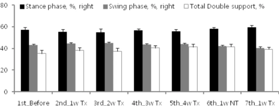
54.90±3.02, 56.57±1.64, 55.95±1.87, 57.88±
1.78, 59.47±1.71로 측정되었다. 유각기(%)는 42.99±2.66, 44.50±2.14, 45.10±3.02, 43.43±
1.64, 44.05±1.87, 42.12±1.78, 40.53±1.71로 측정되었다. 양하지 지지기(%)는 35.31±2.92, 38.18±2.47, 37.30±3.04, 40.48±2.07, 41.49±
2.41, 41.70±2.18, 39.20±1.91으로 측정되었다.
6. 마비된 측 다리의 체중전달기(loading response, %), 단하지 지지기(single sup- port, %), 전유각기(pre-swing, %)의 변화 보행 주기에서 마비된 측 다리의 입각기 중 체중 전달기, 단하지 지지기, 전유각기가 차지하는 비율 순서대로 25.72±2.72, 24.50±2.92, 25.44±2.43,
25.71±2.42, 25.20±1.98, 27.82±3.35, 26.42±
2.56으로 측정되었다. 바마비된 측 다리는 유각기에 서 전진하지 못하고 제 자리에 내려놓는 양상을 보 였기 때문에 마이너스 값으로 측정되었다.
5. 마비된 측 다리의 입각기(stance phase,
%)와 유각기(swing phase, %), 양하지 지지기(total double support, %)의 변화 보행 주기에서 마비된 측 다리의 입각기, 유각 기, 양하지 지지기가 보행 주기에서 차지하는 비율 을 비교하였다(Fig. 7). 입각기(%)는 첫번째부터 일 곱번째까지 순서대로 57.00±2.66, 55.50±2.14,
Fig. 7. Stance phase and swing phase of affected lower limb, total double support of gait cycle during walk- ing. Before, before peroneal nerve stimulation treatment(Tx); 1w Tx, after Tx for 1week; 2w Tx, after Tx for 2weeks; 3w Tx, after Tx for 3weeks; 4w Tx, after Tx for 4weeks; 1w NT, after pause of Tx for 1week; 1w RTx, after Re-Tx for 1week.
Fig. 8. Loading response and single support, pre-swing of affected lower limb during stance phase. Before,
before peroneal nerve stimulation treatment(Tx); 1w Tx, after Tx for 1week; 2w Tx, after Tx for 2weeks; 3w
Tx, after Tx for 3weeks; 4w Tx, after Tx for 4weeks; 1w NT, after pause of Tx for 1week; 1w RTx, after Re-Tx
for 1week.
7. 비마비된 측 다리의 체중전달기(loading response, %), 단하지 지지기(single sup- port, %), 전유각기(pre-swing, %)의 변화 보행 주기에서 비마비된 측 다리의 입각기 중 체 중전달기, 단하지 지지기, 전유각기가 차지하는 비 율을 비교하였다(Fig. 9). 체중전달기(%)는 첫번째 부터 일곱번째까지 순서대로 17.38±2.22, 16.89±
1.86, 15.65±2.61, 16.82±1.89, 16.24±1.98, 16.26±2.58, 16.07±1.66으로 측정되었다. 단하 지 지지기(%)는 첫번째부터 일곱번째까지 순서대로 43.06±2.50, 44.62±2.03, 45.45±3.10, 43.36±
1.85, 44.11±2.29, 42.12±1.81, 40.51±2.02로 측 을 비교하였다(Fig. 8). 체중전달기(%)는 첫번째부
터 일곱번째까지 순서대로 17.93±1.90, 21.37±
1.82, 21.76±2.28, 23.69±1.56, 25.27±1.67, 25.47±2.27, 23.15±1.80으로 측정되었다. 단하 지 지지기(%)는 첫번째부터 일곱번째까지 순서대로 21.62±2.04, 17.29±2.07, 17.45±1.83, 16.13±
1.49, 14.43±1.52, 16.18±1.98, 20.20±2.25로 측 정되었다. 전유각기(%)는 첫번째부터 일곱번째까지 순서대로 17.36±2.04, 16.84±1.84, 15.60±2.61, 16.75±1.88, 16.24±2.18, 16.23±2.54, 16.12±
1.68으로 측정되었다.
Fig. 9. Loading response and single support, pre-swing of unaffected lower limb during stance phase. Before, before peroneal nerve stimulation treatment(Tx); 1w Tx, after Tx for 1week; 2w Tx, after Tx for 2weeks; 3w Tx, after Tx for 3weeks; 4w Tx, after Tx for 4weeks; 1w NT, after pause of Tx for 1week; 1w RTx, after Re-Tx for 1week.
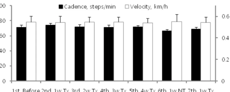
Fig. 10. Cadence and velocity during walking. Before, before peroneal nerve stimulation treatment(Tx); 1w Tx, after Tx for 1week; 2w Tx, after Tx for 2weeks; 3w Tx, after Tx for 3weeks; 4w Tx, after Tx for 4weeks;
1w NT, after pause of Tx for 1week; 1w RTx, after Re-Tx for 1week.
골 신경 자극의 치료 효과를 파악하기 위해 매주 보 행 분석을 실시하였다. 첫 4주 동안은 1주 6회(일요 일 제외) 매일 20분씩 비골 신경을 자극하고, 치료 중단 시 치료 효과가 변하는지 확인하기 위하여 5주 째에는 1주 동안 치료를 중지하였다. 6주째에 1주 동안 치료를 재개하였고, 7번째 보행 분석을 마지막 으로 연구를 종료하였다. 보행 분석을 통해 시공간 적 변수와 운동역학적 변수를 확인하였다. 시공간적 변수로는 1분당 보행 수, 보행 속도, 입각기 비율과 유각기 비율, 양하지 지지기, 한 발짝 시간과 한 걸 음 시간, 한 발짝 길이와 한 걸음 길이를 측정했다.
운동역학적 변수로는 지면 접촉시간 비율과 지면반 발력 최대값을 측정했다. 7번의 검사 결과 상, 시공 간적 변수인 1분당 보행 수, 보행 속도, 입각기 비 율과 유각기 비율, 양하지 지지기, 한 발짝 시간과 한 걸음 시간, 한 발짝 길이와 한 걸음 길이 모두 큰 변화가 없었다. 발이 지면에 닿는 부분을 forefoot, midfoot, heel로 나누었을 때 운동역학적 변수 중 지면 접촉시간 비율은 heel의 결과값이 증가하였고, 정되었다. 전유각기(%)는 첫번째부터 일곱번째까지
순서대로 17.94±2.00, 21.35±1.96, 21.74±2.39, 23.65±1.47, 25.25±1.77, 25.50±2.36, 23.13±
1.92으로 측정되었다.
8. 1분당 보행 수(cadence, steps/min)와 보 행 속도(velocity, km/h)의 변화
1분당 보행 수와 보행 속도의 변화를 비교하였다 (Fig. 10). 1분당 보행수(steps/min) 첫번째부터 일 곱번째까지 순서대로 71.08±3.14, 74.07±2.36, 71.56±3.40, 71.15±3.30, 71.57±2.29, 66.32±
2.50, 68.88±2.56으로 측정되었다. 보행 속도(km/
h)는 첫번째부터 일곱번째까지 순서대로 0.55±
0.06, 0.54±0.06, 0.55±0.05, 0.55±0.04, 0.54±
0.04, 0.55±0.07, 0.55±0.05으로 측정되었다.
o 고찰 및 결론
뇌졸중 후 편마비로 인해 족하수 증상을 보이는 환자에게 6주 동안 비골 신경 자극을 시행하고, 비
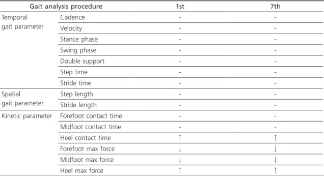
Table 2. Change of Gait Parameters by Peroneal Nerve Stimulation during 6 Weeks.
Gait analysis procedure 1st 7th
Temporal gait parameter
Cadence - -
Velocity - -
Stance phase - -
Swing phase - -
Double support - -
Step time - -
Stride time - -
Spatial gait parameter
Step length - -
Stride length - -
Kinetic parameter Forefoot contact time - -
Midfoot contact time - -
Heel contact time ↑ ↑
Forefoot max force ↓ ↓
Midfoot max force ↓ ↓
Heel max force ↑ ↑
↓(or ↑), Relative decrease(or increase) of gait events compared to previous measurement. -, no change of gait events compared with previous measurement.
환자의 마비된 측 비골 신경 자극은 시공간적 변수 에는 영향이 거의 없으나 운동역학적 변수 중 발뒤 꿈치의 지면접지율과 지면반발력의 최대 값을 증가 시키고 앞발과 중간발의 지면반발력 최대 값을 감소 시켜서 재활치료와 보행능력 향상에 도움이 된다.
참고문헌
01. O’Sullivan SB, Schmitz TJ, Fulk GD. Physical rehabilitation: FA Davis; 2007.
02. Niemi M-L, Laaksonen R, Kotila M, Waltimo O. Quality of life 4 years after stroke. Stroke.
1988;19(9):1101-7.
03. Langhorne P, Bernhardt J, Kwakkel G. Stroke re- habilitation. The Lancet. 2011;377(9778):1693- 702.
04. Olney SJ, Richards C. Hemiparetic gait follow- ing stroke. Part I: Characteristics. Gait & Pos- ture. 1996;4(2):136-48.
05. Ada L, Vattanasilp W, O’Dwyer NJ, Crosbie J.
Does spasticity contribute to walking dysfunc- tion after stroke? Journal of Neurology, Neuro- surgery & Psychiatry. 1998;64(5):628-35.
06. Rao N, Chaudhuri G, Hasso D, D’Souza K, Wening J, Carlson C, et al. Gait assessment during the initial fitting of an ankle foot orthosis in individuals with stroke. Disability & Rehabil- itation: Assistive Technology. 2008;3(4):201-7.
07. Dietz V, Berger W. Interlimb coordination of posture in patients with spastic paresis impaired function of spinal reflexes. Brain.
1984;107(3):965-78.
08. de Wit D, Buurke J, Nijlant J, IJzerman M, Her- mens H. The effect of an ankle-foot orthosis on walking ability in chronic stroke patients.
Co-ordination Patterns & Functional Recovery.
2005;18:79.
09. Granat MH, Maxwell DJ, Ferguson AC, Lees KR, Barbenet JC. Peroneal stimulator: evalu- ation for the correction of spastic drop foot in hemiplegia. Archives of physical medicine and rehabilitation. 1996;77(1):19-24.
10. 홍승홍. 일측 마비환자의 전기자극에 의한 보행기 능의 회복(Electrical Stimulation for the Resto-
지면반발력 최대값은 heel의 결과값이 증가한 반면forefoot과 midfoot의 결과값은 감소하였다(Table 2).
시공간적 변수는 거의 변화하지 않았는데. 이는 뇌졸중 후 편마비 환자의 운동 기능 회복이 발병 후 6개월이 지난 후에는 매우 느려지기 때문15)으로 보 인다. 본 연구에 참여한 환자는 2012년 10월 10일에 뇌출혈이 발병하여 연구를 시작한 시점에서 이미 발 병 후 10개월이 경과하였고, 더 이상의 운동 기능 회 복을 기대하기 어려워 장애진단을 받은 상황이었다.
다만 운동역학적 변수 중 지면 접촉시간 비율과 지면반발력 최대값이 치료를 시행한 4주 동안 꾸준 히 변화하였다. 5주차에는 1주일 간 비골 신경 자극 을 하지 않았으나 보행 분석 상 변화한 결과값이 보 존되었고 마지막 1주일 간 비골 신경 자극을 가하자 운동역학적 변수가 첫 4주와 동일한 양상으로 변화 하였다. 따라서 비골 신경 자극이 뇌졸중 후 편마비 로 인한 족하수가 있는 환자의 보행 양상을 긍정적 인 방향으로 변화시킬 수 있으며, 일정 기간 치료를 한 후에는 치료를 중단하거나 횟수를 줄이더라도 치 료 효과가 잔존하는 것으로 사료된다.
위의 보행 양상 변화가 양방 재활 치료와 침 치료 에 영향을 받았을 가능성을 배제할 수는 없으나, 발 병시점이 6개월을 지난 것을 고려할 때, 기존에 알 려지기로는 재활 치료가 운동 기능 회복에 크게 영 향이 없으므로15) 비골신경 전기자극에 의하여 보행 양상이 변화한 것으로 보인다. 본 연구는 환자 1명 만을 다루었고, 증상이 고착화된 환자를 대상으로 하여 실제적인 재활 치료 기간에서의 효과에 대해 파악하기 힘들다는 제한점이 있다. 그러나 비골신경 전기자극을 통해 운동역학적 변수가 긍정적인 변화 하는 효과를 확인할 수 있었다. 따라서 비골신경 전 기자극을 주기적으로 시행한다면, 보행 양상을 장기 적으로 교정할 수 있을 것이라고 사료된다. 향후 추 가적인 연구와 개발을 통하여 본 연구에서 나타난 제한점을 해결한다면, 비골신경 전기자극은 뇌졸중 후 편마비 환자의 보행기능 향상에 긍정적인 효과를 미칠 것으로 기대한다.
발병 후 6개월이 경과한 뇌졸중 후 만성 편마비