https://doi.org/10.5468/ogs.19242 pISSN 2287-8572 · eISSN 2287-8580
Introduction
The first trimester of pregnancy is a critical period for the continuity and progression of a healthy pregnancy. Approxi- mately 15% of clinically confirmed pregnancies result in spontaneous abortion, and 80% of those occur during the first trimester of pregnancy [1]. The major etiological factors for spontaneous abortions during the first trimester of preg- nancy are genetic, endocrine, anatomical, and immunologi- cal; however, the etiology of 50% of such abortions remains unknown. Regulation of the finely controlled process of trophoblast invasion plays a major role in preventing compli- cations of pregnancy, including early spontaneous abortion [2,3]. The ability of markers such as mean arterial pressure,
Is placental KISS-1 expression associated with first trimester abortion spontaneous?
Eser Colak, MD
1, Emel Ebru Ozcimen, MD
1, Ozgur Hilal Erinanç, MD
2, Yusuf Aytac Tohma, MD
3, Mehmet Ufuk Ceran, MD
1Departments of 1Gynecology and Obstetrics, 2Pathology, Konya Medical and Research Center, Baskent University School of Medicine, Konya;
3Department of Gynecology and Obstetrics, Baskent University School of Medicine, Ankara, Turkey
Objective
Several studies have examined biological markers during the first trimester to predict the maintenance of a healthy pregnancy. One such marker is kisspeptin, which is encoded by the KISS-1 gene. We aimed to determine whether first- trimester pregnancy losses were associated with levels of placental KISS-1 expression.
Methods
This prospective case control study was conducted at a tertiary center. The study group included 27 and 24 patients who underwent dilation and curettage at <10 weeks of gestation, due to first trimester spontaneous pregnancy loss and for elective termination (control), respectively. Placental and decidual tissues from all patients were sectioned and immunohistochemically analyzed for kisspeptin.
Results
Age, gravida status, parity number, gestational week, and number of previous abortions did not significantly differ between the groups. KISS-1 expression levels were significantly lower in the group with spontaneous abortion compared with the group with elective termination. The median staining intensity of KISS-1 expression in the elective and spontaneous termination groups were 3 (strong) and 2 (moderate), respectively (P=0.004). KISS-1 expression levels were significantly lower among patients with previous abortions in the elective termination group (P=0.002).
Conclusion
KISS-1 expression levels were found to be significantly reduced in patients with spontaneous pregnancy loss; KISS-1 plays an important role in the implantation and continuation of pregnancy.
Keywords: Genes, tumor suppressor; Abortion, spontaneous; Pregnancy, unwanted; Placenta
Received: 2019.12.17. Revised: 2020.01.21. Accepted: 2020.02.04.
Corresponding author: Eser Colak, MD
Department of Gynecology and Obstetrics, Konya Medical and Research Center, Baskent University School of Medicine, Hocacihan mah. Saray cad. No:1, Selcuklu, Konya 42030, Turkey E-mail: [email protected]
https://orcid.org/0000-0002-8184-7531
Articles published in Obstet Gynecol Sci are open-access, distributed under the terms of the Creative Commons Attribution Non-Commercial License (http://creativecommons.
org/licenses/by-nc/3.0/) which permits unrestricted non-commercial use, distribution, and reproduction in any medium, provided the original work is properly cited.
Copyright © 2020 Korean Society of Obstetrics and Gynecology
uterine artery pulsatility index, placental growth factor, pregnancy-associated plasma protein A, maternal serum alpha-fetoprotein, and free beta-HCG, in predicting healthy maintenance of pregnancy, have been examined during the first trimester [4,5]. However, studies of markers that could indicate the well-being of pregnancy during the first trimes- ter, are under way.
Recent studies have focused on molecular mechanisms like those of tumor cells because trophoblast migration and inva- sion occur via such molecular mechanisms [6,7]. One such mechanism is that of kisspeptin.
Kisspeptin constitutes a group of peptide fragments encod- ed by the KISS-1 gene (a tumor metastasis suppressor) in hu- mans and each fragment acts on various tissues via numer- ous mechanisms [8]. Kisspeptin-10 (KP-10) might play a role in the regulation of trophoblast invasion, puberty onset, and in the development of the hypothalamic–pituitary–gonadal axis [9,10]. Kisspeptins, especially KP-10, reportedly play im- portant roles in trophoblast invasion through the regulation of placental angiogenesis. Trophoblast invasiveness reduced by KP-10 might induce the downregulation of matrix metal- lopeptidase activity [11]. Kisspeptin expression in syncytio- trophoblasts and cytotrophoblasts is reduced in women who experience recurrent spontaneous abortion compared with controls [12]. Sullivan-Pyke et al. [13] found lower serum kis- speptin levels in cases experiencing spontaneous abortions.
Thus, kisspeptin might serve as a new serum biomarker to discriminate the likelihood of spontaneous abortion from vi- able intrauterine pregnancy [13].
The present study compared the expression of placental KISS-1 that encodes kisspeptin, between first-trimester preg- nancy losses and elective pregnancy terminations to deter- mine the differences at the tissue level.
Materials and methods
1. Study population
This prospective, case control study was conducted at the Department of Gynecology and Obstetrics of a tertiary cen- ter.
The sample size of the study was calculated using the G*Power (G*Power Ver. 3.1.9.2, Franz Faul, Üniversität zu Kiel, Kiel, Germany) statistical package. The required sample sizes for α=0.05 and Zα=1.96, and β=0.10 and Zβ=−1.28
was calculated for each group. The study and control groups included 27 and 24 consecutive patients who underwent dilation and curettage at <10 weeks of gestation due to first trimester spontaneous pregnancy loss and for elective termi- nation, respectively, between July 2017 and January 2018.
Elective pregnancy termination is possible before gestational week 10 in Turkey. Therefore, we included first-trimester pregnancy losses only before gestational week 10.
Spontaneous pregnancy loss was diagnosed via transvagi- nal ultrasound and clinical assessment. We excluded patients with a retroplacental hematoma, active maternal infec- tion, systemic lupus erythematosus, hypertension, diabetes, chronic renal insufficiency, alcohol consumption, a smoking habit (cigarettes), or a history of ≥2 spontaneous abortions.
According to the guidelines of the Royal College of Obstetri- cians and Gynaecologists, recurrent pregnancy loss is defined as a total of ≥3 abortions [14]. We analyzed the karyotypes of all patients, and excluded those with abnormalities to un- mask and avoid possible effects of chromosomal abnormali- ties on kisspeptin expression between the groups. Patients with vaginal bleeding and hematomas were not included due to low risk. As much as possible, we aimed to select only patients with healthy pregnancies for the control group.
Curettage specimens from 27 spontaneous pregnancy loss- es and 24 elective pregnancy terminations histopathologically diagnosed as “decidua showing the Arias-Stella reaction and chorionic villi” at the Faculty of Medicine, Department of Pa- thology between 2001 and 2003 were collected and archival hematoxylin and eosin stained sections were reevaluated.
Kisspeptin was immunohistochemically assessed in tissue samples containing decidua and chorionic villi without areas of hemorrhage and necrosis.
2. Immunohistochemistry
Sections were incubated at 56°C for 24 hours, deparaffinized
in xylene, then rehydrated using a descending alcohol series
(100–70%). Endogenous peroxidase activity was blocked by
immersing the sections for 5 minutes in methanol contain-
ing 0.5% hydrogen peroxide. Antigens were retrieved by
microwave-heating the sections in trisodium citrate buffer
(10 mM sodium citrate, pH 6.0) for 10 minutes. The sections
were rinsed 3 times in de-ionized distilled water, immersed
in 0.3% hydrogen peroxidase for 30 minutes to block en-
dogenous peroxidase activity, then washed in PBS for 2–3
minutes. Non-specific background staining was minimized
using Ultra V Block (Thermo Fisher Scientific Inc., Waltham, MA, USA) for 5 minutes. The sections were then incubated for 1 hour at room temperature with the mouse monoclonal primary antibody to KISS-1, 24-Q (Santa Cruz Biotechnology Inc., Dallas, TX, USA) diluted 1:100. This antibody was raised against a partial recombinant protein mapped within amino acids 46–145 of KISS-1 of human origin. After a wash with PBS for 5 minutes, Goat Anti-Polyvalent (Lab Vision Corp., Fremont, CA, USA) was applied, then the sections were washed in PBS. The sections were incubated for 15 minutes at room temperature with streptavidin peroxidase (Lab Vi- sion Corp.). Immunoreactivity was visualized using the chro- mogen, DAB (Thermo Fisher Scientific Inc.). The sections were counterstained with Mayer’s hematoxylin solution and mounted. Staining specificity was confirmed by comparison with a positive control.
3. Analysis of immunohistochemical expression
Two pathologists who were blinded to the pregnancy stage and outcomes analyzed all sections. Immunohistochemical staining was evaluated by scanning whole sections for anti- bodies using a BX51 light microscope (Olympus Optical Co., Ltd., Tokyo, Japan) at 4×, 10×, 20×, and 40× magnification.
KISS-1 proteins were mainly located in the glandular epithe- lium, decidualized stromal cells, cytotrophoblasts, and syncy- tiotrophoblasts.
The results are expressed as ratios (%) of positivity based on staining intensity scored as 0 (negative), 1+ (low), 2+
(moderate), and 3+ (strong). Ratios (%) of stained cells were:
0 (<10%), 1 (10–25%), 2 (10–50%), 3 (51–80%), and 4
(>80%). The final score was calculated as the product of the ratio of stained cells and staining intensity, resulting in weak (0–2), moderate (3–6), and strong (8) KiSS-1 expression [15].
4. Statistical analysis
Variables were analyzed using SPSS 22.0 (IBM Corp., Ar- monk, NY, USA) and MedCalc 14 (MedCalc Software Ltd., Ostend, Belgium) software. The normality of data distribution was evaluated using Shapiro-Wilk tests. The homogeneity of variances was assessed using Levene tests. Pairs of inde- pendent groups were compared using independent samples
t-tests with Bootstrap results, and Mann-Whitney U-testswith Monte Carlo results. Categorical variables were com- pared using Pearson χ
2tests with exact results. Correlations between KISS-1 and age, number of births, parity, and abor- tus were assessed using Spearman rho tests. The sensitivity and specificity of relationships between classifications based on cutoff values calculated according to the number of abor- tions and KISS-1 variables for the 2 groups of patients, and the actual classification were analyzed using receiver operat- ing characteristics curves. Diagnostic-dependent variables with explanatory variables were estimated using logistic regression analysis, K-Nearest Neighbor, and Decision Tree.
However, the results were not significant and are not shown.
Quantitative variables are expressed as mean±standard devia- tion, range (maximum–minimum), and medians. Categorical variables are expressed as number (%). The variables were analyzed at 95% confidence intervals, and P<0.05 was con- sidered statistically significant.
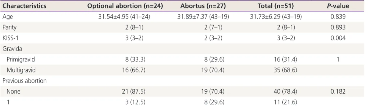
Table 1. Comparison of demographic data and KISS-1 levels between the 2 groups
Characteristics Optional abortion (n=24) Abortus (n=27) Total (n=51) P-value
Age 31.54±4.95 (41–24) 31.89±7.37 (43–19) 31.73±6.29 (43–19) 0.839
Parity 2 (8–1) 2 (7–1) 2 (8–1) 0.893
KISS-1 3 (3–2) 2 (3–2) 3 (3–2) 0.004
Gravida
Primigravid 8 (33.3) 8 (29.6) 16 (31.4) 1
Multigravid 16 (66.7) 19 (70.4) 35 (68.6)
Previous abortion
None 21 (87.5) 19 (70.4) 40 (78.4) 0.182
1 3 (12.5) 8 (29.6) 11 (21.6)
Values are presented as mean±standard deviation, median (maximum–minimum), or number (%). Data were compared using independent samples t-tests (Bootstrap), Mann-Whitney U-tests (Monte Carlo), and Pearson χ2 tests (exact).
Results
Table 1 shows the demographic characteristics of the pa- tients. Mean age (control vs. study group: 31.54±4.95 vs.
31.89±7.37 years), gravida status, parity number, previous abortions and gestational weeks (P=0.072) did not signifi-
cantly differ between the 2 groups. These finding confirmed the homogeneous distribution of demographic characteris- tics within each group.
Median values of placental KISS-1 expression significantly differed between the two groups. Median values for stain- ing intensity indicating KISS-1 expression in the control and study groups were 3 (strong) and 2 (moderate), respectively (P=0.004; Table 1). Fig. 1 shows a loss of KISS-1 in expression in the glandular epithelium and decidualized stromal cells.
Fig. 2 shows strong KISS-1 staining intensity in the glandular epithelium and decidualized stromal cells.
The effects of previous abortions on placental KISS-1 ex- pression were significantly lower in the group with elective termination (P=0.002), but did not significantly differ in the group with spontaneous abortion (Table 2).
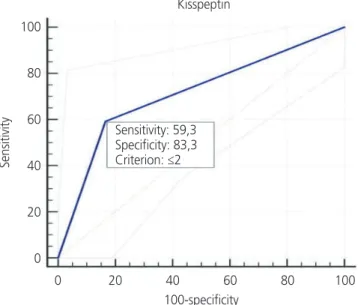
We determined a cutoff value of 2 for KISS-1 expression based on the areas under receiver operator characteris- tics curves. The sensitivity and specificity were 59.3% and 83.3%, respectively (Table 3, Fig. 3).
The expression of KISS-1 was significantly lower in the patients with, than without a history of abortion (P<0.001;
Table 4).
Fig. 1. Weak (+) staining intensity.
Fig. 2. Strong (+++) intensity of staining.
Table 2. Investigation of the relationship between KISS-1 levels and previous abortion in both groups
Previous abortion No. KISS-1 P-value Optional abortion
No 21 3 (3–2) 0.002
Yes 3 2 (2–2)
Abortus
No 19 2 (3–2) 1
Yes 8 2 (3–2)
Values are presented as median (maximum–minimum). Data were compared using Mann-Whitney U-tests (Monte Carlo).
Table 3. Determination of cut-off, sensitivity and specificity values for KISS-1
Cut-off Optional abortion (n=24) Abortus (n=27) AUC (95% CI) P-value
KISS-1
>2 20 (83.3)a) 11 (40.7) 0.713 (0.856–0.570) 0.004
<2 4 (16.7) 16 (59.3)b)
Values are presented as number (%). Data were analyzed using ROC (Honley & Mc Nell - Youden index J).
AUC, area under the ROC curve; CI, confidence interval; ROC, receiver operating curve.
a)Sensitivity; b)Specificity.
Discussion
This study investigated whether placental KISS-1 expression differed between patients with pregnancy losses and those who had elective pregnancy terminations within ≤10 weeks of the first trimester.
Serum kisspeptin values during early gestation are found to be associated with abortive imminence, poor obstetric outcomes such as preterm birth, preeclampsia, and intrauter- ine growth retardation [16]. The same study also associated kisspeptin with recurrent pregnancy losses, indicating that serum kisspeptin could serve as a marker of placental func- tion [12]. However, comparative information about KISS-1 expression between healthy pregnancies and non-recurrent abortion is scarce.
Our results showed significantly lower placental KISS-1 ex- pression in patients with first-trimester pregnancy losses than in those requesting elective terminations. As far as we are aware, this is the first comparison of KISS-1 in materials de-
rived from non-recurrent spontaneous abortions and elective pregnancy terminations.
Kisspeptin has many effects on the physiology of puberty, the neuroendocrine system, reproductive
physiology, and it plays a regulatory role in syncytiotropho- blasts [9,10]. Kisspeptin and its receptor system are expressed during implantation and placentation in the endometrial and placental tissues of various species, including humans other. A study of hysterectomy materials obtained from non-pregnant female dogs and dogs at different stages of pregnancy found a significant increase in KP-10 levels after implantation and around the middle of pregnancy [17]. An- other study found higher levels of KISS-1R expression in the early placenta, where the invasive capacity of trophoblasts needs to be tightly controlled compared with term placenta in humans [18]. These studies suggested a potential role of the kisspeptin/KISS-1R system in the invasive and migratory properties of trophoblasts.
Levels of kisspeptin and its receptor GPR54 were also as- sociated with spontaneous abortions due to their actions on trophoblast invasion. Wu et al. [12] compared the im- munohistochemical expression of kisspeptin in decidua and trophoblasts between women with first-trimester pregnancy loss and those who requested legal termination. Levels of kisspeptin were significantly lower in decidual tissue and trophoblasts from women experiencing first-trimester preg- nancy loss than in those requesting legal elective termina- tion. We compared KISS-1 expression in decidual cells and trophoblasts between women with first-trimester pregnancy loss and those with elective pregnancy termination. Levels of KISS-1 expression were significantly lower in decidua and tro- phoblasts from patients with first-trimester pregnancy losses compared with elective termination. We found significantly lower KISS-1 expression in the group with spontaneous abor- tion, which supports published findings [12,19]. However, unlike the patients in those studies, none of our patients had anamnesis of recurrent pregnancy losses.
Fig. 3. Sensitivity and specificity for KISS-1.
Kisspeptin
Sensitivity: 59,3 Specificity: 83,3 Criterion: ≤2
Sensitivity
100 80 60 40 20
0
0 20 40 60 80 100 100-specificity
Table 4. Correlation analysis for KISS-1
KISS-1 Optional abortion Abortus Total
r P-value r P-value r P-value
Age −0.243 0.252 0.170 0.397 0.005 0.97
Previous abortion −0.845 <0.001 −0.043 0.832 −0.544 <0.001
Data assessed using Spearman’s ρ test.