• 검색 결과가 없습니다.

Delayed Ventricular Septal Rupture Following Blunt Chest TraumaNa Young Kim, MD

N/A
N/A
Protected

Academic year: 2022

Share "Delayed Ventricular Septal Rupture Following Blunt Chest TraumaNa Young Kim, MD"

Copied!
4
0
0

로드 중.... (전체 텍스트 보기)

전체 글

(1)

625

Print ISSN 1738-5520 / On-line ISSN 1738-5555 Copyright © 2011 The Korean Society of Cardiology CASE REPORT

http://dx.doi.org/10.4070/kcj.2011.41.10.625

Open Access

Delayed Ventricular Septal Rupture Following Blunt Chest Trauma

Na Young Kim, MD

1

, Tae Jung Kwon, MD

1

, Jeong Rang Park, MD

1

, Ho Cheol Choi, MD

2

, Yun Hong Cheon, MD

1

, Min Kyu Kang, MD

1

, Seok-Jae Hwang, MD

1

, Yongwhi Park, MD

1

, Young-Hoon Jeong, MD

1

, Choong Hwan Kwak, MD

1

, and Jin-Yong Hwang, MD

1

1

Departments of Internal Medicine and

2

Radiology, Gyeongsang National University Hospital, Jinju, Korea

ABSTRACT

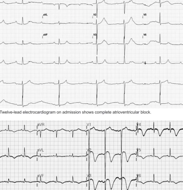
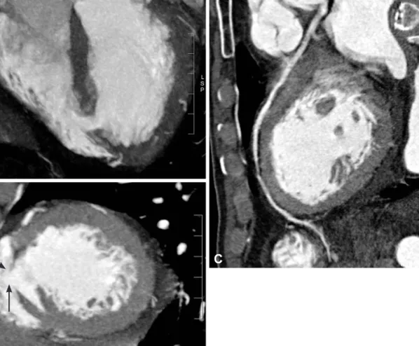
Cardiac injury is a common but occasionally serious complication of blunt chest trauma. A ventricular septal rupture (VSR) is a rare complication and is variable in its presentation, temporal course and severity. Here, we report a rare case of 75-year- old man who developed delayed VSR following blunt chest trauma. (Korean Circ J 2011;41:625-628)

KEY WORDS: Ventricular septal rupture; Thoracic injuries.

Received: December 8, 2010 Revision Received: January 4, 2011 Accepted: January 17, 2011

Correspondence: Jeong Rang Park, MD, Department of Internal Medi- cine, Gyeongsang National University Hospital, 90 Chiram-dong, Jinju 660- 702, Korea

Tel: 82-55-750-8068, Fax: 82-55-755-9078 E-mail: [email protected]

• The authors have no financial conflicts of interest.

cc

This is an Open Access article distributed under the terms of the Cre- ative Commons Attribution Non-Commercial License (http://creativecom- mons.org/licenses/by-nc/3.0) which permits unrestricted non-commer- cial use, distribution, and reproduction in any medium, provided the origi- nal work is properly cited.

Introduction

Cardiac injury is a common complication of blunt chest tr- auma. The common injuries include cardiac contusion, val- vular damage and aortic insufficiency.

1)

A ventricular septal rupture (VSR) is a rare complication following non-pene- trating chest trauma. VSR may be delayed for several hours to several days after trauma and its clinical presentation is va- rying from immediate death to complete spontaneous clo- sure. Therefore, closed observation is essential for the manage- ment of these patients. We report a rare case of delayed VSR with complete atrioventricular (AV) block following blunt chest trauma.

Case

A 75-year-old man was admitted to the regional hospital following a traffic accident. His motorized wheelchair turn- ed over and he suffered bruised left ribs. Before the accident,

he had been treated for essential hypertension. He under- went below-knee amputation of his left leg due to a traffic accident 40 years ago. He was initially diagnosed with frac- tures of the left 3rd to 5th ribs and left scapula. On the second day after hospitalization, he had respiratory distress and was consequently transferred to Gyeongsang National University Hospital. His blood pressure was 70/40 mm Hg; heart rate, 40 beats/min; respiration rate, 30/min; and temperature, 36.9°C.

On physical examination, cardiac murmur was not heard. Ra- les and wheezing sound were heard on both lung fields. Ini- tial chest radiograph showed infiltration on both upper lung fields. The initial electrocardiogram (ECG) showed a compl- ete AV block (Fig. 1). Transthoracic echocardiogram reveal- ed normal ventricular function and mild tricuspid regurgit- ation with moderately elevated pulmonary hypertension. Cr- eatine kinase (CK) isoenzyme levels were raised (CK 2,389 U/L, normal range 0-170 U/L; CK-MB 21.3 ng/mL, normal range 0.6-6.3 ng/mL) and troponin I level was 0.3 ng/mL (nor- mal range 0.0-0.04 ng/mL). After fluid replacement, his blood pressure was restored to 140/90 mm Hg. He received treat- ment for acute lung injury. On the 11th day after the accident, he presented with aggravated dyspnea and tachypnea. A new grade 4/6 holosystolic murmur could be heard at the left ster- nal border. Findings on ECG were changed to normal sinus rhythm and diffuse T inversion on inferior and precordial leads (Fig. 2). CK-MB was 6.7 ng/mL and troponin I was raised to a level of 4.31 ng/mL. Transthoracic echocardiogram reveal- ed septal akinesia and ventricular septal defect (VSD) near the apex (Fig. 3), but the patient had no complaint of chest pain.

On coronary CT angiography, significant stenosis was not

seen and VSD was observed (Fig. 4). Cardiac magnetic reson-

(2)

626 Ventricular Septal Rupture Following Blunt Chest Trauma ance imaging (MRI) to rule out myocardial infarction re- vealed apical septal VSD with normal perfusion of the inter- ventricular septal wall. There were no delayed enhanced le-

sions suggesting myocardial fibrosis (Fig. 5). The Qp/Qs mea- sured by shunt scan was 1.52. After medication with diuretics, dyspnea and pulmonary edema were completely improved. He

Fig. 1. Twelve-lead electrocardiogram on admission shows complete atrioventricular block.

Fig. 2. Electrocardiogram on the 11th day after the accident shows normal sinus rhythm with diffuse T wave inversion on inferior and pre- cordial leads.

Fig. 3. Transthoracic echocardiography. A: apical four chamber view: ventricular septal defect (arrow) is seen near the apex. B: subcostal view: continuous wave Doppler shows left-to-right shunt pattern.

A B

(3)

Na Young Kim, et al. 627

was discharged without surgical correction because he did not want an operation and Qp/Qs was less than 2. Unfortunately, he died due to uncontrolled pneumonia after 1 month.

Discussion

Myocardial contusion is the most frequent cardiac injury fol- lowing blunt chest trauma and occurs in 16-76% of patients involved in motor vehicle accidents.

2)

A VSR is a rare complic- ation following blunt chest trauma.

3)

The first postulated mechanism of VSR is acute compres- sion of the heart with a resultant sudden rise in intracardiac pressure during the end of diastole or isovolumetric systole

3)

and the second postulated mechanism is that myocardial in- jury causes a microvascular disruption leading to infarction and liquefaction of the septum.

4)

The contused myocardium can become necrotic and subsequently perforate. Therefore, VSR may occur several hours to months after blunt trauma.

5)

Traumatic VSR is usually located near the apex, in the mus- cular portion of the septum, in contrast to a congenital VSD that occurs adjacent to the membranous septum. Cleland et al.

6)

reported that presence of fibrosis adjacent to the rup- tured myocardium is helpful for differentiating traumatic VSR from a congenital VSD. Jun et al.

7)

showed that cardiac MRI demonstrated delayed enhancement consistent with fibrotic scars in the patient with VSR after blunt trauma. In our pa- tient, cardiac MRI did not show delayed enhancement and we confirmed normal coronary perfusion. We thought that pre- sence or absence of fibrosis and the amount of fibrosis may differ among the various mechanisms of VSR.

Echocardiography is the method of choice for diagnosis and guiding the management of VSR. However, echocardiogra- phy is sometimes difficult to give enough information. Cardi- ac CT and MRI can help to differentiate the causes of VSD and Fig. 5. Cardiac magnetic resonance imaging revealed apical ven-

tricular septal defect without delayed enhancement.

Fig. 4. Cardiac computed tomography. Ventricular septal rupture and 0.7 cm size shunt (inter-arrow site) is identified at apical four chamber view (A) and short axis view (B). There is no significant stenosis of the left anterior descending artery (C).

A

B

C

(4)

628 Ventricular Septal Rupture Following Blunt Chest Trauma confirm the presence of combined coronary artery disease.

In traumatic VSR, conservative approach has been recom- mended for asymptomatic patients with small VSD when the ratio of pulmonary to systemic flows is clearly less than 2 : 1 because spontaneous closure is possible.

8)

Surgical or percu- taneous closure is necessary in patients with hemodynamic instability and a large defect.

Our patient presented with complete AV block at the time of admission. Arrhythmia as a complication of blunt chest trauma had been reported to occur in up to 70% of patients within the first 3 days of hospitalization.

9)

High degree AV blo- ck is an unusual complication after blunt chest trauma. The suggested mechanisms are a tear in the conduction system and a contusion or hemorrhage near the AV node.

10)

In this case, the development of VSR and conversion to sinus rhythm oc- curred simultaneously. Therefore, we believed that the main mechanism of AV block in our patient may be stretching of the conduction system as a result of edema of the interventri- cular septum. VSR may lead to relieving of tension and re- covering to sinus rhythm.

There were a few cases of VSR following non-penetrating chest trauma in Korea.

11)12)

However, to our knowledge there has not been a case reported of delayed presentation of trau- matic VSR with AV block. Here, we report a case of delayed VSR and high degree AV block following blunt chest trauma.

VSR can occur several days later and VSR should be consid- ered in cases of chest trauma with increased troponin level, new heart murmur, ECG change or hemodynamic instability.

Therefore, watchful physical examination and hemodynamic monitoring are required for patients with chest trauma. Se- rial echocardiographic monitoring should be performed, es-

pecially if cardiac contusion is expected.

REFERENCES

1) Hamdan-Challe M, Godin M, Bouchart F, Doguet F. Isolated ven- tricular septal rupture secondary to blunt trauma. Interact Cardiovasc Thorac Surg 2010;11:667-9.

2) Fang BR, Li CT. Acute myocardial infarction following blunt chest trauma. Eur Heart J 1994;15:705-7.

3) Blasco PB, Comas JG, del Alcazar Munoz R. Spontaneous improve- ment of a haemodynamically significant ventricular septal defect pro- duced by blunt chest trauma in a child. Cardiol Young 2009;19:109-10.

4) Genoni M, Jenni R, Turina M. Traumatic ventricular septal defect. He- art 1997;78:316-8.

5) Dehghani P, Ibrahim R, Collins N, Latter D, Cheema AN, Chisholm RJ. Post-traumatic ventricular septal defects: review of the literature and a novel technique for percutaneous closure. J Invasive Cardiol 2009;21:483-7.

6) Cleland WP, Ellman P, Goodwin J, Hollman A. Repair of ventricular septal defect following indirect trauma. Br J Dis Chest 1961;55:17-22.

7) Jun SL, Chanani NK, Moore P, Higgins CB. Magnetic resonance im- aging of a posttraumatic myocardial infarction and ventricular septal defect with a closure device in place. Circulation 2007;115:e13-5.

8) Pesenti-Rossi D, Godart F, Dubar A, Rey C. Transcatheter closure of traumatic ventricular septal defect: an alternative to surgery. Chest 2003;123:2144-5.

9) Sakka SG, Huettemann E, Giebe W, Reinhart K. Late cardiac ar- rhythmias after blunt chest trauma. Intensive Care Med 2000;26:

792-5.

10) Helling TS, Duke P, Beggs CW, Crouse LJ. A prospective evaluation of 68 patients suffering blunt chest trauma for evidence of cardiac injury. J Trauma 1989;29:961-6.

11) Yun HJ, Jin SW, Ahn YY, et al. A case of isolated ventricular septal rupture following non-penetrating chest trauma. J Korean Soc Echo- cardiogr 2001;9:157-60.

12) Lee WS, Lee IK, Kim KS, Kim YN, Kim KB. A case of traumatic ventricular septal defect secondary to nonpenetrating chest trauma.

Korean Circ J 1987;17:381-7.

수치

Fig. 1. Twelve-lead electrocardiogram on admission shows complete atrioventricular block.
Fig. 4. Cardiac computed tomography. Ventricular septal rupture and 0.7 cm size shunt (inter-arrow site) is identified at apical four chamber  view (A) and short axis view (B)

참조

관련 문서