ISSN: 2233-601X (Print) ISSN: 2093-6516 (Online)
− 211 −
Received: August 4, 2016, Revised: November 2, 2016, Accepted: November 7, 2016, Published online: June 5, 2017
Corresponding author: Weon Yong Lee, Department of Cardiothoracic Surgery, Hallym University Sacred Heart Hospital, 22 Gwanpyeong-ro 170beon-gil, Dongan-gu, Anyang 14068, Korea
(Tel) 82-31-380-3815 (Fax) 82-31-380-4118 (E-mail) [email protected]
© The Korean Society for Thoracic and Cardiovascular Surgery. 2017. All right reserved.
This is an open access article distributed under the terms of the Creative Commons Attribution Non-Commercial License (http://creativecommons.org/
licenses/by-nc/4.0) which permits unrestricted non-commercial use, distribution, and reproduction in any medium, provided the original work is properly cited.
Delayed Repair of Ventricular Septal Rupture
Following Preoperative Awake Extracorporeal Membrane Oxygenation Support
Bong Suk Park, M.D., Weon Yong Lee, M.D., Ph.D., Jung Hyeon Lim, M.D., Yong Joon Ra, M.D., Yong Han Kim, M.D., Hyoung Soo Kim, M.D., Ph.D.
Department of Cardiothoracic Surgery, Hanllym University Sacred Heart Hospital, Hanllym University College of Medicine
Outcomes of ventricular septal rupture (VSR) as a complication of acute myocardial infarction are extremely poor, with an in-hospital mortality rate of 45% in surgically treated patients and 90% in patients managed with medication. Delaying surgery for VSR is a strategy for reducing mortality. However, hemodynamic in- stability is the main problem with this strategy. In the present case, venoarterial extracorporeal membrane oxygenation (ECMO) was used to provide stable hemodynamic support before the delayed surgery. Awake ECMO was also used to avoiding the complications of sedatives and mechanical ventilation. Here, we describe a successful operation using awake ECMO as a bridge to surgery.
Key words: 1. Ventricular septal rupture
2. Extracorporeal membrane oxygenation 3. Awake extracorporeal membrane oxygenation 4. Myocardial infarction
Case report
A 68-year-old woman was admitted to Hallym University Sacred Heart Hospital for chest pain per- sisting for 2 days. An electrocardiogram showed ST-segment elevation in all the precordial leads and Q waves in leads II, III, avF, and V1–V6. Initial labo- ratory results showed increased levels of troponin I, creatinine kinase-myocardial band, and brain natriu- retic peptide. An emergency coronary angiogram showed chronic total occlusion of the mid-right coro- nary artery (RCA) with collaterals and total occlusion of the mid-left anterior descending (LAD) artery.
Percutaneous coronary intervention (PCI) was per- formed by inserting a Resolute Onyx stent (Medtro-
nic Inc., Santa Rosa, CA, USA) into the LAD. The oc- cluded LAD was successfully reperfused.
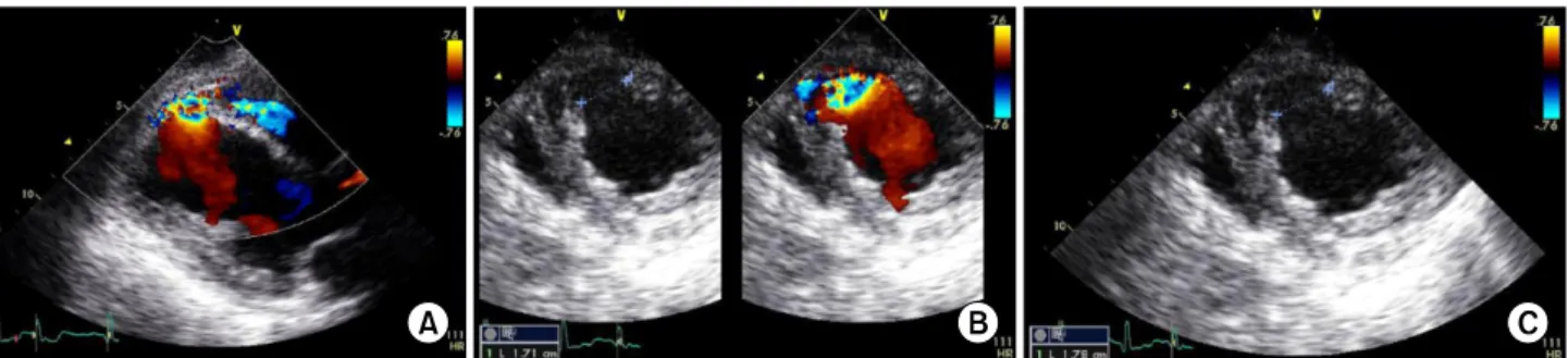
Three hours after PCI, the patient experienced sud- den dizziness with hypotension and tachycardia. A transthoracic echocardiogram (TTE) showed a 1.78-cm ventricular septal rupture (VSR) at the lower muscu- lar part of the ventricular septum, extensive ischemic insult in the LAD territory, and severe right and left ventricular dysfunction with an ejection fraction (EF) of 19% (Fig. 1). Cardiogenic shock was sustained de- spite incremental infusions of norepinephrine and in- tra-aortic balloon pump (IABP) support. Therefore, after 3 hours and 5 minutes of hemodynamic in- stability, venoarterial extracorporeal membrane oxy- genation (ECMO) was administered through right
Korean J Thorac Cardiovasc Surg 2017;50:211-214 □ CASE REPORT □
https://doi.org/10.5090/kjtcs.2017.50.3.211
Bong Suk Park, et al
− 212 −
Fig. 1. 2D echocardiograms. (A) 2D color Doppler image in the parasternal long-axis view. (B) 2D color Doppler image in the short-axis view. (C) Ventricular septal rupture measured 1.78 cm on an echocardiogram. 2D, two-dimensional.
Fig. 2. Simple chest radiographs.
(A) Initial plain chest radiograph obtained after admission. (B) Plain chest radiograph obtained just af- ter extracorporeal membrane oxy- genation insertion. Pulmonary ede- ma is not prominent, despite the increase in the pulmonary vascular markings.
femoral vessel cannulation instead of the IABP. This was promptly followed by mechanical ventilator care and continuous renal replacement therapy. On a plain chest radiograph obtained initially after ECMO, pulmonary edema was not prominent, although the pulmonary vascular parameters increased (Fig. 2).
The mean arterial pressure was maintained at more than 70 mm Hg with a continuous infusion of nor- epinephrine at a dose of 3 μg/kg/min, which was gradually decreased. Sedation was induced with a controlled dose of fentanyl (0.6 μg/kg/hr) and dex- medetomidine HCl (0.2 μg/kg/hr). After 4 days of ECMO support, extubation was performed since the patient was cooperative and had minimal airway secretions. Thereafter, awake ECMO was maintained with the addition of quetiapine (25 mg orally) before bedtime, and oral feeding was started the next day.
On the ninth day of ECMO support, the EF recov-
ered to 27% on a repeat TTE. Therefore, surgery for post-myocardial infarction (MI) VSR was planned.
The VSR was found anteriorly and 2.5 cm away from the apex. It had a rough, irregular margin with ad- jacent necrotic debris and muscular discoloration; it was closed by interrupted 3-0 prolene sutures using a single-layered Gore-Tex patch through left ven- triculotomy, which was performed laterally along the course of the LAD. The distal RCA was bypassed us- ing a greater saphenous vein graft. The left ventricle was repaired with 2-0 prolene sutures buttressed with Teflon felts. Cardiopulmonary bypass (CPB) was weaned according to the patient’s recovery status, and this was followed by the removal of ECMO sup- port and the repair of femoral vessels. The duration of CPB and aortic cross-clamping was 174 minutes and 115 minutes, respectively.
The patient recovered uneventfully and was ex-
Delayed Repair of Ventricular Septal Rupture Following Awake Extracorporeal Membrane Oxygenation Support
− 213 − tubated on the day after the operation. Percutaneous catheter drainage (PCD) was performed on post- operative day 2 because of pleural effusion. Despite the delay due to PCD, she was discharged from the hospital on postoperative day 18 without additional complications. She has been doing well for 6 months without residual or recurrent VSR.
Discussion
VSR as a complication of acute myocardial in- farction (AMI) is uncommon, with an incidence of 1% to 2% among patients with AMI. It typically oc- curs in the first week after infarction, with a mean time of 3 to 5 days from symptom onset. Previous investigations have found that age and female sex are risk factors for the development of VSR; such pa- tients also commonly have no prior angina or MI.
Angiographically, patients with VSR have been noted to have total occlusion of the infarct-related artery with minimal collaterals. Outcomes after the develop- ment of VSR are extremely poor, with an in-hospital mortality rate of 45% in surgically treated patients and 90% in patients managed with medication. Poor prognostic factors in this patient population include the development of cardiogenic shock, right ven- tricular dysfunction, advanced age, and an inferior in- farct location [1].
Labrousse et al. [2] reported that a relatively short interval between septum perforation and surgical re- pair was an important risk factor, with no deaths oc- curring in patients who were operated on 15 days or more after the perforation. T his result can be ex- plained by the fact that a more chronic VSR is easier to repair because the septum is well scarred and the patch can be securely sutured. However, the delay in surgery is directly dependent on the patient’s condition. Frequently, patients with similar conditions undergo an urgent or emergency operation if they have a hemodynamically unstable condition [2,3].
To delay surgery, various strategies such as ad- equate fluid therapy, high-dose inotropic drugs, and IABP and mechanical ventilator support have been used. Recently, ECMO has been widely administered to hemodynamically compromised patients, with the advent of a long-running oxygenator, heparin-coated tubing, a new centrifugal blood pump and generator, and percutaneous catheters. Nevertheless, conven-
tional ECMO requires general anesthesia and mechan- ical ventilation, which are associated with risks such as pulmonary barotrauma, respiratory infections, and hemodynamic collapse [4]. The concept of awake ECMO has been introduced to avoid these compli- cations. Awake ECMO prevents critically ill patients from losing muscle power due to long-term mechan- ical ventilation [5]. In addition, a recent study has re- ported the benefit of active physical therapy, includ- ing ambulation for patients on veno-arterial ECMO [6].
T he indications for ECMO in patients with post-MI VSR are crucial. Patients can be placed on ECMO if their cardiac function can be resolved with primary repair or transplantation, the medical means of stabi- lization are unsuccessful, and emergency surgery is considered to have a prohibitive risk.
In the present case, we used awake ECMO to avoid the complications of mechanical ventilation and to delay surgery in order to improve the patient’s like- lihood of survival. Awake ECMO may be a good alter- native as a bridge to surgery in hemodynamically compromised patients with post-MI VSR. However, ECMO can cause complications such as hemolysis, bleeding, systemic inflammation and infection, throm- boembolism, neurological sequelae, and vascular prob- lems associated with the cannulation sites. Therefore, excessive use of ECMO can be dangerous. Thus, care- ful consideration is required to determine the opti- mal timing of surgical interventions in patients with post-MI VSR with ECMO support.
Conflict of interest
No potential conflict of interest relevant to this ar- ticle was reported.
References
1. Crenshaw BS, Granger CB, Birnbaum Y, et al. Risk factors, angiographic patterns, and outcomes in patients with ven- tricular septal defect complicating acute myocardial infarction. GUSTO-I (Global Utilization of Streptokinase and TPA for Occluded Coronary Arteries) Trial Investi- gators. Circulation 2000;101:27-32.
2. Labrousse L, Choukroun E, Chevalier JM, et al. Surgery for post infarction ventricular septal defect (VSD): risk fac- tors for hospital death and long term results. Eur J Cardiothorac Surg 2002;21:725-31.
Bong Suk Park, et al
− 214 −
3. Deja MA, Szostek J, Widenka K, et al. Post infarction ven- tricular septal defect: can we do better? Eur J Cardiothorac Surg 2000;18:194-201.
4. Alozie A, Kische S, Birken T, et al. Awake extracorporeal membrane oxygenation (ECMO) as bridge to recovery after left main coronary artery occlusion: a promising concept of haemodynamic support in cardiogenic shock. Heart Lung Circ 2014;23:e217-21.
5. Schmidt F, Sasse M, Boehne M, et al. Concept of “awake venovenous extracorporeal membrane oxygenation” in pe- diatric patients awaiting lung transplantation. Pediatr Transplant 2013;17:224-30.
6. Abrams D, Javidfar J, Farrand E, et al. Early mobilization of patients receiving extracorporeal membrane oxygen- ation: a retrospective cohort study. Crit Care 2014;18:
R38.