853 Copyright © 2012 The Korean Society of Cardiology
Korean Circulation Journal
Introduction
A paradoxical embolism (PDE) is defined as a systemic arterial embolism that enters from the venous thrombi into the arterial cir- culatory system through a right-to-left shunt. The most common right-to-left shunt, associated with PDEs, is patent foramen ovale (PFO), which has been reported as an important cause of crypto- genic strokes.
1-3) In addition to the isolated cryptogenic strokes, re- lated to PFOs, the coexistence of pulmonary thromboembolisms (PTE) and deep vein thromboses (DVT) have also been reported,
4-6) as rare cases of arterial systemic embolic involvement through PFO.
7-9)
Here, we report a rare case of cryptogenic stroke, caused by a PDE through PFO, which is complicated with massive PTE, DVT, and renal infarctions.
Case Report
http://dx.doi.org/10.4070/kcj.2012.42.12.853
Print ISSN 1738-5520 • On-line ISSN 1738-5555
A Case of Cryptogenic Stroke Associated with Patent Foramen Ovale Coexisting with Pulmonary Embolisms, Deep Vein Thromboses, and Renal Artery Infarctions
Moon-Sik Park, MD, Jong-Pil Park, MD, So-Hee Yun, MD, Jae-Un Lee, MD, Joong-Keun Kim, MD, Na-Eun Lee, MD, Ji-Eun Song, MD, Shin-Eun Lee, MD, Sung-Hee John, MD, Ji-Hyun Lim, MD, and Jay-Young Rhew, MD
Division of Cardiology, Department of Internal Medicine, Presbyterian Medical Center, Jeonju, Korea
A paradoxical embolism is defined as a systemic arterial embolism requiring the passage of a venous thrombus into the arterial circulato- ry system through a right-to-left shunt, and is commonly related to patent foramen ovale (PFO). However, coexisting pulmonary embo- lisms, deep vein thromboses (DVT), and multipe systemic arterial embolisms, associated with PFO, are rare. Here, we report a patient who had a cryptogenic ischemic stroke, associated with PFO, which is complicated with a massive pulmonary thromboembolism, DVT, and re- nal infarctions, and subsequently, the patient was treated using a thrombolytic therapy. (Korean Circ J 2012;42:853-856)
KEY WORDS: Paradoxical embolism; Stroke; Patent foramen ovale; Pulmonary thromboembolism; Kidney disease.
Received: April 6, 2012
Revision Received: May 10, 2012 Accepted: May 12, 2012
Correspondence: Jong-Pil Park, MD, Division of Cardiology, Department of Internal Medicine, Presbyterian Medical Center, 365 Seowon-ro, Wansan- gu, Jeonju 560-750, Korea
Tel: 82-63-230-8677, Fax: 82-63-230-1344 E-mail: [email protected]
• The authors have no financial conflicts of interest.
This is an Open Access article distributed under the terms of the Creative Commons Attribution Non-Commercial License (http://creativecommons.
org/licenses/by-nc/3.0) which permits unrestricted non-commercial use, distribution, and reproduction in any medium, provided the original work is properly cited.
Case
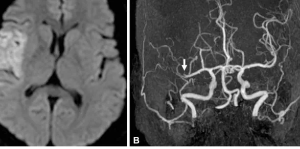
A 52-year-old woman was transferred to our emergency room (ER) with a 10-hour history of left-side weakness, and a sudden onset of slurred speech, with a clear mental state. Her previous medical, me- dication, and familial histories were unremarkable, and she was a non-smoker. Upon arrival in the ER, she was hemodynamically sta- ble and her electrocardiogram showed a normal sinus rhythm. How- ever, a magnetic resonance diffusion image of her brain revealed an infarction of the right middle cerebral artery territory (Fig. 1A), and a magnetic resonance angiogram of the cerebral and carotid arteries showed a total obstruction of the genu branch of the right middle cerebral artery (Fig. 1B). She was immediately admitted to the neu- rologic intensive care unit, with a diagnosis of acute cerebral infarc- tion. Further studies were then ordered, since the etiology of the ce- rebral infarction was unclear. To evaluate the intracardiac source of the embolism, a transthoracic echocardiogram (TTE) and transeso- phageal echocardiogram (TEE) were performed after 1 week. The TTE revealed no evidence of embolism; however, the TEE revealed separ- ation of the septum primum from the septum secundum that con- stitutes a PFO, up to 6 mm, according to the pressure variance (Fig.
2A), and an agitated saline injection test was used to identify the
right-to-left microbubble shunting, during a Valsalva maneuver
used to confirm the PFO presence (Fig. 2B). Based on these tests, we
subsequently recommended an anticoagulation therapy based on
warfarinization.