Hypoparathyroidism and Subclinical Hypothyroidism with Secondary Hemochromatosis
Hyung Ki Jeong, Joon Hwan An, Hyoung Sang Kim, Eun Ae Cho, Min Gui Han, Jung Sik Moon, Hee Kyung Kim, Ho-Cheol Kang
Department of Internal Medicine, Chonnam National University Medical School, Hwasun, Korea
Hemochromatosis is an inherited genetic disorder of iron metabolism which can also occur as a secondary result of iron-overload.
It leads to organ damage such as cardiomyopathy, liver cirrhosis, hypogonadism, and diabetes. This paper discusses a case of sec- ondary hemochromatosis associated with repeated transfusions, presenting as asymptomatic hypoparathyroidism and subclinical hypothyroidism with multiple organ involvement. The 29-year-old female, who had severe aplastic anemia, received multiple transfusions totaling approximately 1,400 units of red blood cells over 15 years. During her routine laboratory examination, hy- pocalcemia was detected with decreased intact parathyroid hormone and increased thyroid stimulating hormone. Serum ferritin, iron, and total iron binding capacity had increased to 27,583.03 ng/mL, 291 μg/dL, and 389 μg/dL, respectively. She had unusu- ally bronze skin and computed tomography revealed iron deposition in the thyroid, liver, and heart. Multiorgan involvement as seen in this case is rare in hemochromatosis associated with secondary transfusions. To the best of the author’s knowledge, this is the first case report in Korea of hypoparathyroidism and subclinical hypothyroidism due to iron deposition in the parathyroid and thyroid gland.
Keywords: Hypoparathyroidism; Hypothyroidism; Hemochromatosis
INTRODUCTION
Hemochromatosis is a hereditary or secondary disorder result- ing from iron-overload and iron-impregnation in the organs, leading to organ damage [1]. Hereditary hemochromatosis is an autosomal recessive disease common in Caucasians and caused by a gene mutation related to iron absorption in the bowels. Some case reports from Korea suggest that end-stage renal disease patients with hemodialysis or patients with aplas- tic anemia had secondary hemochromatosis caused by repeat- ed transfusions [2]. Unlike hereditary hemochromatosis where iron is impregnated in parenchymal cells, repeated transfusion
induced-iron-overload mainly impregnates iron into reticulo- endothelial cells which has been considered to be less harmful organ failure than parenchymal iron accumulation [3]. The au- thors report a case of a patient with aplastic anemia who re- ceived a total of approximately 1,400 units of red blood cells (RBCs) over 15 years. She was admitted for asymptomatic hypocalcaemia and diagnosed with secondary hemochromato- sis accompanied by hypoparathyroidism, hypothyroidism, he- patic dysfunction, and cardiac dysfunction. To the best of the author’s knowledge, this is the first case report of secondary hemochromatosis presenting as hypoparathyroidism and hy- pothyroidism.
Received: 12 February 2013, Accepted: 15 May 2013 Corresponding author: Hee Kyung Kim
Department of Internal Medicine, Chonnam National University Hwasun Hospital, Chonnam National University Medical School, 322 Seoyang-ro, Hwasun 519-763, Korea
Tel: +82-61-379-7621, Fax: +82-61-379-7628, E-mail: [email protected]
Copyright © 2014 Korean Endocrine Society
This is an Open Access article distributed under the terms of the Creative Com- mons Attribution Non-Commercial License (http://creativecommons.org/
licenses/by-nc/3.0/) which permits unrestricted non-commercial use, distribu- tion, and reproduction in any medium, provided the original work is properly cited.
CASE REPORT
A 29-year-old female admitted to our hospital for evaluation of asymptomatic hypocalcemia. Fifteen years ago she was di- agnosed with aplastic anemia, treated with antilymphocyte globulin and antithymocyte globulin, and had routine transfu- sions of RBCs of 2 units per week (about 1,400 units over 15 years). In addition, starting 10 years ago, selective iron-chelat- ing agent (deferasirox 1,500 mg/day) was administered due to increased serum ferritin levels. Continued hypocalcaemia was detected in blood tests at regular follow-up visits and the pa- tient visited the endocrinologic department as an outpatient for additional examinations and treatment. She has no histories of cervical irradiation, thyroid disease or related family history.
She had a blood pressure of 120/80 mm Hg, pulse rate of 78 beats per minute, temperature of 36.8°C, and a respiratory rate of 20 breaths per minute. Her skin was unusually dark in color with conjunctiva pallor. Chest examination showed that breath- ing was clear, heartbeat was regular and no unusual sounds were detected. Her abdomen was flat and soft with no unusual sounds, and the liver and spleen were not palpated. Neither leg had pitting edema. Neurological tests showed no local neuro- logical symptoms, Chvostek sign or Trousseau sign.
In the laboratory findings, peripheral blood test indicated a white blood cell count of 700/mm3 (neutrophil 150), hemoglo- bin level of 8.0 g/dL, and platelet count of 37,000/mm3. A se- rum biochemical test reported blood urea nitrogen levels of 10.1 mg/dL, serum creatinine of 0.7 mg/dL, albumin levels of 3.9 g/dL, aspartate aminotransferase levels of 53 IU/L, alanine aminotransferase levels of 60 IU/L, alkaline phosphatase of 94 IU/L, and total bilirubin of 0.5 mg/dL. In a serum electrolyte test, sodium level was 140 mEq/L and potassium level was 4.3 mEq/L, both of which are in the normal range. However, total calcium was 6.7 mEq/dL (albumin corrected calcium 6.78
mEq/dL; normal range, 8.4 to 10.2), and ionized calcium was 1.78 mEq/L (normal range, 2.2 to 2.6). Serum ferritin levels were 27,583.03 ng/mL (normal range, 4.63 to 274.6), iron lev- els were 291 μg/dL (normal range, 65 to 157), and total iron binding capacity was 389 μg/dL (normal range, 256 to 426) which showed an increase. The transferrin saturation level was high to 74.8% (normal range, 22% to 46%).
She was diagnosed with hypocalcaemia induced by hypo- parathyroidism based on test results showing intact parathy- roid hormone (iPTH) levels of 31 pg/mL (normal range, 9 to 55), 25(OH)VitD of 7.03 ng/mL (normal range, 4.8 to 52.8), and 1,25(OH)2VitD levels of 36.2 pg/mL (normal range, 25.1 to 66.1). Although 1,000 mg of calcium carbonate and 0.5 μg of alfacalcidol were given twice a day, hypocalcaemia contin- ued with increased levels of 24 hours urine calcium (700 mg/
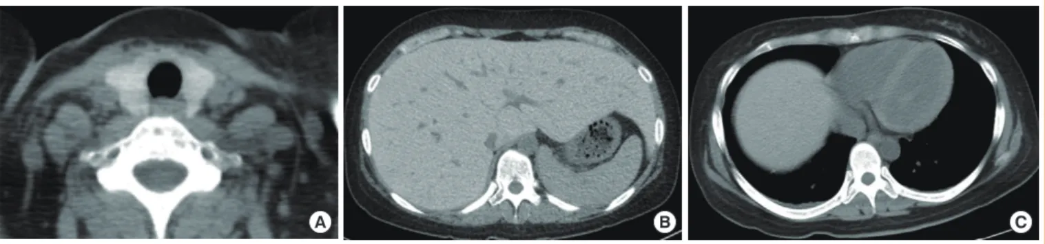
day) and 24 hours urine creatinine (0.99 g/day); thiazide 12.5 mg was also administered to maintain the total calcium at 7.5 to 8.0 mEq/dL and ionized calcium at 1.9 to 2.2 mEq/L. Tests were performed to determine if the hemochromatosis had spread to other organs; chest X-rays showed no signs of car- diomegaly, but computed tomography (CT) before contrast en- hancement showed thyroid (Hounsfield unit 157.0) and liver (Hounsfield unit 136.0), as well as a increased signal intensity associated with the cardiac muscle and enlarged inferior vena cava and hepatic veins (Fig. 1). The T2-weighted magnetic res- onance imaging (MRI) of the abdomen revealed a dark signal intensity in the liver, spleen, bone marrow, and pancreas, lead- ing to the secondary diagnosis of hemochromatosis (Fig. 2).
Organs showing signs of iron impregnation were tested for function; the thyroid function test showed elevated levels of thyroid stimulating hormone (TSH, 17.18 μIU/mL; normal range, 0.4 to 4.8) with free thyroxine levels of 1.28 ng/dL (nor- mal range, 0.8 to 1.71), and triiodothyronine levels of 1.43 ng/
mL (normal range, 0.6 to 1.6). Thyroid peroxidase antibody
A B C
Fig. 1. Nonenhanced computed tomography scan revealed the high intensity of (A) the thyroid gland, (B) liver parenchyma with dilated inferior vena cava and intrahepatic vain, and (C) myocardium.
and thyroglobulin antibody were both negative which resulted in apparent hypothyroidism caused by hemochromatosis. The patient was receiving low-dose of thyroid hormone (levothy- roxine 50 μg/day), but her TSH levels were still over 10 μIU/
mL at follow-up. Therefore the dose of the hormone was grad- ually increased. The most recent test result showed TSH levels of 8.12 μIU/mL, and the dose of thyroid hormone was in- creased to 150 μg/day for follow-up observation. Echocardiog- raphy revealed dilated left ventricular chamber size (left ven- tricular end diastolic diameter 54 mm) with global hypokinesia, and a moderate to severe left ventricular systolic dysfunction (ejection fraction 23%), which suggests congestive heart fail- ure with dilated cardiomyopathy and restrictive diastolic dys- function induced by hemochromatosis. The patient is medicat- ed with digoxin 0.1875 mg, candesartan 8 mg, bisoprolol 2.5 mg, and spiractone 25 mg without any significant symptoms and is on follow-up observation as an outpatient.
DISCUSSION
With this patient, secondary hemochromatosis occurred, in- duced by iron over-absorption due to repeated transfusion for aplastic anemia. While on follow-up observation, she was di- agnosed with hypocalcaemia caused by hypoparathyroidism, subclinical hypothyroidism, hepatic failure, and cardiac dys- function.
Excessive iron absorption is known to cause various organ failures due to iron accumulation of the multiple organs, but it is rare to find clinically severe organ failure cases that involve secondary hemochromatosis induced by transfusion [3]. He- mochromatosis is a genetic or secondary disorder caused by damaged cells and organ failure after iron overload within the
parenchyma of the organ [1]. Patients with symptoms and signs such as increased levels of iron concentration in the blood, iron-binding capacity, and serum ferritin concentration, hepatomegaly, pigmented skin, diabetes mellitus, heart dis- ease, arthritis, and lower urinary tract symptoms can be diag- nosed if the patient has a secondary cause that induces iron overload or if iron impregnation is confirmed by tissue biopsy- with HFE genetic mutation [4]. Normally the human body maintains iron levels at 3 to 4 g. One milligram of iron for males and 1.5 mg for menstruating females are lost every day, but equal amounts of iron are absorbed by intestinal mucosa [5]. However, in the case of hemochromatosis, 4 mg of iron are absorbed per day, which increases iron levels in the blood, leading to increased blood ferritin levels and iron-impregna- tion of the organs. Hereditary hemochromatosis was transmit- ted as an autosomal recessive trait associated with the HLA-A locus on the short arm of chromosome 6. It accounts for 80%
to 90% of all cases of hereditary hemochromatosis [4]. Sec- ondary factors include over-absorption of iron due to inade- quate production of RBCs in cases of Mediterranean anemia and sideroblastic anaemia. In addition, as in this case, repeated transfusions can cause hemochromatosis [2]. When 1 unit of RBC is transfused it contains 200 to 250 mg of iron. If RBC production is inadequate because of bone marrow failure such as aplastic anemia, infused iron cannot be used to produce RBCs and instead is captured by macrophages. When this iron-capturing behavior of macrophages reaches its limit, overloaded iron is impregnated into multiple organs such as the liver, heart, spleen, pancreas, and bone marrow, which re- sults in multiple organ failures [3].
In the early stages of hemochromatosis, fatigue or weakness can be accompanied by arthritis or skin pigmentation. In- creased levels of iron impregnation in organs such as the liver, heart, pancreas, thyroid, parathyroid, and pituitary gland can cause functional failures [6]. One of the organs commonly im- pregnated is the liver, and impregnation of iron in the paren- chyma of the liver can cause hepatomegaly, which is associat- ed with increased levels of hepatic enzyme and can cause of liver cirrhosis and hepatocellular carcinoma. Hepatocellular carcinoma, cardiomyopathy, conduction disturbance, and con- gestive heart failure can increase mortality in patients with he- mochromatosis. Regular check-ups with follow-up visits are necessary to prevent these issues [7,8].
Diabetes mellitus is the most common disease associated with endocrine system abnormalities, and is due to reduced in- sulin resistance and secretion induced by siderosis in liver and Fig. 2. T2-weighted magnetic resonance imaging revealed the
dark signal intensity of liver, pancreas, spleen, and bone marrow.
pancreas β-cells [6]. Gonadotropin deficiency commonly in- cludes the entire pituitary hormone, resulting in impotency, menstrual irregularities, and decreased libido [9]. Siderosis can also cause functional failures in the adrenal, parathyroid, and thyroid, although these issues are less common than diabetes or gonadotropin deficiency [10,11].
There are not a sufficient number of studies on the correla- tion between multiple organ damage and degree and period of siderosis. In 2001, Kwon et al. [12] reported a correlation be- tween total volume of blood transfusion and increased serum ferritin, but there was no correlation between total volume of blood transfusions and endocrine system complications in 14 pediatric patients with repeated transfusions. Chern and Lin [13] reported that 10.7% (3/28) of patients who received blood transfusions over an extended period acquired hypoparathy- roidism, which is often accompanied by other issues such as hypogonadism and diabetes mellitus. In the case study being presented here, the patient was diagnosed with hypoparathy- roidism from hypocalcaemia without markedly increased iPTH, and organ examinations confirmed iron accumulation to the thyroid, liver, and cardiac muscle. This indicates that hemo- chromatosis patients with the rare complication of hypothy- roidism and hypoparathyroidism need to be tested for other or- gan functions and treated accordingly. In the future, larger- scale prospective studies are necessary to determine correla- tions between symptoms and the volume and period of blood transfusions that accompany multiple organ failures.
Hemochromatosis can be correctly diagnosed if siderosis is confirmed from a liver tissue biopsy. However, as shown in this case, for a patient with low platelet levels, even with re- peated platelet transfusions, an invasive examination is difficult to perform. Instead of a liver tissue biopsy, CT before contrast enhancement and MRI can be useful to examine iron impreg- nation. Bell et al. [14] reported a correlation between blood fer- ritin levels and signal intensity of the liver in CT, and suggested that if serum ferritin levels are above 1,000 ng/mL. CT can re- place invasive biopsy and be helpful in diagnosing hemochro- matosis. However, although CT has a high specificity (96%), its sensitivity is relatively low (63%) for diagnosing secondary hemochromatosis [15,16]. Meanwhile, MRI has both high specificity and sensitivity for diagnosing hemochromatosis and evaluating the treatment progress [17]. In this case study, CT before contrast enhancement showed markedly elevated Houn- sfield units in the liver (136.0) compared to the normal range (61.0±9.0) [14], and a T2-weighted liver MRI revealed darker sections of liver, spleen, and bone marrow, which also suggest
hemochromatosis. In the case presented here, hypothyroidism related to other factors was excluded since the patient had mul- tiple functional failures of the organs caused by hemochroma- tosis. The CT results included elevated Hounsfield units of the thyroid (157) compared to normal range (118.1±12.2), Graves disease level of 69.5±17.6, and Hashimoto thyroiditis of 61.4±
9.1 [18]. The elevated Hounsfield units led to the patient’s diag- nosis of siderosis-induced hypothyroidism. In terms of hypo- parathyroidism, previous studies also reported hemochromato- sis in the study subjects. Due to difficulties with imaging and biopsy examinations, hypoparathyroidism induced by hemo- chromatosis was diagnosed although the patients presented with hypocalcemia without increased iPTH [10,11,13,19,20].
Patients with secondary hemochromatosis induced from re- peated transfusions may have irreversible functional disorders with severe impacts or consequences on their daily life [13].
Endocrine failure can cause delayed development and second- ary sexual characteristics in children. Therefore, it is critical to restrict transfusions and iron chelate agents as needed, and schedule patients for regular follow-up visits. Furthermore, it is necessary to establish screening examinations each period, with a larger-scale study requiring repeated transfusions to clarify the effects of the amount and period of blood transfu- sions on endocrine failure.
CONFLICTS OF INTEREST
No potential conflict of interest relevant to this article was re- ported.
REFERENCES
1. Burt MJ, George DK, Powell LW. Haemochromatosis: a clinical update. Med J Aust 1996;164:348-51.
2. Kim MK, Lim DJ, Baek KH, Song KH, Kang MI, Lee KW, Lee JW. A case of transfusion-associated hemochro- matosis involving multiple organs. Korean J Med 2008;75:
709-13.
3. Schafer AI, Cheron RG, Dluhy R, Cooper B, Gleason RE, Soeldner JS, Bunn HF. Clinical consequences of acquired transfusional iron overload in adults. N Engl J Med 1981;
304:319-24.
4. Allen KJ, Gurrin LC, Constantine CC, Osborne NJ, De- latycki MB, Nicoll AJ, McLaren CE, Bahlo M, Nisselle AE, Vulpe CD, Anderson GJ, Southey MC, Giles GG, English DR, Hopper JL, Olynyk JK, Powell LW, Gertig
DM. Iron-overload-related disease in HFE hereditary he- mochromatosis. N Engl J Med 2008;358:221-30.
5. Beard JL, Dawson H, Pinero DJ. Iron metabolism: a com- prehensive review. Nutr Rev 1996;54:295-317.
6. Hahn JU, Steiner M, Bochnig S, Schmidt H, Schuff-Wer- ner P, Kerner W. Evaluation of a diagnostic algorithm for hereditary hemochromatosis in 3,500 patients with diabe- tes. Diabetes Care 2006;29:464-6.
7. Adams PC, Deugnier Y, Moirand R, Brissot P. The rela- tionship between iron overload, clinical symptoms, and age in 410 patients with genetic hemochromatosis. Hepa- tology 1997;25:162-6.
8. Rivers J, Garrahy P, Robinson W, Murphy A. Reversible cardiac dysfunction in hemochromatosis. Am Heart J 1987;
113:216-7.
9. Fujisawa I, Morikawa M, Nakano Y, Konishi J. Hemochro- matosis of the pituitary gland: MR imaging. Radiology 1988;
168:213-4.
10. Himoto Y, Kanzaki S, Nomura H, Araki T, Takahashi Y, Seino Y. Hypothyroidism and hypoparathyroidism in an 11 year old boy with hemochromatosis secondary to aplastic anemia. Acta Paediatr Jpn 1995;37:534-6.
11. Shirota T, Shinoda T, Aizawa T, Mizukami T, Katakura M, Takasu N, Yamada T. Primary hypothyroidism and multi- ple endocrine failure in association with hemochromatosis in a long-term hemodialysis patient. Clin Nephrol 1992;
38:105-9.
12. Kwon HJ, Joo SW, Kook JH, Rha JY, Kook H, Woo YJ, Hwang TJ. Endocrinopathy in hemochromatosis patients multi-transfused for aplastic anemia. Korean J Pediatr He- matol Oncol 2001;8:181-8.
13. Chern JP, Lin KH. Hypoparathyroidism in transfusion-de- pendent patients with beta-thalassemia. J Pediatr Hematol
Oncol 2002;24:291-3.
14. Bell H, Rostad B, Raknerud N, Try K. Computer tomogra- phy in the detection of hemochromatosis. Tidsskr Nor Lae- geforen 1994;114:1697-9.
15. Howard JM, Ghent CN, Carey LS, Flanagan PR, Valberg LS. Diagnostic efficacy of hepatic computed tomography in the detection of body iron overload. Gastroenterology 1983;84:209-15.
16. Guyader D, Gandon Y, Deugnier Y, Jouanolle H, Loreal O, Simon M, Bourel M, Carsin M, Brissot P. Evaluation of computed tomography in the assessment of liver iron over- load. A study of 46 cases of idiopathic hemochromatosis.
Gastroenterology 1989;97:737-43.
17. Alustiza JM, Artetxe J, Castiella A, Agirre C, Emparanza JI, Otazua P, Garcia-Bengoechea M, Barrio J, Mujica F, Recondo JA; Gipuzkoa Hepatic Iron Concentration by MRI Study Group. MR quantification of hepatic iron con- centration. Radiology 2004;230:479-84.
18. Iida Y, Konishi J, Harioka T, Misaki T, Endo K, Torizuka K.
Thyroid CT number and its relationship to iodine concen- tration. Radiology 1983;147:793-5.
19. Tanimoto K, Okubo Y, Harada C, Saito H, Sata A, Ni- shikawa A, Ohwada R, Tsuiki M, Yamamoto M, Hashimo- to E, Sato K, Takano K. Latent hypoparathyroidism in an osteoporotic patient with multiple endocrinopathies and secondary hemochromatosis due to multiple blood transfu- sions, unmasked by alendronate and glucocorticoid at ad- renal crisis. Intern Med 2008;47:515-20.
20. Mautalen CA, Kvicala R, Perriard D, Bugnard E, Rossi E, Duhart J. Case report: hypoparathyroidism and iron stor- age disease. Treatment with 25-hydroxy-vitamin D3. Am J Med Sci 1978;276:363-8.