ORIGINAL ARTICLE
위암에서 Helicobacter pylori CagA에 따른 RUNX3의 메틸화 및 발현의 소실과 임상병리학적 특성과의 관계
나윤주1,3, 심기남1, 주양희1, 김성은1, 정혜경1, 정성애1, 조민선2
이화여자대학교 의학전문대학원 내과학교실1, 병리학교실2, 삼성서울병원 건강의학본부3
RUNX3 Methylation, Loss of RUNX3 Expression and Clinicopathologic Findings according to Helicobacter pylori CagA in Gastric Carcinoma
Yoon Ju Na1,3, Ki-Nam Shim1, Yang Hee Joo1, Seong-Eun Kim1, Hye-Kyung Jung1, Sung-Ae Jung1, and Min Sun Cho2
Departments of Internal Medicine1 and Pathology2, Ewha Womans University School of Medicine, Center for Health Promotion, Samsung Medical Center3, Seoul, Korea
Background/Aims: Helicobacter pylori cytotoxin-associated gene A (CagA) has been suggested to be involved in the inactivation of Runt-related transcription factor 3 (RUNX3), a known gastric carcinoma tumor suppressor gene. It remains unclear how H. pylori CagA initiates or maintains RUNX3 promoter methylation and inactivates its protein expression in gastric carcinoma.
Methods: RUNX3 promoter methylation status, RUNX3 expression, and H. pylori CagA were investigated in 76 sample pairs of gastric carcinoma tissue. The patients’ medical records were reviewed. The association between RUNX3 methylation or loss of RUNX3 expression and clinicopathologic variables according to H. pylori CagA status were investigated.
Results: In gastric carcinoma patients with H. pylori CagA-positive infection, RUNX3 methylation did not show association with lymphatic invasion, venous invasion, and TNM stages. However RUNX3 methylation was observed more frequently in poorly differentiated adenocarcinoma and signet ring cell carcinoma (77.8% vs. 20.0%, p=0.023) in early stage. In gastric carcinoma patients with H. pylori CagA-positive infection, loss of RUNX3 expression did not show association with lymphatic invasion, venous invasion, and TNM stages. However loss of RUNX3 expression was observed more frequently in early gastric carcinoma than in advanced gastric carcinoma (84.2% vs. 75.0%, p=0.51), but this difference was not significant.
Conclusions: In gastric carcinoma patients with H. pylori CagA-positive infection, RUNX3 methylation or loss of RUNX3 expression did not show correlation with lymphovascular invasion and TNM stages. In early gastric carcinoma patients with H. pylori CagA-positive infection, RUNX3 methylation was observed more in poorly differentiated adenocarcinoma and signet ring cell carcinoma. (Korean J Gastroenterol 2015;66:75-84)
Key Words: Helicobacter pylori; CagA; RUNX3; Methylation; Carcinoma
Received February 2, 2015. Revised June 16, 2015. Accepted June 26, 2015.
CC This is an open access article distributed under the terms of the Creative Commons Attribution Non-Commercial License (http://creativecommons.org/licenses/
by-nc/4.0) which permits unrestricted non-commercial use, distribution, and reproduction in any medium, provided the original work is properly cited.
Copyright © 2015. Korean Society of Gastroenterology.
교신저자: 심기남, 07985, 서울시 양천구 안양천로 1071, 이화여자대학교 의학전문대학원 내과학교실
Correspondence to: Ki-Nam Shim, Department of Internal Medicine, Ewha Womans University School of Medicine, 1071 Anyangcheon-ro, Yangcheon-gu, Seoul 07985, Korea. Tel: +82-2-2650-2632, Fax: +82-2-2655-2076, E-mail: [email protected]
Financial support: None. Conflict of interest: None.
INTRODUCTION
Helicobacter pylori infection occurs in over 80% of all gas- tric carcinoma patients1 with the bacterium classified as a group I human carcinogen for gastric adenocarcinoma.2 H.
pylori infection is more associated with early gastric carcino- ma patients than with non-neoplastic lesions or advanced gastric carcinoma patients.3 Cytotoxin-associated gene A (CagA) is a significant virulence factor of H. pylori.4 The CagA pathogenicity island also encodes a type IV secretion system
for injection of Cag A into epithelial cells of the stomach, where it causes disruption of the cell cytoskeleton, apical junction complex, and other intracellular activities.5,6 Infection with CagA-positive strains of H. pylori further in- creases the risk for non-cardiac gastric cancer as compared to the risk associated with H. pylori infection alone.7
Since 2002, Runt-related transcription factor 3 (RUNX3) has been recognized as a tumor suppressor gene for gastric cancer.8 RUNX3 is frequently inactivated in gastric cancer through the following four mechanisms: aberrant methyl- ation of the gene’s promoter, protein mislocalization, histone modification, and hemizygous deletion.8,9 RUNX3 inactiva- tion by methylation occurs at the CpG site by the addition of a methyl group, subsequently converting to 5-methylcytosine.
In a recent meta-analysis, an association between methyl- ation of the RUNX3 promoter and gastric carcinoma was re- ported, confirming the role of RUNX3 as a tumor suppressor gene.10 The same study found that the RUNX3 promoter methylation status was not correlated with either TNM stag- ing or lymphatic and venous invasion.10 However, RUNX3 in- activation by protein mislocalization is a marker for the loss of tumor suppression and impairment of transforming growth factor- signaling in gastric carcinoma.9
The relationship between H. pylori CagA and RUNX3 meth- ylation or protein mislocalization has only recently been eluci- dated in early gastric cancer tissue. H. pylori 16S RNA may be an independent risk factor for RUNX3 methylation in patients with early gastric carcinoma.11 The protein mislocalization of RUNX3, because of which it moves from the nucleus to the cytoplasm, may be a major mechanism underlying the asso- ciation between H. pylori infection and gastric carcinoma development.9 RUNX3 inactivation occurs early during the progression to malignancy.10,12,13
In the current study, we investigated the association be- tween RUNX3 methylation and various clinicopathological variables in gastric carcinoma patients according to their H.
pylori CagA-positive infection status. We also examined the relationship between RUNX3 protein expression and the clin- icopathological variables according to CagA-positive H. pylori infection.
SUBJECTS AND METHODS
1. Patients
Seventy-six patients with primary gastric carcinoma were included in the current study. All patients underwent curative or palliative subtotal or total gastrectomy. None of them re- ceived chemotherapy or radiotherapy preoperatively. For the analysis of the methylation status by methylation specific PCR, endoscopic biopsy of macroscopically neoplastic and non-neoplastic mucosa were collected, snap-frozen, and stored at −70oC for isolation of genomic DNA. For im- munohistochemical staining of the RUNX3 protein, formal- in-fixed and paraffin-embedded operative samples were pre- pared from the tissues of the 76 patients. The patients’ in- formation including age, gender, tumor site, lymphatic in- vasion, venous invasion, and TNM tumor classification ac- cording to the American Joint Committee on Cancer/Union for International Cancer Control staging system for gastric cancer (7th edition, 2010) were documented. Histological tu- mor differentiation statuses were determined on the basis of the World Health Organization classification of pathology and the genetics of tumors of the digestive system.14 Rapid ure- ase test, H&E stain, and modified Giemsa stain were per- formed for evaluation of H. pylori infection status. This study was approved by the Institutional Human Research Board of Ewha Woman’s University Mokdong Hospital, Seoul, Korea (ECT-12-22A-23). Informed consent was obtained from all patients.
2. Methylation-specific PCR for RUNX3
Methylation-specific PCR (MSP) was performed using the bisulfite-modified DNA templates obtained from the endo- scopic specimen of human gastric carcinoma tissues. Endo- scopic samples from neoplastic and non-neoplastic gastric mucosa were used. The macroscopic non-neoplastic gastric mucosa was defined to be at least 5 cm away from the neo- plastic gastric mucosa. Genomic DNA was obtained by protei- nase K digestion and extraction using the LaboPassTM Tissue Mini kit (COSMO Genetech, Seoul, Korea). The genomic DNA concentration and purity were measured using a spectropho- tometer (purity=1.7−1.9 at 260/280 nm) and the DNA was treated with sodium bisulfite according to the manufac- turer’s instructions. Thereafter, 2 g of the genomic DNA was denatured with 2 M NaOH and modified with 3 M sodium bi-
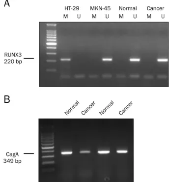
Fig. 1. Representative results of methylation-specific PCR for RUNX3 and PCR for CagA in human normal and gastric cancer tissue samples. (A) Methylation specific PCR for RUNX3. Positive control, negative control, molecular weighted marker for RUNX3 and un-methylated bands (U) were seen in cancer tissue. (B) PCR for CagA. Presence of band in normal gastric mucosa or gastric carcinoma tissue was considered positive for Helicobacter pylori CagA.
sulfite for 16 h. The DNA samples were then purified using a DNA purification kit (Intron®, Seongnam, Korea) with 3 M NaOH, precipitated with ethanol, and resuspended in 20 L distilled water. RUNX3 was amplified using the GeneAmp PCR system 9600 (Perkin-Elmer, Wellesley, MA, USA); the PCR reaction mixture (10 L) consisted of 2 L of the DNA template, 0.1 L of TaKaRa hotstart Taq polymerase, 0.2 mM of deoxynucleotide triphosphates, 0.5 M of each primer (sense and antisense), 1 L of the primer 10× PCR buffer, and 5.6 L of distilled water. The PCR conditions were as follows:
95oC for 1 min and 40 cycles of denaturation at 95oC for 30 s, annealing at 66oC (methylated) or 63oC (unmethylated) for 30 s, and final 30 s extension at 72oC, followed by a final 5-min extension at 72oC. The PCR products were loaded onto a 2% agarose gel and visualized under ultraviolet (UV) transillumination. Irrespective of the methylation status in normal tissues, the presence of methylated bands and parti- ally methylated bands was considered to indicate that the carcinoma tissues were positive for RUNX3 methylation. The primer sequences for the methylated RUNX3 gene were as follows: 5′-TACGAGGGGCGGTCGTACGCGGG-3′ and 5′-AAAAC GACCGACGCGAACGCCTCC-3′ (64,970-65,189 bp). The pri- mer sequences for the unmethylated RUNX3 gene were as follows: 5′-TTATGAGGGGTGGTTGTATGTGGG-3′ and 5′-AAAAC AACCAACACAAACACCTCC-3′ (64,970-65,189 bp). The primers for GAPDH were as follows: 5′-GCCTCAAGATCATCAGCAAT-3′
(530-549 bp) and 5′-TCCAGCTCAGGGATGACCTT-3′. Valida- tion of the primer sets had been previously performed by bi- sulfate genomic sequencing of the MSP products in the carci- noma cell lines. DNA from the HT-29 (Korean Cell Line Bank [KCLB] No. 30038) and MKN-45 (KCLB No. 80103) cell lines were used as positive controls for methylated and un- methylated RUNX3, respectively (Fig. 1A). Distilled water was used as the negative control.
3. PCR for H. pylori CagA
The amplification reaction mixture (20 L) contained 2 L DNA template, 0.1 L GoTaq polymerase, 0.2 mM dNTPs, 0.5
M of each primer (sense and antisense), 4 L of the primer 5× PCR buffer, and 5.6 L distilled water. The primers for H.
pylori CagA15,16 were as follows: 5′-GATAACA GGCAAGCTTTTG AGG-3′ and 5′- CTGCAAAAGATTGTTTGGCAGA-3′ (1,228-1,556 bp). The PCR conditions were as follows: 94oC for 1 min fol- lowed by 40 cycles of 94oC for 1 min, 55oC for 1 min, 72oC for
1 min, and a final 5-min extension at 72oC.15 The PCR prod- ucts were loaded onto a 2% agarose gel and visualized under UV transillumination. The presence of bands in normal or car- cinoma tissues indicated positivity for CagA (Fig. 1B). Endo- scopic samples from neoplastic and non-neoplastic gastric mucosa were used.
4. Immunohistochemical staining for RUNX3
Immunohistochemical analysis was performed using the BondTM Polymer DAB Detection Kit (Leica Microsystems, Weltzar, Germany) and Bond-X automated immunohisto- chemistry slide staining system (Leica Microsystems).
Formalin-fixed and paraffin-embedded serial sections using four micron-thick samples were deparaffinized and de- hydrated. The sections were heated in citrate buffer (pH 6.0, 10 mM) in a microwave for 20 min for retrieval of antigens.
Endogenous peroxidase activity was blocked with 0.3% hy- drogen peroxide for 10 min followed by incubation with the primary antibodies: mouse monoclonal anti-RUNX3 antibody
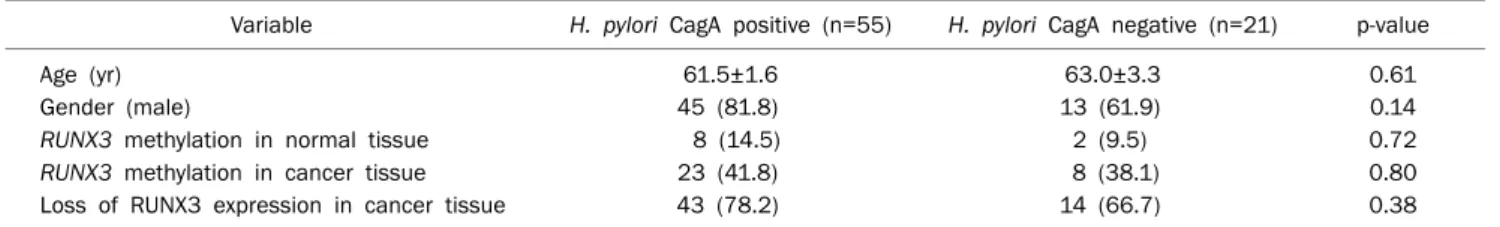
Fig. 2. Immunohistochemical stain for RUNX3 protein. (A) Adjacent mucosa surrounded by tumor showed cytoplasmic positive reaction in goblet cells (black arrowhead). The lymphocytes in lamina propria showed a nuclear positive reaction of lymphocytes (blue arrowhead) (×200). (B) The tumor cells showed a negative reaction (×200). (C) The tumor cells showed a strong brownish nuclear positive reaction (×200). (D) The tumor cells showed no nuclear positive reaction but a cytoplasmic positive reaction (cytoplasmic mislocalization) (black arrowheads) (×400).
(eBioscience, San Diego, CA, USA). Sections were then wash- ed in phosphate-buffered saline and incubated with horse- radish-peroxidase polymer for 30 min. The reaction was vi- sualized after incubation with 3,3-diaminobenzidine tetrahy- drochloride for 5 min. The samples were then counterstained with Mayer’s hematoxylin.
RUNX3 staining showed positive staining in the nuclei of mononuclear inflammatory cells and in the cytoplasmic re- gions of goblet cells of gastric glands with intestinal meta- plasia (Fig. 2A). When no staining or only cytoplasmic staining was detected, the sample was considered negative for RUNX3 expression or was considered as showing loss of nu- clear RUNX3 expression (Fig. 2B). Nuclear staining alone or both nuclear and cytoplasmic staining indicated positive RUNX3 expression. This determination was made by a
pathologist.
5. Statistical analysis
The relationship between RUNX3 methylation status and clinicopathological variables was investigated according to CagA-positive H. pylori infection in gastric carcinoma patients.
The results of immunohistochemical staining for RUNX3 were analyzed according to H. pylori CagA status and the sta- tus of RUNX3 methylation. The association between RUNX3 expression and clinicopathological variables was analyzed.
Statistical analyses were performed using a two-tailed Fisher’s exact test or a linear test. The results of the analyses of continuous variables are expressed as mean±standard deviation, as determined using Student’s t-test. Data analy- ses were performed using the STATA version 12.0 (Stata
Table 1. Baseline Characteristics in Gastric Carcinoma Patients
Variable Total (n=76)
Age (yr) 61.9±1.5
Gender (male) 57 (75.0)
H. pylori positive 59 (77.6)
H. pylori CagA positive 55 (72.4) Differentiation
W/D, M/D 27 (35.5)
P/D, SRC 49 (64.5)
Location of tumor
Upper 12 (15.8)
Middle 17 (22.4)
Lower 45 (59.2)
Whole 2 (2.6)
Lymphatic invasion
Negative 26 (34.2)
Positive 50 (65.8)
Venous invasion
Negative 54 (71.1)
Positive 22 (28.9)
Depth of invasion
T1a (mucosa) 10 (13.2)
T1b (submucosa) 14 (18.4)
T2, 3, 4 52 (68.4)
Nodal status
N0 26 (34.2)
N1, 2, 3 50 (65.8)
Distant metastasis
M0 61 (80.3)
M1 15 (19.7)
Stage
I 22 (28.9)
II 15 (19.7)
III 24 (31.7)
IV 15 (19.7)
Values are presented as mean±SD or n (%).
W/D, tubular adenocarcinoma, well differentiated; M/D, tubular adenocarcinoma moderately differentiated; P/D, tubular adeno- carcinoma, poorly differentiated; SRC, signet ring cell cancer.
Table 2. Correlation between Helicobacter pylori CagA Status and Clinicopathologic Variables in Gastric Carcinoma
Variable H. pylori CagA positive (n=55) H. pylori CagA negative (n=21) p-value
Age (yr) 61.5±1.6 63.0±3.3 0.61
Gender (male) 45 (81.8) 13 (61.9) 0.14
RUNX3 methylation in normal tissue 8 (14.5) 2 (9.5) 0.72
RUNX3 methylation in cancer tissue 23 (41.8) 8 (38.1) 0.80
Loss of RUNX3 expression in cancer tissue 43 (78.2) 14 (66.7) 0.38
Values are presented as mean±SD or n (%).
Corp., College Station, TX, USA), and p<0.05 was considered to indicate a statistically significant difference.
RESULTS
1. Baseline characteristics in gastric carcinoma patients The enrolled patients included 57 men and 19 women with a mean age of 61.9±1.5 years (range, 35-90 years). All 76 participants were pathologically confirmed to have adeno- carcinoma or signet ring cell carcinoma, with 49 participants (64.5%) diagnosed with poorly differentiated adenocarcinoma or signet ring cell carcinoma. Fifty-nine (77.6%) patients showed H. pylori infection. Twenty-two (28.9%) patients showed venous invasion and 50 (65.8%) showed lymphatic invasion. Twenty-two (28.9%), 15 (19.7%), 24 (31.7%), and 15 (19.7%) patients had stage I, stage II, stage III, and stage IV disease, respectively (Table 1).
2. Correlation between H. pylori CagA status and RUNX3 inactivation
H. pylori CagA status did not show correlation with RUNX3 methylation or a loss of nuclear RUNX3 expression in cancer tissue. H. pylori CagA status also did not show correlation with RUNX3 methylation in normal tissues (Table 2). However, CagA-positive H. pylori infection was observed in 74.2% of RUNX3 methylation occurrences (23/31 cases), and 75.4%
of the cases had a loss of nuclear RUNX3 expression (43/57 cases).
3. Correlation between RUNX3 methylation status and clinicopathological variables according to H. pylori CagA status in gastric carcinoma patients
The methylation status of the RUNX3 promoter was exam- ined in all 76 enrolled patients, including 24 early stage gas- tric carcinoma patients. RUNX3 methylation was found in 31 (40.8%) cases. No correlation was found between RUNX3
methylation status and age, gender, or H. pylori CagA status.
Similarly, the RUNX3 methylation status did not show correla- tion with histological differentiation, lymphatic invasion, ve- nous invasion, T stage, N stage, distant metastasis, or tumor stage in any of the gastric carcinoma cases (data not shown).
Table 3. Correlation between RUNX3 Methylation and Clinicopathologic Variables according to Helicobacter pylori CagA Status in Gastric Carcinoma (n=76)
Variable
H. pylori CagA positive (n=55) H. pylori CagA negative (n=21) RUNX3 methylation
positive (n=23)
RUNX3 methylation
negative (n=32) p-value RUNX3 methylation positive (n=8)
RUNX3 methylation negative (n=13) p-value
Differentiation 1.00 1.00
W/D, M/D 9 (39.1) 13 (40.6) 2 (25.0) 3 (23.1)
P/D, SRC 14 (60.9) 19 (59.4) 6 (75.0) 10 (76.9)
Lymphatic invasion 0.78 0.047
Negative 8 (34.8) 13 (40.6) 4 (50.0) 1 (7.7)
Positive 15 (65.2) 19 (59.4) 4 (50.0) 12 (92.3)
Venous invasion 0.55 0.046
Negative 15 (65.2) 24 (75.0) 8 (100) 7 (53.8)
Positive 8 (34.8) 8 (25.0) 0 (0) 6 (46.2)
Depth of invasion 0.40 0.24
T1a 5 (21.7) 3 (9.3) 2 (25.0) 0 (0.0)
T1b 4 (17.4) 7 (21.9) 1 (12.5) 2 (15.4)
T2, 3, 4 14 (60.9) 22 (68.8) 5 (62.5) 11 (84.6)
Nodal status 1.00 0.012
N0 9 (39.1) 13 (40.6) 4 (50.0) 0 (0)
N1, 2, 3 14 (60.9) 19 (59.4) 4 (50.0) 13 (100)
Distant metastasis 1.00 1.00
M0 19 (82.6) 27 (84.4) 6 (75.0) 9 (69.2)
M1 4 (17.4) 5 (15.6) 2 (25.0) 4 (30.8)
Stage 0.55 0.29
I 6 (26.1) 11 (34.4) 3 (37.5) 2 (15.4)
II 8 (34.8) 6 (18.8) 1 (12.5) 0 (0)
III 5 (21.7) 10 (31.2) 2 (25.0) 7 (53.8)
IV 4 (17.4) 5 (15.6) 2 (25.0) 4 (30.8)
Values are presented as n (%).
W/D, tubular adenocarcinoma, well differentiated; M/D, tubular adenocarcinoma, moderately differentiated; P/D, tubular adenocarcinoma, poorly differentiated; SRC, signet ring cell cancer.
Patients were divided into two groups according to their H.
pylori CagA status, and the association between RUNX3 methylation and pathologic variables was investigated. In gastric carcinoma patients with H. pylori CagA-positive in- fection, no correlation was found between RUNX3 methyl- ation and lymphatic invasion, venous invasion, T stage, N stage, distant metastasis, or tumor stage (Table 3).
In H. pylori CagA-negative gastric carcinoma patients, those without RUNX3 methylation showed lymphatic in- vasion (92.3% vs. 50.0%, p=0.047), venous invasion (46.2%
vs. 0%, p=0.046), and lymph node metastasis (100% vs.
50.0%, p=0.012) more frequently than those with RUNX3 methylation (Table 3). However, in early gastric carcinoma pa- tients with CagA-positive H. pylori infection, RUNX3 methyl- ation occurred more often in cases of poorly differentiated adenocarcinoma or signet ring cell carcinoma than in cases of well or moderately differentiated adenocarcinoma (77.8%
vs. 20.0%, p=0.023) (Table 4).
4. Correlation between RUNX3 expression and clinic- opathological variables according to H. pylori CagA status in gastric carcinoma patients
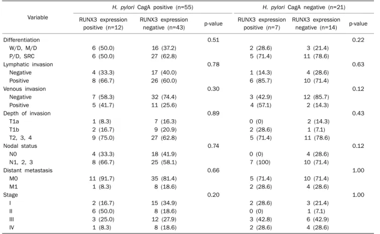
Immunohistochemical staining for RUNX3 was performed on samples obtained from all 76 patients. A loss of nuclear RUNX3 expression (negative expression) was found in 57 (75.0%) patients, and nuclear RUNX3 expression (positive expression) was found in 19 (25.0%) patients (Fig. 2C).
Three cases of cytoplasmic mislocalization were observed in two patients with early gastric cancer and one patient with advanced gastric cancer (Fig. 2D). Of the 31 patients with RUNX3 methylation, 26 (83.9%) had a loss of RUNX3 ex- pression, and 31 (68.9%) of the 45 patients without RUNX3 methylation had a loss of RUNX3 expression (p=0.14).
Loss of nuclear RUNX3 expression (negative expression) did not show correlation with histological differentiation, lym- phatic invasion, venous invasion, T stage, N stage, distant
Table 4. Correlation between RUNX3 Methylation and Clinicopathologic Variables according to Helicobacter pylori CagA Status in Early Gastric Carcinoma (n=24)
Variable
H. pylori CagA positive (n=19) H. pylori CagA negative (n=5) RUNX3 methylation
positive (n=9)
RUNX3 methylation
negative (n=10) p-value RUNX3 methylation positive (n=3)
RUNX3 methylation negative (n=2) p-value
Differentiation 0.023 0.40
W/D, M/D 2 (22.2) 8 (80.0) 0 (0) 1 (50.0)
P/D, SRC 7 (77.8) 2 (20.0) 3 (100) 1 (50.0)
Lymphatic invasion 0.65 0.10
Negative 5 (55.6) 7 (70.0) 3 (100) 0 (0)
Positive 4 (44.4) 3 (30.0) 0 (0) 2 (100)
Venous invasion 0.58 0.40
Negative 7 (77.8) 9 (90.0) 3 (100) 1 (50.0)
Positive 2 (22.2) 1 (10.0) 0 (0) 1 (50.0)
Depth of invasion 0.37 0.40
1a 5 (55.6) 3 (30.0) 2 (66.7) 0 (0)
1b 4 (44.4) 7 (70.0) 1 (33.3) 2 (100)
Nodal status 0.65 0.10
N0 5 (55.6) 7 (70.0) 3 (100) 0 (0)
N1, 2, 3 4 (44.4) 3 (30.0) 0 (0) 2 (100)
Stage 0.63 1.00
I 6 (66.7) 8 (80.0) 3 (100) 2 (100)
II 3 (33.3) 2 (20.0) 0 (0) 0 (0)
Values are presented as n (%).
W/D, tubular adenocarcinoma, well differentiated; M/D, tubular adenocarcinoma, moderately differentiated; P/D, tubular adenocarcinoma, poorly differentiated; SRC, signet ring cell cancer.
metastasis, or tumor stage in any of the gastric carcinoma pa- tients (data not shown). In gastric carcinoma patients with CagA-positive H. pylori infection too, no association was found between the loss of RUNX3 expression and histological differentiation, lymphatic invasion, venous invasion, T stage, N stage, distant metastasis, or tumor stage (Table 5). In pa- tients with H. pylori CagA infection, loss of RUNX3 expression in early gastric carcinoma patients was more than that in ad- vanced gastric carcinoma patients (84.2% [16/19] vs.
75.0% [27/36]) (p=0.51), but the differences were not significant.
DISCUSSION
In our study, in gastric carcinoma patients with CagA-pos- itive H. pylori infection, RUNX3 methylation status or loss of RUNX3 expression was not found to show association with lymphatic invasion, venous invasion, or TNM stages. However, RUNX3 methylation is more significantly found in poorly dif- ferentiated adenocarcinoma and signet ring cell carcinoma in the early stages. Loss of RUNX3 expression was found more often in early gastric carcinoma than in advanced gas- tric carcinoma.
The RUNX family includes RUNX1, RUNX2, and RUNX3, with each member exerting different regulatory functions in intracellular processes.17 RUNX3, with its encoding gene lo- cated on chromosome 1p36, is involved in gastric epithelial growth8,12 and T-cell differentiation.18 Mislocalization of RUNX3 leading to its activation has been reported to result in a loss of nuclear RUNX3 expression in 44% of gastric carci- noma cases and cytoplasmic mislocalization in 38% of cases.9 Inactivation of RUNX3 by promoter methylation was reported in 45-65% of all gastric carcinomas.10,19,20
Most reported cases of gastric carcinoma in East Asia are associated with CagA-positive H. pylori infection. In compar- ison to the Western CagA-positive H. pylori strain, CagA in East Asia more often causes severe gastric mucosal in- flammation and is associated with severe atrophic gastritis and gastric adenocarcinoma.21,22 In a meta-analysis, the per- centage of patients sero-positive for H. pylori CagA in East Asia varied from 61% to 96.6% of the total number of gastric cancer patients.23 This positive result included both current and past infections. In another meta-analysis, the rate of CagA infection, determined using PCR techniques, varied from 79% to 100% in gastric cancer.24 The rate of the East-Asian type H. pylori CagA was 83%.24 In this study, the
Table 5. Correlation between RUNX3 Expression and Clinicopathologic Variables according to Helicobacter pylori CagA Status in Gastric Carcinoma (n=76)
Variable
H. pylori CagA positive (n=55) H. pylori CagA negative (n=21) RUNX3 expression
positive (n=12)
RUNX3 expression
negative (n=43) p-value RUNX3 expression positive (n=7)
RUNX3 expression negative (n=14) p-value
Differentiation 0.51 0.22
W/D, M/D 6 (50.0) 16 (37.2) 2 (28.6) 3 (21.4)
P/D, SRC 6 (50.0) 27 (62.8) 5 (71.4) 11 (78.6)
Lymphatic invasion 0.78 0.63
Negative 4 (33.3) 17 (40.0) 1 (14.3) 4 (28.6)
Positive 8 (66.7) 26 (60.0) 6 (85.7) 10 (71.4)
Venous invasion 0.30 0.12
Negative 7 (58.3) 32 (74.4) 3 (42.9) 12 (85.7)
Positive 5 (41.7) 11 (25.6) 4 (57.1) 2 (14.3)
Depth of invasion 0.89 0.43
T1a 1 (8.3) 7 (16.3) 0 (0) 2 (14.3)
T1b 2 (16.7) 9 (20.9) 2 (28.6) 1 (7.1)
T2, 3, 4 9 (75.0) 27 (62.8) 5 (71.4) 11 (78.6)
Nodal status 0.74 0.12
N0 4 (33.3) 18 (41.9) 0 (0) 4 (28.6)
N1, 2, 3 8 (66.7) 25 (58.1) 7 (100) 10 (71.4)
Distant metastasis 0.66 1.00
M0 11 (91.7) 35 (81.4) 5 (71.4) 10 (71.4)
M1 1 (8.3) 8 (18.6) 2 (28.6) 4 (28.6)
Stage 0.20 1.00
I 2 (16.7) 15 (34.9) 2 (28.6) 3 (21.4)
II 6 (50.0) 8 (18.6) 0 (0) 1 (7.1)
III 3 (25.0) 12 (27.9) 3 (42.8) 6 (42.9)
IV 1 (8.3) 8 (18.6) 2 (28.6) 4 (28.6)
Values are presented as n (%).
W/D, tubular adenocarcinoma, well differentiated; M/D, tubular adenocarcinoma, moderately differentiated; P/D, tubular adenocarcinoma, poorly differentiated; SRC, signet ring cell cancer.
rate of CagA-positive H. pylori infection determined using PCR techniques16 was 86.4%. In the subset of early gastric carci- noma patients, RUNX3 methylation was observed at a higher frequency among cases of poorly differentiated adenocar- cinoma or signet ring cell carcinoma. Other studies have shown that a higher rate of RUNX3 methylation may be re- lated to gastric carcinoma with undifferentiated histopathol- ogy10 and poorly differentiated colorectal carcinoma.25,26 There are possible explanations regarding the mechanism of H. pylori-induced RUNX3 methylation. Nitric oxide (NO) has been suggested to play important roles in RUNX3 methyla- tion. H. pylori CagA might increase the level of NO or other in- flammatory cytokines.27,28 NO, along with lipopolysac- charide, then induces RUNX3 methylation in early gastric carcinoma.29 In this study, RUNX3 methylation did not show association with loss of RUNX3 expression. The rate of loss of RUNX3 expression in patients with RUNX3 methylation was 83.9%, and 68.9% in patients without RUNX3 methyla-
tion. In patients without RUNX3 methylation, protein mis- localization or other factors such as hypoxia30 may be related to the loss of RUNX3 expression.
Although the pathogenesis of CagA infection has not been completely elucidated, we hypothesize that CagA infection has two probable roles in cell signaling. Phosphorylation-de- pendent cell signaling of CagA is known to be associated with apoptosis, cell motility, elongation, proliferation, and inflam- mation. Phosphorylation-independent cell signaling of CagA is known to induce the disruption of cellular junctions.31 Recent studies have shown that H. pylori or H. pylori CagA in- fection plays an important role in RUNX3 expression. H. pylori CagA can result in a loss of RUNX3 protein expression in a phosphorylation-dependent or independent manner.32,33 In the former, CagA undergoes phosphorylation by the pro- to-oncogenic tyrosine-protein kinases Src and ABL1.34 Phosphorylated H. pylori CagA subsequently induce a loss of nuclear RUNX3 expression via the Src/MEK/ERK or MEK/
P38/MAPK pathways in gastric epithelial cells.32 In a phos- phorylation-independent manner, H. pylori CagA directly in- duce the ubiquitination and degradation of the RUNX3 pro- tein in the cytoplasm of gastric carcinoma cells.35 In addition, RUNX3 protein mislocalization or loss of nuclear RUNX3 ex- pression is a known mechanism of RUNX3 inactivation in ear- ly-stage carcinoma in both the colon and the breast.36,37
In this study, cytoplasmic mislocalization was observed in three cases. Cytoplasmic mislocalization may be a mecha- nism of RUNX3 inactivation in early and advanced gastric carcinoma. Although the correlations between the loss of RUNX3 expression and advanced tumor stage or lymph node metastasis have been reported in gastric carcinoma,38 in the current study, loss of RUNX3 expression was not found to show association with histological differentiation, lymphatic invasion, venous invasion, T stage, N stage, distant meta- stasis, or tumor stage. When classified according to H. pylori CagA status, loss of RUNX3 expression was not associated with clinicopathological variables. However, in patients with CagA-positive H. pylori infection, 43 of 55 patients showed loss of nuclear RUNX3 expression (78.2%). Loss of RUNX3 ex- pression was observed more in early stages than advanced stages (84.2% vs. 75.0%) of gastric carcinoma. Clinically, it has been shown that the loss of RUNX3 expression correlates with poor survival.39,40 This analysis was not performed in the current study.
There are a few limitations of this study. First, the sample size was small. However, this cross-sectional, descriptive study provided evidence of the importance of H. pylori CagA and RUNX3 in gastric carcinoma in the early stages. Second, there was no long-term follow-up data.
In conclusion, in H. pylori CagA-positive patients, RUNX3 methylation or loss of RUNX3 expression did not show corre- lation with lymphatic invasion, venous invasion, or TNM stages. In early gastric carcinoma patients with CagA-positive H. pylori infection, RUNX3 methylation was observed more frequently in poorly differentiated adenocarcinoma or signet ring cell carcinoma.
REFERENCES
1. Houghton J, Wang TC. Helicobacter pylori and gastric cancer: a new paradigm for inflammation-associated epithelial cancers.
Gastroenterology 2005;128:1567-1578.
2. IARC Working Group on the Evaluation of Carcinogenic Risks to
Humans. Schistosomes, liver flukes and Helicobacter pylori.
IARC Monogr Eval Carcinog Risks Hum 1994;61:1-241.
3. Wang C, Yuan Y, Hunt RH. The association between Helicobacter pylori infection and early gastric cancer: a meta-analysis. Am J Gastroenterol 2007;102:1789-1798.
4. Covacci A, Censini S, Bugnoli M, et al. Molecular characterization of the 128-kDa immunodominant antigen of Helicobacter pylori associated with cytotoxicity and duodenal ulcer. Proc Natl Acad Sci U S A 1993;90:5791-5795.
5. Asahi M, Azuma T, Ito S, et al. Helicobacter pylori CagA protein can be tyrosine phosphorylated in gastric epithelial cells. J Exp Med 2000;191:593-602.
6. Backert S, Selbach M. Role of type IV secretion in Helicobacter pylori pathogenesis. Cell Microbiol 2008;10:1573-1581.
7. Huang JQ, Zheng GF, Sumanac K, Irvine EJ, Hunt RH. Meta-analy- sis of the relationship between cagA seropositivity and gastric cancer. Gastroenterology 2003;125:1636-1644.
8. Li QL, Ito K, Sakakura C, et al. Causal relationship between the loss of RUNX3 expression and gastric cancer. Cell 2002;109:
113-124.
9. Ito K, Liu Q, Salto-Tellez M, et al. RUNX3, a novel tumor sup- pressor, is frequently inactivated in gastric cancer by protein mislocalization. Cancer Res 2005;65:7743-7750.
10. Fan XY, Hu XL, Han TM, et al. Association between RUNX3 pro- moter methylation and gastric cancer: a meta-analysis. BMC Gastroenterol 2011;11:92.
11. Kitajima Y, Ohtaka K, Mitsuno M, et al. Helicobacter pylori in- fection is an independent risk factor for Runx3 methylation in gastric cancer. Oncol Rep 2008;19:197-202.
12. Ito K, Chuang LS, Ito T, et al. Loss of Runx3 is a key event in induc- ing precancerous state of the stomach. Gastroenterology 2011;140:1536-1546.
13. Cinghu S, Goh YM, Oh BC, et al. Phosphorylation of the gastric tumor suppressor RUNX3 following H. pylori infection results in its localization to the cytoplasm. J Cell Physiol 2012;227:
1071-1080.
14. Hamilton S, Aaltonen L. World Health Organization classification of tumours. Pathology and genetics of tumours of the digestive system. Lyon: IARC Press, 2000.
15. Lee SH, Kim TO, Lee DH, et al. Helicobacter pylori cagA, vacA, iceA Gene and Interleukin-1beta and Interleukin-1 receptor an- tagonist gene polymorphisms in gastric carcinoma. Korean J Med 2006;71:24-37.
16. Yamaoka Y, Kodama T, Gutierrez O, Kim JG, Kashima K, Graham DY. Relationship between Helicobacter pylori iceA, cagA, and vacA status and clinical outcome: studies in four different countries. J Clin Microbiol 1999;37:2274-2279.
17. Chuang LS, Ito Y. RUNX3 is multifunctional in carcinogenesis of multiple solid tumors. Oncogene 2010;29:2605-2615.
18. Woolf E, Xiao C, Fainaru O, et al. Runx3 and Runx1 are required for CD8 T cell development during thymopoiesis. Proc Natl Acad Sci U S A 2003;100:7731-7736.
19. Kim TY, Lee HJ, Hwang KS, et al. Methylation of RUNX3 in various types of human cancers and premalignant stages of gastric carcinoma. Lab Invest 2004;84:479-484.
20. Song HJ, Shim KN, Joo YH, Kim SE, Jung SA, Yoo K. Methylation
of the tumor suppressor gene RUNX3 in human gastric carcinoma. Gut Liver 2008;2:119-125.
21. Blaser MJ, Perez-Perez GI, Kleanthous H, et al. Infection with Helicobacter pylori strains possessing cagA is associated with an increased risk of developing adenocarcinoma of the stomach. Cancer Res 1995;55:2111-2115.
22. Shimoyama T, Fukuda S, Tanaka M, Mikami T, Munakata A, Crabtree JE. CagA seropositivity associated with development of gastric cancer in a Japanese population. J Clin Pathol 1998;
51:225-228.
23. Shiota S, Matsunari O, Watada M, Yamaoka Y. Serum Helicobac- ter pylori CagA antibody as a biomarker for gastric cancer in east-Asian countries. Future Microbiol 2010;5:1885-1893.
24. Sahara S, Sugimoto M, Vilaichone RK, et al. Role of Helicobacter pylori cagA EPIYA motif and vacA genotypes for the development of gastrointestinal diseases in Southeast Asian countries: a meta-analysis. BMC Infect Dis 2012;12:223.
25. Imamura Y, Hibi K, Koike M, et al. RUNX3 promoter region is spe- cifically methylated in poorly-differentiated colorectal cancer.
Anticancer Res 2005;25:2627-2630.
26. Hibi K, Nakao A. Highly-methylated colorectal cancers show poorly-differentiated phenotype. Anticancer Res 2006;26:
4263-4266.
27. Li CQ, Pignatelli B, Ohshima H. Increased oxidative and nitrative stress in human stomach associated with cagA+ Helicobacter pylori infection and inflammation. Dig Dis Sci 2001;46:836- 844.
28. Wang YF, Guo CL, Zhao LZ, Yang GA, Chen P, Wang HK. Effect of Helicobacter pylori infection on gastric mucosal pathologic change and level of nitric oxide and nitric oxide synthase. World J Gastroenterol 2005;11:5029-5031.
29. Katayama Y, Takahashi M, Kuwayama H. Helicobacter pylori causes runx3 gene methylation and its loss of expression in gas- tric epithelial cells, which is mediated by nitric oxide produced by macrophages. Biochem Biophys Res Commun 2009;388:
496-500.
30. Lee SH, Kim J, Kim WH, Lee YM. Hypoxic silencing of tumor sup- pressor RUNX3 by histone modification in gastric cancer cells.
Oncogene 2009;28:184-194.
31. Tegtmeyer N, Wessler S, Backert S. Role of the cag-pathogenicity island encoded type IV secretion system in Helicobacter pylori pathogenesis. FEBS J 2011;278:1190-1202.
32. Liu Z, Xu X, Chen L, et al. Helicobacter pylori CagA inhibits the ex- pression of Runx3 via Src/MEK/ERK and p38 MAPK pathways in gastric epithelial cell. J Cell Biochem 2012;113:1080-1086.
33. Tsang YH, Lamb A, Chen LF. New insights into the inactivation of gastric tumor suppressor RUNX3: the role of H. pylori infection.
J Cell Biochem 2011;112:381-386.
34. Tammer I, Brandt S, Hartig R, König W, Backert S. Activation of Abl by Helicobacter pylori: a novel kinase for CagA and crucial mediator of host cell scattering. Gastroenterology 2007;132:
1309-1319.
35. Tsang YH, Lamb A, Romero-Gallo J, et al. Helicobacter pylori CagA targets gastric tumor suppressor RUNX3 for protea- some-mediated degradation. Oncogene 2010;29:5643-5650.
36. Subramaniam MM, Chan JY, Soong R, et al. RUNX3 inactivation in colorectal polyps arising through different pathways of colonic carcinogenesis. Am J Gastroenterol 2009;104:426-436.
37. Subramaniam MM, Chan JY, Soong R, et al. RUNX3 inactivation by frequent promoter hypermethylation and protein mislocaliza- tion constitute an early event in breast cancer progression.
Breast Cancer Res Treat 2009;113:113-121.
38. Hsu PI, Hsieh HL, Lee J, et al. Loss of RUNX3 expression corre- lates with differentiation, nodal metastasis, and poor prognosis of gastric cancer. Ann Surg Oncol 2009;16:1686-1694.
39. Wei D, Gong W, Oh SC, et al. Loss of RUNX3 expression sig- nificantly affects the clinical outcome of gastric cancer patients and its restoration causes drastic suppression of tumor growth and metastasis. Cancer Res 2005;65:4809-4816.
40. Oshimo Y, Oue N, Mitani Y, et al. Frequent loss of RUNX3 ex- pression by promoter hypermethylation in gastric carcinoma.
Pathobiology 2004;71:137-143.