Received:April 2, 2018, Revised:May 24, 2018, Accepted:June 9, 2018 Corresponding to:Yoon-Kyoung Sung, http://orcid.org/0000-0001-6691-8939
Department of Rheumatology, Hanyang University Hospital for Rheumatic Diseases, 222 Wangsimni-ro, Seongdong-gu, Seoul 04763, Korea. E-mail:[email protected]
Copyright ⓒ 2018 by The Korean College of Rheumatology. All rights reserved.
This is a Open Access article, which permits unrestricted non-commerical use, distribution, and reproduction in any medium, provided the original work is properly cited.
Antiphospholipid Antibody Positivity and the Clinical Outcomes of Patients with Systemic Lupus Erythematosus
Seoung Wan Nam1, Soo-Kyung Cho1, Dam Kim1, Kyung-Eun Lee2, Dong-Jin Park2, Shin-Seok Lee2, Yoon-Kyoung Sung1
1Department of Rheumatology, Hanyang University Hospital for Rheumatic Diseases, Seoul, 2Division of Rheumatology, Department of Internal Medicine, Chonnam National University Medical School and Hospital, Gwangju, Korea
Objective. To identify the prevalence of antiphospholipid antibodies (aPL) in systemic lupus erythematosus (SLE) patients and determine the relationship between aPL and the clinical outcomes. Methods. SLE patients with aPL test results within 2 years of enrollment were selected from Korean lupus network study. They were classified into two groups: aPL (+) group, patients positive for at least one aPL, and aPL (−) group, patients without an aPL. The clinical characteristics of the two groups were com- pared and the role of aPL in the risk of chronic kidney disease (CKD) in SLE patients was examined. Results. Among the 469 SLE patients, 69 (14.7%) had at least one aPL. The prevalence of cerebrovascular disease and CKD was higher in the aPL (+) group than in the aPL (−) group (10.1% vs. 1.8% and 13.8% vs. 5.1%, p<0.05). Multivariable regression analysis showed that the aPL positivity (odds ratio=3.93, 95% confidence interval=1.48∼10.47) was associated with the risk of CKD after adjusting for age, disease duration, and lupus nephritis history. Conclusion. The prevalence of aPL in Korean SLE patients is 14.7%, and it is associated with a higher prevalence of cerebrovascular disease and CKD in SLE patients. The aPL positivity is independently associated with the risk of CKD in SLE patients. (J Rheum Dis 2018;25:239-247)
Key Words. Antiphospholipid antibody, Systemic lupus erythematosus, Chronic kidney disease
INTRODUCTION
Antiphospholipid syndrome (APS) is a systemic auto- immune disease characterized by recurrent thrombosis and pregnancy morbidity in the presence of anti- phospholipid antibodies (aPL) [1,2]. Although APS is of- ten associated with other autoimmune diseases, mainly systemic lupus erythematosus (SLE), it can also be seen in patients without a definable rheumatologic condition (primary APS), or it can accompany other conditions such as infection, drug use, or malignancy [1]. According to the Euro-Phospholipid project which included 1,000 patients who met the classification criteria for APS, 53.1% had pri- mary APS, 36.2% had APS associated with SLE, and 10.7% were described to have APS associated with other diseases (5.0% with lupus-like syndrome and 5.7% with other diseases) [3].
aPL are present in 1%∼5% of the general population [2]. On the contrary, up to 40% of patients with SLE have aPL, but fewer than 40% of them will eventually experi- ence thrombotic events [4]. It has been estimated that APS could develop in 50%∼70% of patients with both SLE and aPL after 20 years of follow up. Although SLE pa- tients frequently harbor aPL, which significantly affects their clinical manifestations and prognosis, information about the characteristics of aPL positivity in SLE patients is insufficient. Previous studies of aPL in SLE might not have reflected the true prevalence of aPL, and their results were inconsistent [2]. Many SLE patients produce aPL in- termittently, and their levels vary depending on SLE dis- ease activity, hindering accurate estimation of the preva- lence of aPL. Also, the methods used to measure serum aPL levels and the cut off values for positivity could have varied among studies, and one study result suggested that
Figure 1. Patient selection flow. SLE: systemic lupus eryth- ematosus, aPL: antiphospholipid antibody.
ethnic differences could influence the prevalence of aPL [4]. One of the main causes of mortality in SLE patients with aPL is related to thrombosis, and the prognosis of SLE appears to be significantly influenced by the presence of aPL [5-7]. Most previous studies on aPL in SLE pa- tients focused on the relationship with thrombotic or ob- stetric complications [8-11]. In the renal aspect, previous studies demonstrated associations of aPL with aPL-asso- ciated nephropathy (APS nephropathy), which is charac- terized by acute or chronic renal vascular lesions [12-15].
In addition, studies of aPL on renal outcomes in SLE pa- tients were mostly confined to patients with lupus neph- ritis (LN) [16-19]. APS nephropathy was associated with increased risk of end-stage renal disease in SLE patients [14]. Although LN is a well-known risk factor for adverse renal outcome [20], studies of the impact of aPL on renal outcomes in patients with LN showed inconsistent re- sults [16-19]. Currently, we do not have enough data to determine the association of aPL with either LN or ad- verse renal outcomes in SLE patients.
In the current study, we identified the prevalence of pa- tients with positive aPL in SLE patients with stricter cut off values in order to reflect revised classification criteria for APS published in 2006 [21], and we determined the relationships between the presence of aPL and clinical outcomes including renal outcomes, in Korean SLE patients.
MATERIALS AND METHODS
Study population
This study was performed as part of KORean lupus NETwork (KORNET), a nationwide prospective lupus co- hort study in South Korea. KORNET is a multicenter, hos- pital-based registry in 12 University Hospitals in South Korea [22]. KORNET allows studies of the clinical char- acteristics, treatment, and prognosis of Korean SLE patients. The ultimate goal of KORNET is to develop treatment guidelines for South Korean SLE patients and to investigate the characteristics of Korean SLE at the na- tional level. We registered Korean SLE patients who met the 1997 update of the 1982 American College of Rheumatology (ACR) revised criteria for the classi- fication of SLE beginning in September 2014 [23]. All subjects who agreed to enroll in this registry provided written informed consent. The KORNET registry con- tains demographics; socioeconomics; obstetrical history;
comorbidities; symptoms and signs since diagnosis; med- ications; laboratory, imaging, and pathologic findings;
disease activity; physiological damage; and health-related quality of life. Annual follow up of subjects using a com- mon case report form is compulsory at each study site. As of November 2016, 640 Korean SLE patients had been en- rolled and followed up in this registry. This study was ap- proved by the Institutional Review Board/Ethics Committee (IRB no. CNUH-2015-250).
Among the 505 SLE patients registered in KORNET be- tween September 2014 and December 2015, we selected 469 patients who underwent aPL tests within 2 years of enrollment. Using the laboratory values in the revised classification criteria for APS [21], these patients were classified into two groups: 1) the aPL (+) group contained patients positive for at least one aPL, immunoglobulin G (IgG) or IgM anticardiolipin antibody (aCL > 40 GPLU/ml), IgG or IgM anti-β2 glycoprotein I (aβ2GPI > 40 SGU/ml), and lupus anticoagulant (LAC, detected ac- cording to the guidelines of the International Society on Thrombosis and Haemostasis); 2) the aPL (−) group contained patients without any aPL (Figure 1).
Definition of clinical features
Data collected from the registry were epidemiologic and clinical characteristics, including comorbidities asso- ciated with thrombosis and obstetric complications. The thrombotic comorbidities collected were those charac- terized by the occurrence of thrombosis (arterial or ve- nous): ischemic heart disease, peripheral vascular dis- order, and cerebrovascular disease. The obstetric compli- cations associated with APS were also collected that in- clude history of spontaneous abortion, number of sponta- neous abortions, and history of stillbirths. Information on the history of LN and its histological classification by the International Society of Nephrology and the Renal
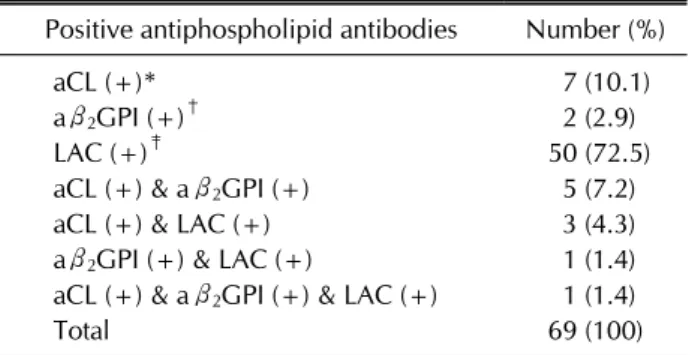
Table 1. Distribution of antiphospholipid antibody positivity patterns in antiphospholipid antibody positive patients
Positive antiphospholipid antibodies Number (%)
aCL (+)* 7 (10.1)
aβ2GPI (+)† 2 (2.9)
LAC (+)‡ 50 (72.5)
aCL (+) & aβ2GPI (+) 5 (7.2)
aCL (+) & LAC (+) 3 (4.3)
aβ2GPI (+) & LAC (+) 1 (1.4) aCL (+) & aβ2GPI (+) & LAC (+) 1 (1.4)
Total 69 (100)
aCL: anticardiolipin antibody, aβ2GPI: anti-β2-glycoprotein I antibody, LAC: lupus anticoagulant, Ig: immunoglobulin.
*IgG or IgM aCL > 40 GPLU/ml. †IgG or IgM aβ2GPI > 40 SGU/ml. ‡LAC, detected according to the guidelines of the International Society on Thrombosis and Haemostasis.
Pathology Society (ISN/RPS) 2003 classification of LN were also gathered from the registry [24]. Among them, LN class III and above were categorized as severe LN [25].
Laboratory studies
The prevalence of chronic kidney disease (stage ≥3) is defined as estimated glomerular filtration rate (eGFR)
<60 mL/min/1.73 m2 calculated by the Modification of Diet in Renal Disease Study Equation, which estimates GFR based on serum creatinine level and patient charac- teristics including age, sex, and race [26-28]. It was a ma- jor outcome of this study that we compared between the two groups of Korean SLE patients defined by aPL positivity. We also investigated the factors associated with increased risk of CKD including aPL.
Statistical analysis
The prevalence of aPL was estimated, and a descriptive analysis of the distribution of different aPL patterns was performed. After dividing patients into aPL (+) and aPL (−) groups, we determined the significance of the differ- ences in clinical characteristics, including thrombotic complications, using the chi-square test or Fisher’s exact test, independent t-test or Mann-Whitney U-test as appropriate. Analysis of covariance (ANCOVA) was used to determine the significance of differences in the preva- lence of vascular comorbidities, renal manifestations, and obstetric complications between two groups. However, ANCOVA was not performed to determine the sig- nificance of difference in serum creatinine levels between the two groups due to the presence of heteroscedasticity.
To examine the factors associated with increased risk of CKD, we performed univariable and multivariable logis- tic regression analyses using various independent clinical variables including aPL. For multivariable regression analysis, variables with p-value less than 0.1 in uni- variable analysis were selected. A two-sided p<0.05 was considered statistically significant. All statistical analyses were performed using IBM SPSS Statistics 21.0 for Windows (IBM Co., Armonk, NY, USA).
RESULTS
Prevalence of aPL in SLE patients
Among the 469 SLE patients enrolled in this study, 69 (14.7%) were positive for any aPL (Figure 1). The dis- tribution of antiphospholipid antibody positivity patterns is shown in Table 1; only one patient (1.4%) was positive
for all three aPL. Only LAC positivity was the most com- mon aPL positivity pattern (72.5%) in this study. In con- trast, both aCL and aβ2GPI more commonly accompanied other aPL rather than showing single positivity patterns.
Among the mixed aPL positivity patterns, aCL positivity combined with aβ2GPI positivity was the most common.
Clinical characteristics of SLE patients with pos- itive aPL
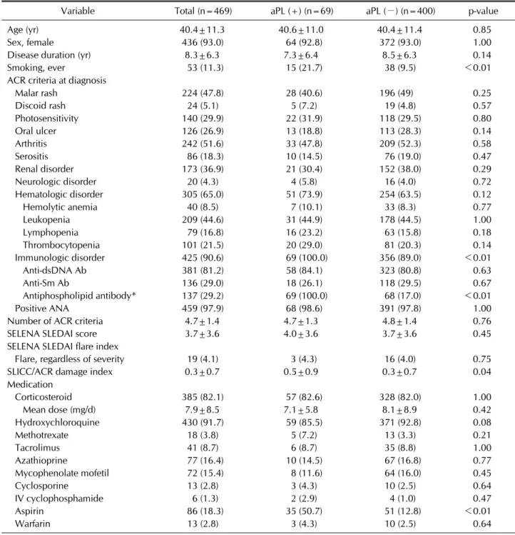
The baseline characteristics of each group at study en- rollment and the ACR criteria met for the classification of SLE at the time of diagnosis are shown in Table 2. We found no significant differences between the groups in age, sex, or autoantibody profile such as ANA and an- ti-dsDNA. However, the aPL (+) group showed a sig- nificantly higher number of patients with smoking his- tory (21.7% in aPL (+) group vs. 9.5% in aPL (−) group, p<0.01). There were no significant differences in the Safety of Estrogens in Lupus Erythematosus National Assessment-SLE Disease Activity Index (SELENA SLEDAI) and the proportions of patients with SLE flares measured by SELENA SLEDAI flare index between the two groups. The Systemic Lupus International Collaborating Clinics/American College of Rheumatology Damage Index, which reflects accumulated damage of SLE pa- tients, was significantly higher in the aPL (+) group (0.5±0.9 in aPL (+) group vs. 0.3±0.7 in aPL (−) group, p=0.04). Hydroxychloroquine and corticosteroid were the most commonly used medications, with a prevalence of more than 80% (91.7% and 82.1%, respectively).
Aspirin was significantly more frequently used in the aPL
Table 2. Demographic and clinical characteristics of patients with or without antiphospholipid antibody
Variable Total (n=469) aPL (+) (n=69) aPL (−) (n=400) p-value
Age (yr) 40.4±11.3 40.6±11.0 40.4±11.4 0.85
Sex, female 436 (93.0) 64 (92.8) 372 (93.0) 1.00
Disease duration (yr) 8.3±6.3 7.3±6.4 8.5±6.3 0.14
Smoking, ever 53 (11.3) 15 (21.7) 38 (9.5) <0.01
ACR criteria at diagnosis
Malar rash 224 (47.8) 28 (40.6) 196 (49) 0.25
Discoid rash 24 (5.1) 5 (7.2) 19 (4.8) 0.57
Photosensitivity 140 (29.9) 22 (31.9) 118 (29.5) 0.80
Oral ulcer 126 (26.9) 13 (18.8) 113 (28.3) 0.14
Arthritis 242 (51.6) 33 (47.8) 209 (52.3) 0.58
Serositis 86 (18.3) 10 (14.5) 76 (19.0) 0.47
Renal disorder 173 (36.9) 21 (30.4) 152 (38.0) 0.29
Neurologic disorder 20 (4.3) 4 (5.8) 16 (4.0) 0.72
Hematologic disorder 305 (65.0) 51 (73.9) 254 (63.5) 0.12
Hemolytic anemia 40 (8.5) 7 (10.1) 33 (8.3) 0.77
Leukopenia 209 (44.6) 31 (44.9) 178 (44.5) 1.00
Lymphopenia 79 (16.8) 16 (23.2) 63 (15.8) 0.18
Thrombocytopenia 101 (21.5) 20 (29.0) 81 (20.3) 0.14
Immunologic disorder 425 (90.6) 69 (100.0) 356 (89.0) <0.01
Anti-dsDNA Ab 381 (81.2) 58 (84.1) 323 (80.8) 0.63
Anti-Sm Ab 136 (29.0) 18 (26.1) 118 (29.5) 0.67
Antiphospholipid antibody* 137 (29.2) 69 (100.0) 68 (17.0) <0.01
Positive ANA 459 (97.9) 68 (98.6) 391 (97.8) 1.00
Number of ACR criteria 4.7±1.4 4.7±1.3 4.8±1.4 0.76
SELENA SLEDAI score 3.7±3.6 4.0±3.6 3.7±3.6 0.45
SELENA SLEDAI flare index
Flare, regardless of severity 19 (4.1) 3 (4.3) 16 (4.0) 0.75
SLICC/ACR damage index 0.3±0.7 0.5±0.9 0.3±0.7 0.04
Medication
Corticosteroid 385 (82.1) 57 (82.6) 328 (82.0) 1.00
Mean dose (mg/d) 7.9±8.5 7.1±5.8 8.1±8.9 0.42
Hydroxychloroquine 430 (91.7) 59 (85.5) 371 (92.8) 0.08
Methotrexate 18 (3.8) 5 (7.2) 13 (3.3) 0.21
Tacrolimus 41 (8.7) 6 (8.7) 35 (8.8) 1.00
Azathioprine 77 (16.4) 10 (14.5) 67 (16.8) 0.77
Mycophenolate mofetil 72 (15.4) 8 (11.6) 64 (16.0) 0.45
Cyclosporine 13 (2.8) 3 (4.3) 10 (2.5) 0.64
IV cyclophosphamide 6 (1.3) 2 (2.9) 4 (1.0) 0.47
Aspirin 86 (18.3) 35 (50.7) 51 (12.8) <0.01
Warfarin 13 (2.8) 3 (4.3) 10 (2.5) 0.64
Values are presented as mean±standard deviation or number (%). aPL: antiphospholipid antibody, ACR: American College of Rheumatology, Anti-dsDNA Ab: anti-double stranded DNA antibody, Anti-Sm Ab: anti-Smith antibody, ANA: antinuclear antibody, SELENA SLEDAI: Safety of Estrogens in Lupus Erythematosus National Assessment–Systemic Lupus Erythematosus Disease Activity Index, SLICC: Systemic Lupus International Collaborating Clinics, IV: intravenous, Ig: immunoglobulin, LAC:
lupus anticoagulant. *Positive finding of antiphospholipid antibodies based on 1) IgG aCL ≥10 GPLU/ml or IgM aCL ≥7 MPLU/ml, 2) LAC, detected according to the guidelines of the International Society on Thrombosis and Haemostasis, or 3) a false positive serologic test for syphilis known to be positive for at least 6 months and confirmed by Treponema pallidum immobilization of fluorescent treponemal antibody absorption test.
(+) group (50.7% in aPL (+) group vs. 12.8% in aPL (−) group, p<0.01), but use of other medications did not dif- fer between the groups.
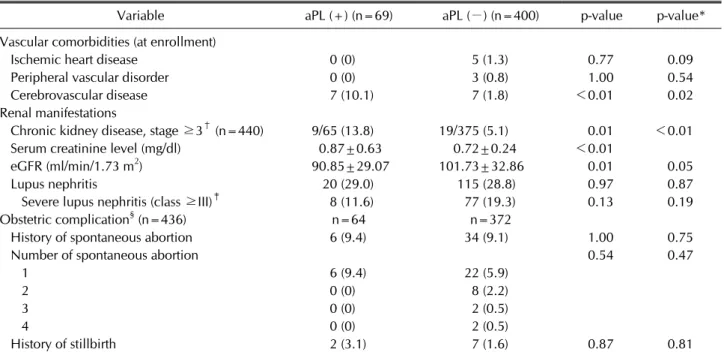
Comorbidities and obstetric complications known to be associated with APS and renal manifestations were com- pared according to aPL positivity, as shown in Table 3.
Table 3. Prevalence of vascular comorbidities, renal manifestations, and obstetric complications associated with antiphospholipid antibody
Variable aPL (+) (n=69) aPL (−) (n=400) p-value p-value*
Vascular comorbidities (at enrollment)
Ischemic heart disease 0 (0) 5 (1.3) 0.77 0.09
Peripheral vascular disorder 0 (0) 3 (0.8) 1.00 0.54
Cerebrovascular disease 7 (10.1) 7 (1.8) <0.01 0.02
Renal manifestations
Chronic kidney disease, stage ≥3† (n=440) 9/65 (13.8) 19/375 (5.1) 0.01 <0.01
Serum creatinine level (mg/dl) 0.87±0.63 0.72±0.24 <0.01
eGFR (ml/min/1.73 m2) 90.85±29.07 101.73±32.86 0.01 0.05
Lupus nephritis 20 (29.0) 115 (28.8) 0.97 0.87
Severe lupus nephritis (class ≥III)‡ 8 (11.6) 77 (19.3) 0.13 0.19
Obstetric complication§ (n=436) n=64 n=372
History of spontaneous abortion 6 (9.4) 34 (9.1) 1.00 0.75
Number of spontaneous abortion 0.54 0.47
1 6 (9.4) 22 (5.9)
2 0 (0) 8 (2.2)
3 0 (0) 2 (0.5)
4 0 (0) 2 (0.5)
History of stillbirth 2 (3.1) 7 (1.6) 0.87 0.81
Values are presented as number (%) or mean±standard deviation. aPL: antiphospholipid antibody, eGFR: estimated glomerular filtration rate. *p-value after adjustment for smoking and aspirin; †Defined as estimated glomerular filtration rate <60 ml/min/1.73 m2. ‡Defined as lupus nephritis International society of Nephrology/Renal Pathology Society (ISN/RPS) class III, IV, and V;
§Obstetric complications were only evaluated in female patients (n=436).
Two independent variables (smoking and aspirin use his- tory) with significant differences at baseline were se- lected as covariates for adjustment to remove the possible confounding effects. More patients in the aPL (+) group experienced cerebrovascular disease than in the aPL (−) group (10.1% vs. 1.8%, p<0.01, p after adjustment=
0.02). However, no statistical difference was found in his- tory of ischemic heart disease, peripheral vascular dis- order, spontaneous abortion, or stillbirth.
Association of positive aPL and risk of CKD in SLE patients
Chronic kidney disease (stage ≥3) was more frequent in the aPL (+) group (13.8% vs. 5.1%, p before adjust- ment=0.01, p after adjustment<0.01), while the pro- portion of underlying LN and severe LN (ISN/RPS class III and above) was comparable between the two groups (Table 3). The serum creatinine level was higher (0.87±
0.63 vs. 0.72±0.24, p<0.01) and eGFR was lower (90.85±29.07 vs. 101.73±32.86, p before adjustment=
0.01, p after adjustment=0.05) in the aPL (+) group compared to aPL (−) group. However, the difference in eGFR showed statistically borderline significance after adjusting for aspirin and smoking (p=0.05) (Table 3).
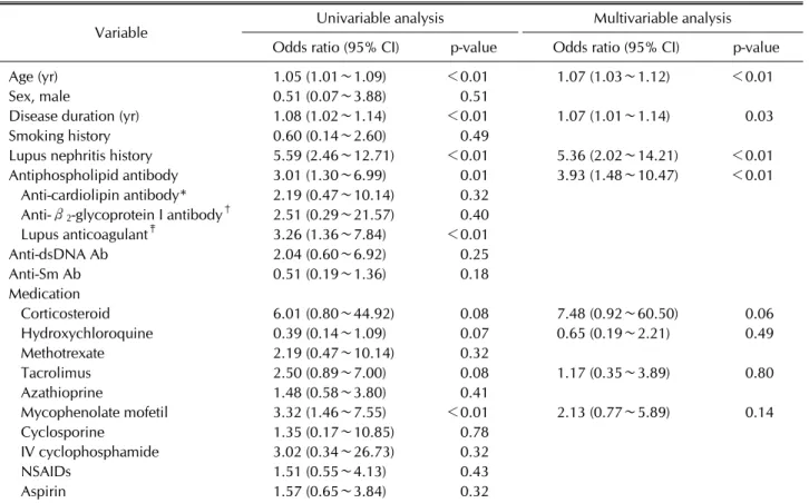
Various clinical variables were evaluated for the associa- tion with increased risk of CKD, as shown in Table 4.
Multivariable analysis showed that age, disease duration, LN history, and aPL positivity were significantly asso- ciated with increased CKD risk (p=0.03 for disease dura- tion and p<0.01 for others). Especially, LN history and aPL positivity showed substantially high odds ratios (odds ratio [OR]=5.36, 95% confidence interval [95%
CI]=2.02∼14.21; OR=3.93, 95% CI=1.48∼10.47, re- spectively).
DISCUSSION
In this study, we identified the prevalence of aPL in SLE patients and evaluated the relationships between the presence of aPL and clinical outcomes. According to our study results, 14.7% of Korean SLE patients showed aPL positivity (positivity for LAC, aCL, or aβ2GPI). In pa- tients with positive aPL, cerebrovascular disease and CKD were more common than they were in aPL (−) patients. Older age, longer disease duration, history of LN, and aPL positivity were factors associated with in- creased risk of CKD in our study. These key findings sug- gest that aPL positivity in SLE patients is associated with
Table 4. Univariable and multivariable logistic regression analyses of the variables associated with increased risk of chronic kidney disease (stage ≥3)
Variable Univariable analysis Multivariable analysis
Odds ratio (95% CI) p-value Odds ratio (95% CI) p-value
Age (yr) 1.05 (1.01∼1.09) <0.01 1.07 (1.03∼1.12) <0.01
Sex, male 0.51 (0.07∼3.88) 0.51
Disease duration (yr) 1.08 (1.02∼1.14) <0.01 1.07 (1.01∼1.14) 0.03
Smoking history 0.60 (0.14∼2.60) 0.49
Lupus nephritis history 5.59 (2.46∼12.71) <0.01 5.36 (2.02∼14.21) <0.01
Antiphospholipid antibody 3.01 (1.30∼6.99) 0.01 3.93 (1.48∼10.47) <0.01
Anti-cardiolipin antibody* 2.19 (0.47∼10.14) 0.32 Anti-β2-glycoprotein I antibody† 2.51 (0.29∼21.57) 0.40
Lupus anticoagulant‡ 3.26 (1.36∼7.84) <0.01
Anti-dsDNA Ab 2.04 (0.60∼6.92) 0.25
Anti-Sm Ab 0.51 (0.19∼1.36) 0.18
Medication
Corticosteroid 6.01 (0.80∼44.92) 0.08 7.48 (0.92∼60.50) 0.06
Hydroxychloroquine 0.39 (0.14∼1.09) 0.07 0.65 (0.19∼2.21) 0.49
Methotrexate 2.19 (0.47∼10.14) 0.32
Tacrolimus 2.50 (0.89∼7.00) 0.08 1.17 (0.35∼3.89) 0.80
Azathioprine 1.48 (0.58∼3.80) 0.41
Mycophenolate mofetil 3.32 (1.46∼7.55) <0.01 2.13 (0.77∼5.89) 0.14
Cyclosporine 1.35 (0.17∼10.85) 0.78
IV cyclophosphamide 3.02 (0.34∼26.73) 0.32
NSAIDs 1.51 (0.55∼4.13) 0.43
Aspirin 1.57 (0.65∼3.84) 0.32
CI: confidence interval, Anti-dsDNA Ab: anti-double stranded DNA antibody, Anti-Sm Ab: anti-Smith antibody, IV: intravenous, NSAIDs: nonsteroidal anti-inflammatory drugs, Ig: immunoglobulin, aCL: anticardiolipin antibody, aβ2GPI: anti-β2-glycoprotein I antibody. *IgG or IgM aCL >40 GPLU/ml. †IgG or IgM aβ2GPI >40 SGU/ml. ‡LAC, detected according to the guidelines of the International Society on Thrombosis and Haemostasis, LAC: lupus anticoagulant.
poor clinical outcomes, especially in the renal aspect.
The prevalence of aPL positivity in our study seems sub- stantially low compared to results from previous studies on SLE patients, even though our results include aβ2GPI positivity [2,29,30]. However, our results are not com- parable to the results from previous studies because we used stricter cut off values (moderate or high titer of aPL), which can lower the apparent prevalence. Also, we classi- fied the aPL (+) group using two-year data at study en- rollment, which could include laboratory values from times with low disease activity. Previous studies have shown that the presence and titers of aPL related to dis- ease activity in SLE and the titers of aPL decreased by treatment [31,32]. aPL positivity as reported in this study could reflect concrete positivity that persisted through the use of corticosteroids, immunosuppressants, or anti- coagulants because all of our study population had been followed and treated properly by rheumatologists for years. According to the SELENA SLEDAI flare index that
was developed for reliable distinction of flares in SLE ac- tivity, most patients in our study (95.9% of the total study population) did not show a disease flare status [33]. In a prospective cohort of 625 SLE patients that compared risk factors of thromboembolism among three ethnic groups (Chinese, African Americans, Caucasians), pres- ence of aPL was less frequent in Chinese patients (29% in Chinese vs. 42% in African Americans vs. 46% in Caucasians, p<0.001) [4]. Those results suggest that in- terethnic differences exist in the prevalence of aPL pos- itivity in SLE patients. Our results further support the idea that aPL positivity could be less frequent in Asian SLE patients than in other ethnic groups. Additional study of genetic factors will be necessary to account for this racial difference.
We found no significant differences in most of the base- line characteristics between the two groups. The most in- teresting result is the relationship between aPL positivity and smoking history. The aPL (+) group had a sig-
nificantly higher prevalence of smoking history, which is a well-known risk factor for increasing thrombotic vas- cular events [34]. However, our logistic regression analy- sis data showed that smoking history was not associated with increased risk of CKD in SLE patients. According to a study analyzing the association between smoking his- tory and SLE-associated autoantibodies, smoking was positively associated only with aPL, not with other auto- antibodies [35]. The underlying mechanism and tempo- rality between smoking and aPL needs further invest- igation. aPL (+) patients used aspirin or warfarin more frequently than aPL (−) patients, though the difference in warfarin use was not statistically significant. Among patients without aPL, 10 (2.5%) used warfarin at study enrollment. The causes for using warfarin in those pa- tients included cerebrovascular accidents, peripheral vas- cular disorders, and pulmonary thromboembolism. Four of them had aPL positivity of low titer or previous history of aPL positivity. However, as only 13 patients (2.8% of the total study population) used warfarin at study enroll- ment, further study with a larger sample is needed for proper comparison between the two groups.
Though the International APS Classification Criteria provide a uniform approach to APS research, aPL can cor- relate with a spectrum of clinical manifestations, some of which are not included in those criteria including renal microangiopathy [1,36]. aPL-associated nephropathy (APS nephropathy) is clinically characterized as a syn- drome of vascular nephropathy associated with hyper- tension, renal insufficiency, proteinuria, and inconstant hematuria that can present differently depending on pathophysiology including thrombotic microangiopathy, arterial fibrous intimal hyperplasia, tubular thyroid-like appearance, arteriosclerosis, arteriolar occlusion, and fo- cal cortical atrophy [37]. The prevalence of APS nephrop- athy in SLE patients was reported as 32% and 23.2%, re- spectively, in two prior studies [12,13]. A cross-sectional study conducted on 112 SLE patients with renal involve- ment and who underwent renal biopsies revealed the as- sociation between APS nephropathy pathology and pres- ence of aPL. Their results indicated that APS nephropathy is a separate renal disease entity with different underlying pathogenic mechanisms compared to LN. Therefore, the importance of conducting renal biopsy to differentiate those two conditions was emphasized [14]. According to a recently reported study encompassing a prospective co- hort of 64 biopsy-proven active LN patients without con- comitant APS nephropathy and cross-sectional analysis
of 498 SLE patients with or without LN, both aPL pos- itivity and level were similar in patients with active LN and non-renal SLE. However, IgG aCL or IgG aβ2GPI pos- itive patients had higher creatinine levels compared with patients without those IgG aPL both at active LN and after induction therapy, but not in the long-term data analyses.
The authors concluded that aPL are not associated with occurrence of LN, and IgG aPL might contribute to an im- paired renal function during a LN flare in the absence of APS nephropathy, probably due to their own pathogenic roles. But, no association was found between IgG aPL and additional renal function deterioration in the long-term follow up [17]. Our results also showed no difference in the prevalence of LN between aPL (+) and aPL (−) groups further supporting the idea that aPL is not asso- ciated with LN. Previous studies revealed that patients with severe LN (WHO class III and above) more likely progress to end-stage kidney disease [25]. Even though the proportion of severe LN was higher in aPL (−) group (without statistical significance), CKD was more preva- lent in aPL (+) patients in our study. In addition, both LN history and aPL positivity were independent factors asso- ciated with increased CKD risk in SLE patients. Taken to- gether with previous reports, the aPL seems to increase the risk of CKD in SLE patients by a mechanism different from that of LN. However, we are unsure if the mecha- nism leading to deteriorating renal function is related with APS nephropathy or other pathogenic mechanism of aPL is concerned if not biopsy proven in this study. aPL is believed to have pathogenic roles not limited to thrombo- sis, mediating several clinical manifestations of APS [34].
Previous studies about the impact of aPL on renal out- come were mostly confined to patients with LN and have shown conflicting results [16-19]. Ünlü et al. [38] sug- gested aPL as a marker to predict renal prognosis in SLE patients by referring to an abstract report on meta-analy- sis of 1,820 patients. But, we still did not have sufficient evidence to clarify the role of aPL as a distinct factor asso- ciated with poor renal outcome in SLE patients excluding the impact of other influential factors such as old age, lon- ger disease duration, or presence of LN. History of LN and aPL positivity were the most influential clinical factors as- sociated with increased risk of CKD even after adjusting for other confounding factors in multivariable logistic re- gression analysis (OR=5.36 and 3.93, respectively]. The most important findings of this study were the poor renal outcome in aPL (+) SLE patients and the clarified role of aPL as an independent factor associated with increased
risk of CKD.
We note three limitations of this study. First, though we used the revised classification criteria for APS to generate our aPL (+) group, we defined positivity as those con- firmed to be positive for aPL at one point in the past two years instead of confirming aPL positivity twice with an at least six-week interval between tests. This approach might increase the possibility of false positives for aPL.
On the other hand, because we used stricter laboratory cut off values and did not include data from more than 2 years before enrollment, the possibility of false positives for aPL seems low. In addition, as all patients in the aPL (+) group met the criteria for aPL positivity in the ACR revised criteria for the classification of SLE at study en- rollment, the possibility of clinically significant firm aPL positivity in this group is high. However, we had limited information on which specific criterion for aPL positivity was satisfied at the time of diagnosis. Second, the pres- ence of APS nephropathy must have been noteworthy when discussing adverse renal outcomes regarding aPL positivity in SLE patients. However, because our clinical data were insufficient to define the presence of APS nephropathy without renal histology results, we could not discuss correlations with the presence of APS nephr- opathy or other renal pathologic features. Succeeding re- searches need to overcome this limitation to more clearly explain how aPL positivity influences renal outcomes in SLE patients. Last, even though we collected data from a prospective cohort study, our results were produced us- ing cross-sectional data collected at enrollment.
Therefore, this study has limitations in explaining long term prognosis including renal prognosis in the study population. We expect to explain the long-term relation- ship more clearly using accumulated clinical data as we proceed further through this cohort study.
CONCLUSION
The prevalence of aPL is 14.7% in Korean SLE patients.
The aPL is associated with higher prevalence of cere- brovascular disease and CKD in SLE patients. And, aPL positivity is independently associated with the risk of CKD in SLE patients.
ACKNOWLEDGMENTS
We thank the patients and their families for their partic- ipation in this study.
CONFLICT OF INTEREST
No potential conflict of interest relevant to this article was reported.
REFERENCES
1. George D, Erkan D. Antiphospholipid syndrome. Prog Cardiovasc Dis 2009;52:115-25.
2. Petri M. Epidemiology of the antiphospholipid antibody syndrome. J Autoimmun 2000;15:145-51.
3. Cervera R, Serrano R, Pons-Estel GJ, Ceberio-Hualde L, Shoenfeld Y, de Ramón E, et al. Morbidity and mortality in the antiphospholipid syndrome during a 10-year period: a multicentre prospective study of 1000 patients. Ann Rheum Dis 2015;74:1011-8.
4. Mok CC, Tang SS, To CH, Petri M. Incidence and risk factors of thromboembolism in systemic lupus erythematosus: a comparison of three ethnic groups. Arthritis Rheum 2005;
52:2774-82.
5. Pons-Estel GJ, Andreoli L, Scanzi F, Cervera R, Tincani A.
The antiphospholipid syndrome in patients with systemic lupus erythematosus. J Autoimmun 2017;76:10-20.
6. Ruiz-Irastorza G, Egurbide MV, Ugalde J, Aguirre C. High impact of antiphospholipid syndrome on irreversible organ damage and survival of patients with systemic lupus erythematosus. Arch Intern Med 2004;164:77-82.
7. Broder A, Mowrey WB, Kim M, Murakhovskaya I, Billett H, Neugarten J, et al. Association between antiphospholipid antibodies and all-cause mortality among end-stage renal disease patients with and without SLE: a retrospective co- hort study. Rheumatology (Oxford) 2016;55:817-25.
8. Yelnik CM, Urbanski G, Drumez E, Sobanski V, Maillard H, Lanteri A, et al. Persistent triple antiphospholipid antibody positivity as a strong risk factor of first thrombosis, in a long-term follow-up study of patients without history of thrombosis or obstetrical morbidity. Lupus 2017;26:163-9.
9. Horbach DA, van Oort E, Donders RC, Derksen RH, de Groot PG. Lupus anticoagulant is the strongest risk factor for both venous and arterial thrombosis in patients with systemic lupus erythematosus. Comparison between differ- ent assays for the detection of antiphospholipid antibodies.
Thromb Haemost 1996;76:916-24.
10. Wahl DG, Guillemin F, de Maistre E, Perret C, Lecompte T, Thibaut G. Risk for venous thrombosis related to anti- phospholipid antibodies in systemic lupus erythematosus-- a meta-analysis. Lupus 1997;6:467-73.
11. Empson M, Lassere M, Craig J, Scott J. Prevention of re- current miscarriage for women with antiphospholipid anti- body or lupus anticoagulant. Cochrane Database Syst Rev 2005;(2):CD002859.
12. Daugas E, Nochy D, Huong DL, Duhaut P, Beaufils H, Caudwell V, et al. Antiphospholipid syndrome nephropathy in systemic lupus erythematosus. J Am Soc Nephrol 2002;13:42-52.
13. Tektonidou MG, Sotsiou F, Nakopoulou L, Vlachoyiannopoulos PG, Moutsopoulos HM. Antiphospholipid syndrome nephr- opathy in patients with systemic lupus erythematosus and antiphospholipid antibodies: prevalence, clinical associa-
tions, and long-term outcome. Arthritis Rheum 2004;50:
2569-79.
14. Gerhardsson J, Sundelin B, Zickert A, Padyukov L, Svenungsson E, Gunnarsson I. Histological antiphos- pholipid-associated nephropathy versus lupus nephritis in patients with systemic lupus erythematosus: an observational cross-sectional study with longitudinal follow-up. Arthritis Res Ther 2015;17:109.
15. Zheng H, Chen Y, Ao W, Shen Y, Chen XW, Dai M, et al.
Antiphospholipid antibody profiles in lupus nephritis with glomerular microthrombosis: a prospective study of 124 cases. Arthritis Res Ther 2009;11:R93.
16. Moroni G, Ventura D, Riva P, Panzeri P, Quaglini S, Banfi G, et al. Antiphospholipid antibodies are associated with an in- creased risk for chronic renal insufficiency in patients with lupus nephritis. Am J Kidney Dis 2004;43:28-36.
17. Parodis I, Arnaud L, Gerhardsson J, Zickert A, Sundelin B, Malmström V, et al. Antiphospholipid antibodies in lupus nephritis. PLoS One 2016;11:e0158076.
18. Mehrani T, Petri M. IgM anti-β2 glycoprotein I is protective against lupus nephritis and renal damage in systemic lupus erythematosus. J Rheumatol 2011;38:450-3.
19. Frampton G, Hicks J, Cameron JS. Significance of anti-phos- pholipid antibodies in patients with lupus nephritis. Kidney Int 1991;39:1225-31.
20. Costenbader KH, Desai A, Alarcón GS, Hiraki LT, Shaykevich T, Brookhart MA, et al. Trends in the incidence, demographics, and outcomes of end-stage renal disease due to lupus nephritis in the US from 1995 to 2006. Arthritis Rheum 2011;63:1681-8.
21. Miyakis S, Lockshin MD, Atsumi T, Branch DW, Brey RL, Cervera R, et al. International consensus statement on an update of the classification criteria for definite anti- phospholipid syndrome (APS). J Thromb Haemost 2006;
4:295-306.
22. Lee JW, Park DJ, Kang JH, Choi SE, Yim YR, Kim JE, et al.
The rate of and risk factors for frequent hospitalization in systemic lupus erythematosus: results from the Korean lu- pus network registry. Lupus 2016;25:1412-9.
23. Hochberg MC. Updating the American College of Rheumatology revised criteria for the classification of sys- temic lupus erythematosus. Arthritis Rheum 1997;40:1725.
24. Weening JJ, D’Agati VD, Schwartz MM, Seshan SV, Alpers CE, Appel GB, et al. The classification of glomerulonephritis in systemic lupus erythematosus revisited. Kidney Int 2004;
65:521-30.
25. Maroz N, Segal MS. Lupus nephritis and end-stage kidney disease. Am J Med Sci 2013;346:319-23.
26. Levey AS, Bosch JP, Lewis JB, Greene T, Rogers N, Roth D.
A more accurate method to estimate glomerular filtration rate from serum creatinine: a new prediction equation.
Modification of Diet in Renal Disease Study Group. Ann
Intern Med 1999;130:461-70.
27. Levey AS, Coresh J, Balk E, Kausz AT, Levin A, Steffes MW, et al. National Kidney Foundation practice guidelines for chronic kidney disease: evaluation, classification, and stratification. Ann Intern Med 2003;139:137-47.
28. Stevens PE, Levin A; Kidney Disease: Improving Global Outcomes Chronic Kidney Disease Guideline Development Work Group Members. Evaluation and management of chronic kidney disease: synopsis of the kidney disease: im- proving global outcomes 2012 clinical practice guideline.
Ann Intern Med 2013;158:825-30.
29. Skare T, Borba EA, Utiyama SR, Nisihara R. Lymphocytope- nia is associated with anti-Beta-2 glycoprotein-1 in patients with 220 systemic lupus erythematosus. Acta Reumatol Port 2016;41:220-5.
30. Taraborelli M, Leuenberger L, Lazzaroni MG, Martinazzi N, Zhang W, Franceschini F, et al. The contribution of anti- phospholipid antibodies to organ damage in systemic lupus erythematosus. Lupus 2016;25:1365-8.
31. Sarabi ZS, Sahebari M, Rezaie AE, Norouzi MT, Hashemza- deh K, Mirfeizi Z. The relationship between systemic lupus erythematosus activity and persistent positive antiphos- pholipid antibodies. Curr Rheumatol Rev 2018;14:145-52.
32. Alarcón-Segovia D, Delezé M, Oria CV, Sánchez-Guerrero J, Gómez-Pacheco L, Cabiedes J, et al. Antiphospholipid anti- bodies and the antiphospholipid syndrome in systemic lu- pus erythematosus. A prospective analysis of 500 consec- utive patients. Medicine (Baltimore) 1989;68:353-65.
33. Buyon JP, Petri MA, Kim MY, Kalunian KC, Grossman J, Hahn BH, et al. The effect of combined estrogen and proges- terone hormone replacement therapy on disease activity in systemic lupus erythematosus: a randomized trial. Ann Intern Med 2005;142:953-62.
34. Meroni PL, Borghi MO, Raschi E, Tedesco F. Pathogenesis of antiphospholipid syndrome: understanding the antibodies.
Nat Rev Rheumatol 2011;7:330-9.
35. Gustafsson JT, Gunnarsson I, Källberg H, Pettersson S, Zickert A, Vikerfors A, et al. Cigarette smoking, anti- phospholipid antibodies and vascular events in Systemic Lupus Erythematosus. Ann Rheum Dis 2015;74:1537-43.
36. Abreu MM, Danowski A, Wahl DG, Amigo MC, Tektonidou M, Pacheco MS, et al. The relevance of “non-criteria” clinical manifestations of antiphospholipid syndrome: 14th International Congress on Antiphospholipid Antibodies Technical Task Force Report on Antiphospholipid Syndrome Clinical Features. Autoimmun Rev 2015;14:401-14.
37. Sciascia S, Cuadrado MJ, Khamashta M, Roccatello D. Renal involvement in antiphospholipid syndrome. Nat Rev Nephrol 2014;10:279-89.
38. Ünlü O, Zuily S, Erkan D. The clinical significance of anti- phospholipid antibodies in systemic lupus erythematosus.
Eur J Rheumatol 2016;3:75-84.