J Korean Ophthalmol Soc 2018;59(11):1097-1102 ISSN 0378-6471 (Print)⋅ISSN 2092-9374 (Online)
https://doi.org/10.3341/jkos.2018.59.11.1097
Case Report
클로르페나피르 음독 후 발생한 독성 시신경병증 1예
Toxic Optic Neuropathy Caused by Chlorfenapyr Poisoning
박수진1,2⋅정재욱1,2⋅강용구1,2⋅전보영1,2⋅손병재1,2
Su Jin Park, MD1,2, Jae Uk Jung, MD1,2, Yong Koo Kang, MD1,2, Bo Young Chun, MD, PhD1,2, Byeong Jae Son, MD1,2
경북대학교 의과대학 안과학교실1, 경북대학교병원 안과2
Department of Ophthalmology, School of Medicine, Kyungpook National University1, Daegu, Korea Department of Ophthalmology, Kyungpook National University Hospital2, Daegu, Korea
Purpose: To report a case of toxic optic neuropathy caused by chlorfenapyr ingestion accompanied by central nervous system involvement.
Case summary: A 44‐year‐old female visited our clinic complaining of reduced visual acuity in both eyes for 7 days. She had in- gested a mouthful of chlorfenapyr for a suicide attempt 2 weeks prior to the visit. Gastric lavage was performed immediately after ingestion at the other hospital. Her best‐corrected visual acuity was finger count 30 cm in the right eye and hand motion in the left eye. Both pupils were dilated by 5.0 mm and the response to light was sluggish in both eyes. A relative afferent pupillary defect was detected in her left eye. Funduscopy revealed optic disc swelling in both eyes. Magnetic resonance imaging of the brain showed a symmetric hyper‐intense signal in the white matter tract including the internal capsule, corpus callosum, middle cer- ebellar peduncle, and brainstem. The patient was diagnosed with toxic optic neuropathy induced by chlorfenapyr ingestion, and underwent high‐dose intravenous corticosteroid pulse therapy. Three days later, the best‐corrected visual acuity was no light perception in both eyes. Three months later, optic atrophy was observed in both eyes. Optical coherence tomography revealed a reduction in the thicknesses of the retinal nerve fiber layer and ganglion cell and inner plexiform layer in the macular area.
Conclusions: Ingestion of even a small amount of chlorfenapyr can cause severe optic nerve damage through the latent period, despite prompt lavage and high‐dose steroid treatment.
J Korean Ophthalmol Soc 2018;59(11):1097-1102
Keywords: Central nervous system involvement, Chlorfenapyr poisoning, Toxic optic neuropathy
■Received: 2018. 6. 7. ■ Revised: 2018. 7. 13.
■Accepted: 2018. 10. 18.
■Address reprint requests to Byeong Jae Son, MD
Department of Ophthalmology, Kyungpook National University Hospital, #130 Dongdeok‐ro, Jung‐gu, Daegu 41944, Korea Tel: 82‐53‐200‐5806, Fax: 82‐53‐426‐6552
E-mail: [email protected]
*Conflicts of Interest: The authors have no conflicts to disclose.
ⓒ2018 The Korean Ophthalmological Society
This is an Open Access article distributed under the terms of the Creative Commons Attribution Non-Commercial License (http://creativecommons.org/licenses/by-nc/3.0/) which permits unrestricted non-commercial use, distribution, and reproduction in any medium, provided the original work is properly cited.
독성 시신경병증은 영양부족, 독소, 환경적 요인 등 다양 한 원인에 의해 전방시각경로의 손상이 발생하여 유두황반 다발의 손상, 중심 혹은 중심부근 암점, 색각 장애 등이 나
타나는 양측성 진행성 시신경질환이다.1,2 독성 시신경병증 의 예후는 환자에 따라 다양하며 약물의 종류, 사용기간, 진단 시기에 따라 다르다.1.2
클로르페나피르(chlorfenapyr)는 케톤 냄새가 나는 연황갈 색 분말 형태의 피롤 계열 농약으로 주로 살충제로 널리 사 용되고 있다.3 클로르페나피르 중독 후 메스꺼움, 구토, 발열, 시력장애, 전신쇠약, 횡문근융해 등 전신적인 영향뿐만 아니 라 중추신경계 손상이 발생한 경우가 보고되어 있으며, 대부 분 사망에 이르는 치명적인 결과를 나타냈다.4‐7 저자들은 클 로르페나피르 음독 후 중추신경계 손상을 동반한 독성 시신 경병증 1예를 경험하였기에 이를 보고하고자 한다.
A
B
C
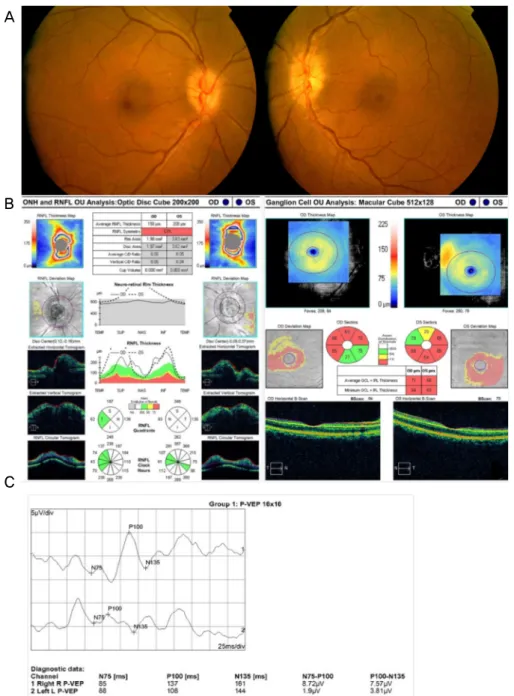
Figure 1. Ocular findings at initial presentation: fundus photographs, optical coherence tomography (OCT), and pattern visual
evoked potential (VEP). (A) Optic disc swelling is observed in both eyes on color fundus photography. (B) OCT shows optic disc swelling and thinning of the ganglion cell and inner plexiform layers in both eyes. (C) Pattern VEP shows delayed P100 latency in the right eye and decreased P100 amplitude in the left eye. ONH = optic nerve head; RNFL = retinal nerve fiber layer; OD = ocu- lus dexter; OS = oculus sinister; OU = oculus uterque; TEMP = temporal; SUP = superior; NAS = nasal; INF = inferior; S = superior; N = nasal; I = inferior; T = temporal; IPL = inner plexiform layer; C/D = cup/disc; GCL = ganglion cell layer.증례보고
44세 여자가 일주일 전부터 시작된 양안 시력저하를 주 소로 내원하였다. 내원 2주 전 자살 목적으로 클로르페나피 르(농도는 알 수 없음) 한 모금(약 10‐20 mL)을 음독하였 다. 음독 직후 근처 병원을 방문하여 위세척을 시행하였고, 시술 이후 환자는 별다른 증상 없이 일상생활을 지속하였
다. 만성질환, 알코올이나 약물 남용, 그 외 시력에 영향을 줄 수 있는 다른 병력은 없었다.
내원 당시 혈압, 심박수, 산소포화도, 체온 등의 활력 징 후는 정상이었다. 발음이 약간 어눌하였으나 그 외 발한, 통증, 의식변화, 사지쇠약(weakness) 등의 신경학적 문제는 없었다. 혈액 및 소변검사 결과상 특이 소견은 없었다. 최 대교정시력은 우안 안전 수지 30 cm, 좌안 안전수동이었다.
A B
Figure 2. Orbit and brain magnetic resonance image at initial presentation. (A) Edematous optic nerve, especially optic nerve sheath
enhancement is demonstrated in axial T1‐weighted fat‐suppressed gadolinium‐enhanced image and coronal T2‐weighted fat‐sup- pressed image. (B) Axial diffusion‐weighted image shows hyperintense signal in white matter tract including internal capsule, corpus callosum, middle cerebellar peduncle and brainstem.양안 동공크기는 5.0 mm로 커져 있었으며, 빛에 대한 반응 은 느렸고, 좌안에서 상대구심동공운동장애가 관찰되었다.
안압은 양안 모두 정상이었으며, 세극등현미경검사에서 특 이 소견은 없었다. 안저검사에서 양안 시신경부종이 관찰 되었으며(Fig. 1A), 빛간섭단층촬영에서 양안의 시신경유 두부종과 황반부 신경절세포‐내망상세포층(ganglion cell and inner plexiform layers) 두께의 감소(우안: 71 μm, 좌안: 68 μm) 가 관찰되었다(Fig. 1B). 문양 시유발전위검사에서 우안 P100의 반응도달시간이 지연되어 있었고 좌안 P100의 진 폭이 감소되어 있었다(Fig. 1C). 뇌자기공명영상 T1 강조영 상과 T2 강조영상에서 시신경과 시신경집의 신호 증강이 관찰되었다(Fig. 2A). 확산강조영상(diffusion‐weighted im- age)에서 속섬유막(internal capsule), 뇌량(corpus callosum), 중소뇌각(middle cerebellar peduncle), 뇌간(brainstem) 등 의 백질 신경로를 따라 대칭적인 고강도 신호가 관찰되었 다(Fig. 2B). 그러나 뇌파검사는 정상이었다.
클로르페나피르에 의한 독성 시신경병증으로 진단하였 고, 하루에 methylprednisolone (Solu-Medrol®, Pfizer, Inc., Puurs, Belgium) 1 g을 3일간 투여하는 고용량 스테로이드 정맥주사요법을 시행하였다. 3일 후, 양안의 최대교정시력 은 광각무로 악화되었고, 양안 동공은 여전히 산동되어 있 었으며, 대광반사는 없었다. 2주 후에도 양안 시력은 광각 무였으며, 양안의 시신경 부종 및 망막신경섬유층 두께도 처음과 큰 차이가 없었다.
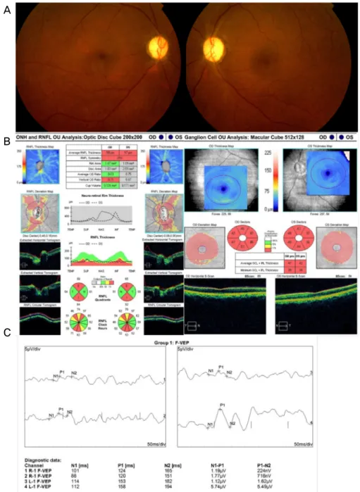
3개월 후, 안저검사에서 양안 시신경위축이 관찰되었고 (Fig. 3A), 빛간섭단층촬영에서 위쪽, 아래쪽 망막신경섬유 층 두께의 감소와 전 구간에서 신경절세포‐내망상세포층 두 께의 감소(우안: 47 μm 좌안: 42 μm)가 관찰되었다(Fig. 3B).
섬광시유발전위검사에서 양안의 P100 진폭의 감소가 관찰 되었다(Fig. 3C). 그 이후에도 최대교정시력, 시유발전위검
사, 빛간섭단층촬영 등의 검사에서 별다른 변화는 없었다.
고 찰
독성 시신경병증은 영양부족, 환경적 요인, 독소 등 다양한 원인에 의해 유발된다.1.2 특히 에탐부톨(ethambutol), 아미오 다론(amiodarone), 에탄올, 클로람페니콜(chloramphenicol), 리네졸리드(linezolid), 에리트로마이신(erythromycin), 스트 렙토마이신(streptomycin), 그리고 항바이러스 약제 등은 미 토콘드리아에서 산화적 인산화 과정을 억제하여 시신경의 손상을 유발할 수 있다.2.8
클로르페나피르 또한 유사한 기전을 보이는데, 전구물질 상태에서 N‐ethoxymethyl 그룹의 산화적 제거를 통해 CL 303268 화합물의 형태가 된다.3 활성화된 클로르페나피르 는 미토콘드리아에서 산화적 인산화 과정을 분리시켜 아데 노신 트리포스페이트 생성을 방해하고 결과적으로 세포사 및 개체의 죽음을 유발한다.5 이러한 2차적 미토콘드리아 기능이상은 시신경과 망막신경절세포의 기능장애를 유발 한다.
지금까지 클로르페나피르 중독에 대한 몇몇 보고들이 있 었으며, Endo et al9은 24명의 클로르페나피르 중독환자 중 8명이나 사망에 이르렀다고 보고하였으며, 심지어 살충제 의 증기 노출만으로도 사망한 증례도 있었다.10 이전 보고 에 의하면 클로르페나피르 중독은 주로 자살시도나 직업적 노출에 의한 것이 대부분이었다. 대부분의 증례에서 초기 7
‐14일간의 잠복기 이후 증상이 빠르게 진행되어 결국 죽음 에 이르게 되었다.4‐7,9‐12 Kang et al7은 클로르페나피르 중독 에서 잠복기가 있는 이유는 전구물질에서 활성화 형태의 독성물질로 변하는데 시간이 필요하기 때문이라고 추정하 였다. 증상과 징후는 메스꺼움, 구토, 발열, 시력장애, 전신
A
B
C
Figure 3. Ocular findings at 3 months after the initial presentation: fundus photographs, optical coherence tomography and flash vis-
ual evoked potential (VEP). (A) Color fundus photography shows both optic discs presenting atrophic change. (B) The thickness of superior and inferior retinal nerve fiber layer is decreased, and ganglion cell and inner plexiform layer thinning is observed in all sectors. (C) Flash VEP shows low P100 amplitude in both eyes. ONH = optic nerve head; RNFL = retinal nerve fiber layer; OD= oculus dexter; OS = oculus sinister; OU = oculus uterque; TEMP = temporal; SUP = superior; NAS = nasal; INF = inferior;
S = superior; N = nasal; I = inferior; T = temporal; IPL = inner plexiform layer; C/D = cup/disc; GCL = ganglion cell layer.
쇠약, 횡문근융해 등으로 다양하며,4,6,9 독성반응이 잠복기 를 지나 중추신경계에 영향을 주는 경우도 관찰되었다.4‐7 본 증례는 클로르페나피르에 노출된 후 중추신경계 손상과 함께 독성 시신경병증으로 주로 나타난 경우로 국내에서는 지금까지 유사한 증례가 보고되지 않았다.
지금까지 보고된 클로르페나피르 중독 사례들을 살펴보
면, 전신적인 증상이 주로 발생하였고 심지어 죽음에 이르 는 경우도 많았으나, 본 사례와 같이 양안의 시력장애가 주 된 증상인 경우는 없었다. 대부분의 증례에서 약 20‐250 mL 정도의 양을 음독했다고 보고하였고, 20 mL 이상을 음독한 경우 대부분 사망하였으며, 20 mL보다 적은 양을 음독한 경우는 대부분 생존하였다.4‐7,9,11,12 본 증례에서는 문진상
약 10‐20 mL정도의 적은 양의 클로르페나피르를 음독하였 고, 음독 후 빠른 위세척이 이루어졌기 때문에 비교적 다른 합병증 없이 생존할 수 있었던 것으로 생각된다. 마찬가지 로 Baek et al5과 Ku et al11은 적은 양의 클로르페나피르 음 독 후 경미한 임상적 증상만 있었던 사례를 보고하였으며, 이러한 결과를 미루어볼 때 클로르페나피르 중독 후 발생 하는 치명적인 결과는 섭취량과 관련된 것으로 보인다. 하 지만 적은 양의 클로르페나피르 음독에도 불구하고 잠복기 를 거쳐 중추신경계 손상이 발생한 경우도 있기 때문에 주 의해야 하겠다.4,7 또한 이 증례와 같이 음독 환자의 경우 약 물 섭취량은 환자의 진술에 의존할 수밖에 없어 정확한 양 을 파악하기 어려운 점이 있으므로 문진 시 자세한 병력 청 취가 중요할 것으로 생각된다.
클로르페나피르 노출 후 중추신경계 손상이 발생한 이전 의 보고들에서 뇌척수 자기공명영상 T2 강조영상에서 뇌, 뇌간, 척수를 포함한 백질 신경로를 따라 광범위하고 대칭 적인 고강도신호 변화가 관찰되었으며, 두드러진 확산제한 이 동반된다고 보고된 바 있다.4‐7 본 증례의 경우도 이전 독성뇌병증 및 독성 시신경병증처럼 전체 백질 신경로 및 시신경에 대칭적인 고강도 신호를 보인다. 백질 신경로를 따라 손상이 발생하는 병리생리학적인 기전은 아직 알려져 있지는 않지만, McKinney et al13은 백질 신경로의 손상이 수초 내 부종, 수초의 액포화 그리고 독성에 의한 직접적인 탈수초화에 의해 발생한다고 생각하였다. 본 증례에서도 이와 비슷한 기전에 의해 시신경 유두 부종, T2 강조영상에 서 시신경 및 시신경집의 신호 증강 등의 소견들이 관찰되 는 것으로 생각된다.
독성 시신경병증의 치료를 위해 원인 물질을 즉시 제거 해야 하며, 이 과정을 통해 손상이 일부 회복될 수 있다. 하 지만 본 증례의 경우 즉각적인 위세척과 고용량 스테로이 드치료를 시행하였음에도 불구하고 최대교정시력 및 시신 경유두부종은 호전되지 않았고, 3개월 후에는 결국 시신경 위축으로 진행하였다. Abrishami et al14은 메탄올 유발 시 신경병증 환자에서 에틸알코올이나 비타민 B1 혹은 B6와 같은 다른 치료없이 스테로이드 치료만으로도 탈수초화 과 정을 억제하여 실명을 예방할 수 있다고 하였다. 하지만 반 대로 스테로이드 치료가 의미 없다는 의견도 있었다.15
본 증례는 중추신경계 손상이 동반된 클로르페나피르 유 발 독성시신경병증의 최초 국내보고이다. 독성 시신경병증 은 흔하지 않은 질환이며, 조기발견과 즉각적인 치료를 통 해 시력상실을 완화하거나 예방하는 것이 중요하다. 하지 만, 본 증례에서는 즉각적인 위세척과 고용량 스테로이드 정맥내주사치료에도 불구하고 최종 최대교정시력은 광각 무로 매우 불량한 결과를 보였다. 결과적으로 클로르페나
피르 음독 후 증상이 발현되기까지 잠복기가 존재하고, 별 다른 전신증상 없이 시력저하만 발생할 수 있으므로 독성 물질노출에 대한 자세한 병력 청취와 지속적인 관찰이 필 요하다는 점을 의료진은 명심해야 하겠다. 더불어 매우 적 은 양이라도 클로르페나피르에 노출되면 적절한 치료에도 불구하고 심한 시신경손상이 발생할 수 있으므로 사용 시 주의해야 하겠다.
REFERENCES
1) Sharma P, Sharma R. Toxic optic neuropathy. Indian J Ophthalmol 2011;59:137‐41.
2) Wang MY, Sadun AA. Drug‐related mitochondrial optic neuropathies.
J Neuroophthalmol 2013;33:172‐8.
3) United States Environmental Protection Agency. Chlorfenapyr- 129093: Health Effects Division Risk Characterization for Use of the Chemical Chlorfenapyr (Alert, EPA File Symbol 5905?GAT) in/on Citrus (6F04623). United States Environmental Protection Agency 1998 Feb. 55p. Report No.: D221320
4) Kwon JS, Kim HY, Han HJ, et al. A case of chlorfenapyr in- toxication with central nervous system involvement. J Clinic Toxicol 2012;2:147.
5) Baek BH, Kim SK, Yoon W, et al. Chlorfenapyr‐induced toxic leu- koencephalopathy with radiologic reversibility: a case report and literature review. Korean J Radiol 2016;17:277‐80.
6) Tharaknath VR, Prabhakar YV, Kumar KS, Babu NK. Clinical and radiological findings in chlorfenapyr poisoning. Ann Indian Acad Neurol 2013;16:252‐4.
7) Kang C, Kim DH, Kim SC, Kim DS. A patient fatality following the ingestion of a small amount of chlorfenapyr. J Emerg Trauma Shock 2014;7:239‐41.
8) DeVita EG, Miao M, Sadun AA. Optic neuropathy in ethambutol‐
treated renal tuberculosis. J Clin Neuroophthalmol 1987;7:77‐86.
9) Endo Y, Tachibana S, Hirano J, et al. Acute chlorfenapyr poisoning.
Chudoku Kenkyu 2004;17:89‐93.
10) Hoshiko M, Naito S, Koga M, et al. Case report of acute death on the 7th day due to exposure to the vapor of the insecticide chlorfenapyr. Chudoku Kenkyu 2007;20:131‐6.
11) Ku JE, Joo YS, You JS, et al. A case of survival after chlorfenapyr intoxication with acute pancreatitis. Clin Exp Emerg Med 2015;2:
63‐6. eCollection 2015 Mar.
12) Choi UT, Kang GH, Jang YS, et al. Fatality from acute chlorfena- pyr poisoning. Clin Toxicol (Phila) 2010;48:458‐9.
13) McKinney AM, Kieffer SA, Paylor RT, et al. Acute toxic leu- koencephalopathy: potential for reversibility clinically and on MRI with diffusion‐weighted and FLAIR imaging. AJR Am J Roentgenol 2009;193:192‐206.
14) Abrishami M, Khalifeh M, Shoayb M, Abrishami M. Therapeutic effects of high‐does intravenous prednisolone in methanol‐induced toxic optic neuropathy. J Ocul Pharmacol Ther 2011;27:261‐3.
15) Sanaei‐Zadeh H. What are the therapeutic effects of high‐dose in- travenous prednisolone in methanol‐induced toxic optic neuro- pathy? J Ocul Pharmacol Ther 2012;28:327‐8.
= 국문초록 =
클로르페나피르 음독 후 발생한 독성 시신경병증 1예
목적: 클로르페나피르 음독 후 중추신경계 손상을 동반한 독성 시신경병증 1예를 보고하고자 한다.
증례요약: 44세 여자가 7일 전부터의 양안 시력저하를 주소로 내원하였다. 환자는 내원 2주 전 자살 목적으로 클로르페나피르 한 모금 을 음독했고, 직후 근처 병원에서 위세척을 시행하였다. 초기 최대교정시력은 우안 안전수지 30 cm, 좌안 안전수동이었다. 양안 동공 은 5.0 mm로 커져 있었고, 빛에 대한 반응은 느렸으며 좌안에는 상대구심동공운동장애가 관찰되었다. 안저검사에서 양안 시신경유두 부종이 관찰되었고, 뇌자기공명영상에서 양안 시신경과 속섬유막, 뇌량, 중소뇌각, 뇌간 등 백질 신경로를 따라 양쪽에 대칭적인 고강 도신호가 관찰되었다. 클로르페나피르 중독으로 인한 독성 시신경병증으로 진단 후, 고용량 스테로이드치료를 3일간 시행하였으나 양안 최대교정시력은 광각무로 악화되었다. 3개월 후, 안저검사에서 양안 시신경위축이 관찰되었고, 빛간섭단층촬영에서 망막신경섬 유층 및 신경절세포‐내망상세포층 두께가 감소하였다.
결론: 매우 적은 양이라도 클로르페나피르에 노출되면 적절한 치료에도 불구하고 잠복기를 거쳐 심각한 시신경손상이 발생할 수 있으 므로 주의해야 하겠다.
<대한안과학회지 2018;59(11):1097-1102>
박수진 / Su Jin Park
경북대학교 의과대학 안과학교실 Department of Ophthalmology,
School of Medicine, Kyungpook National University