Biomedical Science Letters 2016, 22(2): 46~52 http://dx.doi.org/10.15616/BSL.2016.22.2.46 eISSN : 2288-7415
Acute Oral Toxicity of Salicornia herbacea L. Extract in Mice
Hyeong-Seon Lee†
Department of Biomedical Laboratory Science, Jungwon University, Goesan, Chungbuk 28024, Korea
Salicornia herbacea L. (S. herbacea) is an annual herbaceous plant of Chenopodiaceae. It grows in groups on the coast or mud flat of Korea is known to be rich in minerals. S. herbacea has potent anti-cancer, antioxidant, anti-obesity, bowel function improvement. However, pharmacological mechanisms of S. herbacea extract (SHE) remain poorly understood. The aim of this study was to investigate the potential acute toxicity of SHE in ICR mice administered a single oral dose of 0, 500, 1,000, and 2,000 mg/kg by gavage. After administration of the extract, signs of toxicity were observed every day for 14 days. No mortality, abnormal clinical signs, body weight, organ weight or pathological changes were observed compared to a control group, and there were no differences in the body weights of the control and treatment groups. Biological serum activities and histological tests were not significantly changed in the treatment group compared to the control group. Especially, treatment of SHE was significantly decreased of total cholesterol and triglyceride levels.
These results indicated that a single oral administration of SHE does not exerts any toxic effects at a dose of 2,000 mg/kg and that the LD50 of SHE is greater than 2,000 mg/kg. Accordingly, SHE appears to have potential in various functional agents of foods, without toxicity.
Key Words: Acute toxicity, Salicornia herbacea L., Single dose, ICR mice
INTRODUCTION
Salicornia herbacea L. is known as an annual plant of Chenopodiaceae family and called Toongtoong-madi. It is colonizing the shore, mudflat, and salt field and storing vital mineral by absorbing many useful salty ingredients (Jo et al., 2002; Ermawati et al., 2004; Kim et al., 2014). It has been reported that S. herbacea has many physiological active substances such as anti-cancer (Jung et al., 2008), anti-oxidant (Han and Kim, 2003), anti-obesity (Kim et al., 2015), and improvement of bowel function (Kim et al., 2014). However, basic biosafety studies for S. herbacea is not performed clearly yet. Recently, secure of natural pro-
duct originated materials are issued for improvement of national competitiveness and biosafety for human body (Han et al., 2014). Although, some materials have physiological activities but, it cannot be recognized its useful value with- out acquisition of biosafety. Therefore, our study present biosafety of S. herbacea by single dose acute oral toxicity evaluation which is OECD standardized.
MATERIALS AND METHODS Preparation of SHE
S. herbacea was purchased from Haenam-gu, Jeollanam- do was sliced and powdered. The powder of S. herbacea was extracted in water and ethanol, extraction was performed
Original Article
*Received: April 1, 2016 / Revised: May 12, 2016 / Accepted: May 15, 2016
†Corresponding author: Hyeong-Seon Lee. Department of Biomedical Laboratory Science, Jungwon University, Goesan, Chungbuk 28024, Korea.
Tel: +82-43-830-8861, Fax: +82-43-830-8679, e-mail: [email protected]
○CThe Korean Society for Biomedical Laboratory Sciences. All rights reserved.
○CCThis is an Open Access article distributed under the terms of the Creative Commons Attribution Non-Commercial License (http://creativecommons.org/licenses/by-nc/3.0/) which permits unrestricted non-commercial use, distribution, and reproduction in any medium, provided the original work is properly cited.
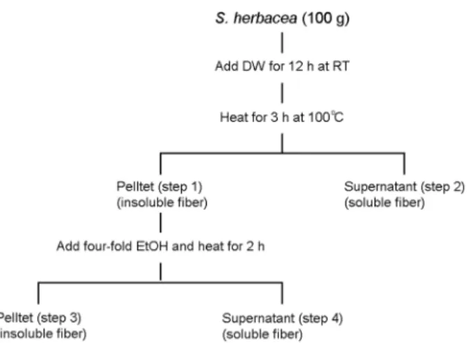
using a method such as Prosky (Prosky et al., 1988), Kim et al. (Kim et al., 1988), and Meas and Delcours (Maes and Delcours, 2002). The extracts obtained through each step of the process (Kim et al., 2014). In this experiment was tested by using the ratio of the highest step of the sample 3 is excellent in the extraction efficiency compared to the total dietary fiber carbohydrate (Fig. 1).
Animal care
Each of the 24 male and female ICR mice (7-weeks) was obtained from Hyochang Science (Korea). Animals were allocated six per polycarbonate cage with stainless steel tops in the animal care facility, where room temperature (20~
25℃), humidity (45~45%), and ventilation were controlled according to international standards. The animals were main-tained in a 12 h light-cycle, and feed and water were supplied free to access. This study was approved by the Animal Ethical Committee of Inje University (Gimhae, Korea) and NIH Guide for the Care and Use of Laboratory Animals and conducted in test guidelines of the Organization for Economic Cooperation and Development (OECD, 2007) and the Korea Food and Drug Administration (KFDA, 2009).
Treatment of SHE
During the acclimatization period of 7d, clinical obser- vations and body weight measurements were conducted to confirm these animals health. The animals were divided into four groups (n=6), with each group having an equal gender ratio and animals of the same weight. The test drug (SHE) was administered orally at dosage levels of 0, 500, 1,000, and 2,000 mg/kg with a volume of 0.1 mL/10 g using sterilized water. The animals were maintained under standard conditions for 7d before the experiment. All groups were orally administrated a single dose and were monitored for 14d. After the completion of the experiment, food and water were removed 12 h prior to sacrifice. The mice were anesthetized by ethyl ether.
General symptoms and mortality
General symptoms and mortality were observed every hour until 12 h after administration of test drug, and con- tinued to next 14d experimental period. During the experi-
mental period, the mortality and type/degree of symptoms, if any, were recorded for each animal.
Autopsy and Hematological analysis
After the overnight fast, the laparotomy was anesthetized with carbon dioxide, blood was collected. After blood col- lection, the liver, heart, kidney, lung, thymus, spleen, and stomach were weight after extirpation as soon as possible.
Mean organ-to-terminal body weight ratios were calculated against fasting body weight of final day. Some of the liver and kidney tissues were fixed in 10% formalin. The tissue are immersed in paraffin blocks and stained with hematoxylin and eosin (H&E stain), and then observed by microscope (×400).
Biochemical analysis
Blood was collected without anticoagulant, and the separ- ated serum was used to assay AST (aspartate amino trans- ferase), ALT (alanine amino transferase), ALP (alkaline phosphatase), BUN (blood urea nitrogen), glucose, albumin, globulin, total cholesterol, and triglyceride levels. All serum sample were stored at -20℃ until analysis, and biochemical values were determined using an automated analyzer (200 FR, TOSIBA, Tokyo, Japan).
Statistical analysis
Data are expressed as mean ± standard deviation (SD).
Fig. 1. Extraction of insoluble fiber compound from Salicornia herbacea L.
All statistical analyses were carried out using SPSS version 22.0 (SPSS Inc, Chicago, Illinois, USA) and Student's t-test
to compare with control. Values of P<0.05 were considered significant.
Table 1. Absolute (g) and relative (%) organ weights of male mice after oral administration of S. herbacea extract
Organs Dose (mg/kg)
Control 500 1,000 2,000
Mean Weight (g)
Kidney (L) 0.291±0.048 0.323±0.023 0.277±0.012 0.304±0.013
Kidney (R) 0.286±0.013 0.303±0.036 0.289±0.022 0.309±0.015
Lung (L) 0.115±0.030 0.103±0.008 0.137±0.007 0.126±0.014
Lung (R) 0.066±0.019 0.090±0.014 0.065±0.005 0.073±0.008
Heart 0.146±0.009 0.137±0.009 0.156±0.008 0.148±0.006
Thymus 0.176±0.070 0.221±0.026 0.217±0.025 0.226±0.012
Spleen 0.123±0.017 0.145±0.024 0.127±0.017 0.135±0.013
Stomach 0.607±0.123 0.613±0.141 0.675±0.107 0.691±0.058
Liver 1.544±0.060 1.648±0.072 1.623±0.058 1.621±0.085
Mean organ-to-terminal body weight ratios (%)
Kidney (L) 0.899±0.147 0.990±0.071 0.880±0.038 0.907±0.039
Kidney (R) 0.882±0.040 0.926±0.110 0.918±0.071 0.920±0.044
Lung (L) 0.355±0.094 0.317±0.024 0.435±0.021 0.374±0.042
Lung (R) 0.203±0.057 0.277±0.043 0.207±0.014 0.217±0.025
Heart 0.450±0.026 0.419±0.026 0.496±0.025* 0.442±0.017
Thymus 0.543±0.217 0.676±0.079 0.690±0.080 0.672±0.037
Spleen 0.381±0.052 0.445±0.074 0.403±0.055 0.401±0.039
Stomach 1.873±0.378 1.876±0.431 2.143±0.338 2.056±0.172
Liver 4.764±0.184 5.046±0.220 5.151±0.186* 4.799±0.254
L, left side; R, right side. Values are expressed as mean ± SD of six mice.
*Significantly different from control at P < 0.05
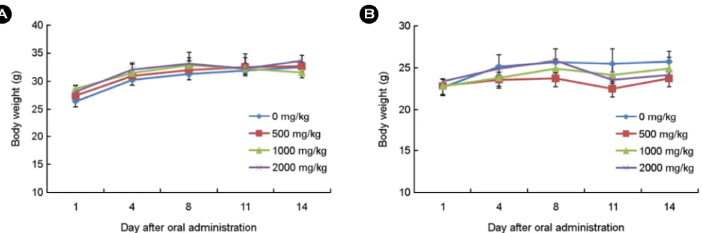
Fig. 2. Changes of body weights in male (A) and female (B) mice after oral administration of S. herbacea extract at dose levels of 0 (◆), 500 (■), 1,000 (▲) and 2,000 (x) mg/kg.
RESULT General symptoms and mortality rate
Upon oral administration toxicity test, there is considered a non-toxic substance in the absence of dead objects from 2,000 mg/kg dose. All the animals died in the test group of male and female during the experiment for SHE was not observed. No physical signs of toxicity such as abnormal breathing, movement, and abnormal stool were observed.
Body weight
The changes in weight of the animals during the experi- mental period are shown in Fig. 2. All groups showed a result that increases than the first. But not a statistically significant changed.
Organ weight
No cellular or organic changes in the animal of male of female were observed. The final weights of organ and organ/
body ratios are shown in Table 1 and 2. Liver weight of 500 mg/kg group of female mice was decreased when compared with those in the control group. However, there is no clinical significance. No significant changes were observed in any of the organ weight between the control group and treated groups.
Serum biochemistry
The biochemical parameters provide an index of the functional changes in the whole body, helping to determine the presence of infection or disease. AST, ALT and ALP are considered specific indicators of hepatocellular necrosis.
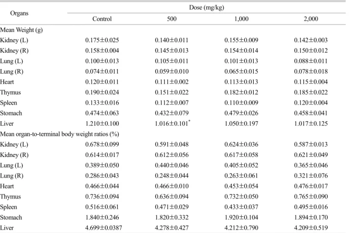
Table 2. Absolute (g) and relative (%) organ weights of female mice after oral administration of S. herbacea extract
Organs Dose (mg/kg)
Control 500 1,000 2,000
Mean Weight (g)
Kidney (L) 0.175±0.025 0.140±0.011 0.155±0.009 0.142±0.003
Kidney (R) 0.158±0.004 0.145±0.013 0.154±0.014 0.150±0.012
Lung (L) 0.100±0.013 0.105±0.011 0.101±0.013 0.088±0.011
Lung (R) 0.074±0.011 0.059±0.010 0.065±0.015 0.078±0.018
Heart 0.120±0.011 0.111±0.002 0.113±0.013 0.115±0.004
Thymus 0.190±0.024 0.151±0.022 0.182±0.012 0.185±0.022
Spleen 0.133±0.016 0.112±0.007 0.110±0.009 0.120±0.004
Stomach 0.474±0.063 0.432±0.079 0.479±0.026 0.458±0.041
Liver 1.210±0.100 1.016±0.101* 1.050±0.197 1.017±0.125
Mean organ-to-terminal body weight ratios (%)
Kidney (L) 0.678±0.099 0.591±0.048 0.624±0.036 0.587±0.013
Kidney (R) 0.614±0.017 0.612±0.056 0.617±0.058 0.621±0.049
Lung (L) 0.389±0.050 0.440±0.046 0.405±0.052 0.365±0.046
Lung (R) 0.286±0.043 0.248±0.044 0.263±0.061 0.321±0.076
Heart 0.466±0.044 0.466±0.010 0.453±0.054 0.476±0.017
Thymus 0.736±0.094 0.636±0.094 0.732±0.050 0.765±0.090
Spleen 0.516±0.061 0.471±0.029 0.433±0.037 0.495±0.016
Stomach 1.840±0.246 1.820±0.332 1.920±0.104 1.894±0.170
Liver 4.699±0.0387 4.278±0.427 4.212±0.790 4.209±0.519
L, left side; R, right side. Values are expressed as mean ± SD of six mice.
*Significantly different from control at P < 0.05
And, BUN levels are used as indicators of renal function (Jo et al., 2008; Kim et al., 2014). There was no significant difference in the AST, ALT, ALP, BUN, albumin, globulin and glucose between the control group and treated group at any dose in mice. Increased in blood fat is deposited in the blood vessel wall inflammation or makes abnormal blood lipid status. After oral administration of the SHE confirm the blood lipid changes, the 2,000 mg/kg treated group of male showed significant decrease from control group in triglyceride. In female mice, triglyceride and total cholesterol was significantly decreased in 1,000 mg/kg and 2,000 mg/
kg dose (Table 3).
Histology
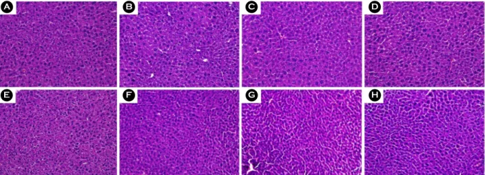
Treatment of toxic substances induces neutrophilic infil- tration and cell death. Therefore, this change was used as an index of necrosis with inflammatory cell infiltration (Kim et al., 2014). Histopathology results of liver and kidney tissue are shown in Fig. 3 and 4. There was no significant difference in liver and kidney between the control group and treated
group at any dose in mice.
DISCUSSION
S. herbacea is an annual plant of Chenopodiaceae family and called Toongtoong-madi and it lives near sea water such as shore, mudflat, and salt field. This plant was used for various traditional remedy, because it absorb many bene- ficial salty ingredients and store (Kim et al., 2014). This study aim for evaluation of biosafety for SHE. Therefore, SHE was produced and performed evaluation of acute oral toxicity in male and female mice. Each 6 mice were grouped and treated various amount of extract; high dose group (2,000 mg/kg), middle dose group (1,000 mg/kg), low dose group (500 mg/kg), and normal group (0 mg/kg). Death rate, normal symptoms, weight change, histopathology diagnosis, biochemistry diagnosis of blood was evaluated for 14d after single dose treatment of SHE in mice. Subsequent single toxicity testing of drugs involves the use of LD50 as an index.
LD50 is the amount of an orally administered test substance Table 3. Biochemical parameters of the male and female mice after oral administration of S. herbacea extract
Parameter Male dose (mg/kg) Female dose (mg/kg)
0 500 1000 2000 0 500 1000 2000
AST (UI/L) 66.33
±4.73 73.33
±3.06 67.67
±2.52 75.33
±9.45 67.67
±7.51 65.33
±7.64 68.00
±3.61 64.00
±2.65
ALT (UI/L) 27.00
±3.00 31.00
±1.00 24.67
±1.53 30.00
±5.20 27.33
±3.21 23.33
±5.03 25.33
±4.16 22.00
±1.00 ALP (UI/L) 111.00
±15.72 96.33
±15.50 87.67
±16.26 83.00
±12.29* 105.00
±9.64 100.67
±15.57 110.33
±17.10 110.67
±9.07 BUN (mg/dL) 33.10
±4.36 29.13
±3.86 27.43
±2.84 26.97
±0.83 21.30
±4.86 21.50
±0.95 20.20
±0.60 21.53
±5.50 T. Chol (mg/dL) 161.33
±26.31 147.33
±23.12 136.33
±10.50 134.33
±18.58 124.67
±7.02 104.33
±23.97 100.00
±14.18* 100.33
±11.06* Triglyceride
(mg/dL)
120.00
±21.79
90.67
±11.72
86.00
±13.11
83.33
±8.96*
121.00
±9.54
138.00
±16.46
126.67
±33.55
79.33
±12.90* Albumin (g/dL) 3.50
±0.10
3.37
±0.12
3.37
±0.06
3.37
±0.12
4.00
±0.10
3.83
±0.15
3.97
±0.25
3.90
±0.17 Globulin (g/dL) 1.60
±0.10
1.50
±0.10
1.60
±0.00
1.70
±0.10
1.40
±0.10
1.33
±0.23
1.27
±0.12
1.30
±0.20 Glucose (g/dL) 203.33
±42.16 220.33
±69.24 247.33
±64.93 190.33
±25.03 134.67
±23.01 108.33
±9.45 110.00
±13.23 131.67
±11.06 AST, aspartate transferase; ALT, alanine transferase; ALP, alkaline phosphatase; BUN, blood urea nitrogen; T. Chol, total cholesterol.
Values are expressed as mean ± SD of six mice.
*Significantly different from control at P < 0.05
that can be expected to cause death in LD50 of animals (Dietrich, 1983). In this study, SHE administration did not affect any clinical symptoms and motility. Thus, the LD50
of orally administered SHE was higher than 2,000 mg/kg in both sexes. During acute toxicity evaluation, died mice, and significant change in normal symptoms, weight change, and histopathology diagnosis were not presented. Various biochemical parameters were used to evaluate the toxicity
of SHE. AST, ALT, ALP, albumin, globulin, BUN, total cholesterol, and triglyceride levels were measured in liver and kidney function test (Johnston, 1999). Biochemistry diagnosis of blood serum is not presented significant change in most of test item, but some lipid components were changed. Total cholesterol and triglyceride were measured as indicators of hyperlipidemia (Kim et al., 2014). Triglyceride was decreased dose-dependently and significant decrease in Fig. 3. Liver tissue stained hematoxylin and eosin (H & E, 400×) showing the effect of S. herbacea extract in mice. The samples (A) through (D) are from male, and samples (E) through (H) are from female. (A) 0 mg/kg; (B) 500 mg/kg; (C) 1,000 mg/kg; (D) 2,000 mg/kg; (E) 0 mg/
kg; (F) 500 mg/kg; (G) 1,000 mg/kg; (H) 2,000 mg/kg.
Fig. 4. Kidney tissue stained hematoxylin and eosin (H & E, 400×) showing the effect of S. herbacea extract in mice. The samples (A) through (D) are from male, and samples (E) through (H) are from female. (A) 0 mg/kg; (B) 500 mg/kg; (C) 1,000 mg/kg; (D) 2,000 mg/kg;
(E) 0 mg/kg; (F) 500 mg/kg; (G) 1,000 mg/kg; (H) 2,000 mg/kg.
high dose group (2,000 mg/kg) was observed. Also, total cholesterol and triglyceride were significantly decreased in female mice. Because of these results, single dose acute toxicity of SHE is higher than 2,000 mg/kg, and the admin- istration of SHE is determined to be effective in reducing lipid contents in the blood. It is determined that there is a value used in the future study for the hyperlipidemia disorder.
Therefore, SHE extract has stable biosafety and expected to use in ingredient of various functional food, drug and physiological activity assessment.
Acknowledgement
This work was supported by the Jungwon University Research Grants.
Conflict of interest
The authors declare that they have no conflict of interests.
REFERENCES
Dietrich L. A new approach to practical acute toxicity testing.
Arch Toxicol. 1983. 54: 275-287.
Ermawati N, Cha JY, Liang YS, Jung MH, Shin DJ, Lee BH, Lee KH, Son DY. Molecular cloning and characterization of outer envelope membrane protein from Salicornia herbacea. Korean J Plant Biotechnol. 2004. 31: 273-278.
Han MH, Kim JW, Kim KY, Kim SG, Yu GJ, Cho YB, Hwang HJ, Kim BW, Kim CM, Choi YH. Single dose oral toxicity of Schisandrae Semen essential oil in ICR mice. J Life Sci. 2014.
24: 191-195.
Han SW, Kim SM. Anti-oxidative effect of Salicornia herbacea L.
grown in closed sea beach. J Korean Soc Food Sci Nutr. 2003.
32: 207-210.
Jo WS, Nam BH, Oh SJ, Choi YJ, Kang EY, Hong SH, Lee SH, Jeong MH. Hepatic protective effect and single-dose toxicity
study of water extract of Cordyceps militaris grown upon Protaetia dreujtatsis. Korean J Food Sci Technol. 2008. 40:
106-110.
Jo YC, Lee KS, Chon SM, Byun DS. Characteristics of growth and germination of Salicornia herbacea L. for the soil salinity and manure condition. Korean J Medicinal Crop Sci. 2002. 10:
100-108.
Johnston DE. Special consideration in interpreting liver function tests. Am Fam Physician. 1999. 59: 2223-2230.
Jung BM, Park JA, Bae SJ. Growth inhibitory and quinone reduc- tase induction activities of Salicornia herbacea L. fractions on human cancer cell lines in vitro. J Korean Soc Food Sci Nutr. 2008. 37: 148-153.
Kim MJ, Young JH, Kim JH. Anti-obesity effect of Korean Hamcho (Salicornia herbacea L.) powon high-fat diet-induced obese rats. J Nutr Health. 2015. 48: 123-132.
Kim SH, Ryu DS, Lee HS, Shin HR, Kwon JH, Lee DS. Acute oral toxicity of the ethyl acetate fraction of Orostachys japonicus in mice. Pharma Biol. 2014. 52: 1345-1350.
Kim SH, Kim SJ, Lee HS. Effect of insoluble dietary fiber ex- tracted from Salicornia herbacea L. on large intestinal function in rats. Korean J Food Sci Technol. 2014. 46: 648-654.
Kim SH, Park HY, Park WK. Determination and physical pro- perties of dietary fiber in seaweed products. J Korean Soc Food Nutr. 1988. 17: 320-325.
Korea Food and Drug Administration. Testing guidelines for safety evaluation of drugs. Notification No. 2009-116, issued by the Korea Food and Drug Administration of August 24, 2009.
Maes C, Delcour JA. Structural characterization of water-extractable and water-unextractable arabinoxylans in wheat bran. J Ceral Sci. 2002. 35: 315-326.
OECD. OECD Guideline for testing of chemicals. Acute oral toxicity-fixed dose procedure. OECD/OEDC. 2007. 420: 1-14 Prosky L, Asp N, Sweizer TF, Deveries J, Furda L. Determination of insoluble, soluble, and total dietary fiber in foods and pro- ducts. J Assoc Off Anal Chem. 1988. 71: 1017-1023.