• 검색 결과가 없습니다.

Gene Expression Analysis from the Normal Stomach Cells Treated with a Cancer Inducer N-methyl-N'-nitro-N-nitrosoguanidine, MNNG

N/A
N/A
Protected

Academic year: 2021

Share "Gene Expression Analysis from the Normal Stomach Cells Treated with a Cancer Inducer N-methyl-N'-nitro-N-nitrosoguanidine, MNNG"

Copied!
4
0
0

로드 중.... (전체 텍스트 보기)

전체 글

(1)

- 30 - Biomedical Science Letters 2017, 23(1): 30~33

http://dx.doi.org/10.15616/BSL.2017.23.1.30 eISSN : 2288-7415

Gene Expression Analysis from the Normal Stomach Cells Treated with a Cancer Inducer N-methyl-N'-nitro-N-nitrosoguanidine, MNNG

Dongju Jung

1,2

, Yoonjung Cho

3

, Tae Ue Kim

3

and Sang-Hee Jeong

1,2,†

1

Biomedical Science Institute,

2

Department of Biomedical Laboratory Science, College of Life and Health Sciences, Hoseo University, Asan, Chungnam 31499, Korea

3

Department of Biomedical Laboratory Science, College of Health Sciences, Yonsei University, Wonju, Kangwon 26493, Korea

N-methyl-N'-nitro-N-nitrosoguanidine (MNNG) is a carcinogen made of modified guanine on which alkyl group is added on 6th oxygen. It has been used for inducing different types of cancers experimentally in vivo and in vitro.

Stomach cancer might be the best well established particular cancer induced with MNNG. Comparative analysis of gene expression between normal stomach cell and MNNG-treated stomach cell could give much information to understand cancer formation in stomach. To this end, normal human stomach cells HS738 were treated with DMSO or MNNG.

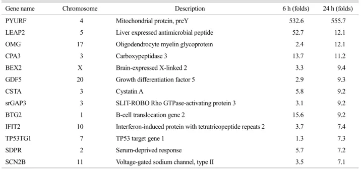
Genetic comparison was conducted with purified RNA from the treated cells for 6 hours or 24 hours. Total 13 genes were selected based on their high induction folds and comprehensible function to cancer formation. Some of the genes were cancer-promoting whereas the others were anti-cancer genes. These results could give important information of genetic changes in stomach cells during MNNG-induced stomach cancer formation.

Key Words: N-methyl-N'-nitro-N-nitrosoguanidine (MNNG), Stomach cancer, Gene expressions

N-methyl-N'-nitro-N-nitrosoguanidine (MNNG) is a car- cinogen made of modified guanine on which alkyl group is added on 6th oxygen. MNNG has been broadly used for inducing cancer formation in normal cells and animals since its cancer-promoting effect on rat stomach was experi- mentally succeeded (Sugimura, 1967). When MMNG is administered to rats, severe inflammation with erosion appears in two weeks and finally adenocarcinoma of the glandular stomach is developed in weeks 35-72 through atypical and regenerative changes (Yamashita, 2002). Cul- tured normal cells were also developed significant cancerous formation after MNNG treatment having changes on their

attachment, morphology, ploidy, mucin secretion, and tyro- sine phosphorylation without complete malignant formation (Malik, 1997). Thus MNNG is a very useful small molecule for inducing artificial cancerous formation in laboratory animals and cultured cells. In addition to this, genetic analysis is a powerful tool to uncover genes that have functions to many different types of cellular processes and cancer for- mation (Jin 2015; Jin 2015). Combining the small molecule that have clear function toward cancer formation and whole RNA genetic analysis could give much information to understand molecular genetic processes in cancer generation.

Here we analyzed the detailed gene inductions by MNNG Brief Communication

*Received: November 29, 2016 / Revised: February 1, 2017 / Accepted: February 2, 2017

Corresponding author: Sang-Hee Jeong. Biomedical Science Institute and Department of Biomedical Laboratory Science, Hoseo University, Asan 31499, Korea.

Tel: +82-41-540-9675, Fax: +82-41-540-9829, e-mail: [email protected]

CThe Korean Society for Biomedical Laboratory Sciences. All rights reserved.

CCThis is an Open Access article distributed under the terms of the Creative Commons Attribution Non-Commercial License (http://creativecommons.org/licenses/by-nc/3.0/) which permits unrestricted non-commercial use, distribution, and reproduction in any medium, provided the original work is properly cited.

(2)

- 31 - treatment in normal stomach cells.

For the gene expression analysis, normal human stomach cell line, HS738 were cultured and treated with MNNG for 6 hr or 24 h. DMSO-treated HS738 cells were used as con- trol. The normal stomach cells were able to maintain viability under 50 M of MNNG for up to 24 h (Fig. 1). Total RNA was purified with Trizol reagent (Thermo scientific) follow- ing manufacturer's instruction. RNA sequencing was con- ducted in the TheragenEtex (Suwon, South Korea). Briefly, 2 μg of total RNA was treated with DNase I, and poly (A) mRNA was enriched via purification with Oligo(dT)-labeled magnetic beads. The purified mRNAs were disrupted into short fragments and used as template for synthesizing double- stranded cDNAs. After end-repair and poly (A) addition, the cDNAs were hybridized with sequencing adapters using

the TruSeq RNA Sample Prep Kit (Illumina, San Diego, CA, USA.) for constructing cDNA library. The library was sequenced using a HiSeq 2500 sequencer (Illumina) in rapid run mode. The sequenced cDNA had highly matched with the genes in chromosome (Fig. 2). Cluster generation was performed, followed by 2×100 cycle sequencing reads separated by a paired-end turnaround. Image analysis was performed using the HiSeq control software version 1.8.4.

MNNG-induced genes were selected based on expression increase that was higher than 7-folds than that form the DMSO-treated cells after 24 h treatment. The most increased gene was a mitochondrial protein, preY (Table 1). The gene expression was induced 557-folds in HS738 cells by MNNG treatment but it seems that there might be no clear relation- ship between preY and cancer formation. LEAP2 induction was higher in 6 hr than that in 24 hr. LEAP2 is acronym of 'liver expressed antimicrobial peptide 2'. Although it was highly induced by MNNG, LEAP2 induction seems not have profound contribution to cancer formation. OMG represent 'oligodendrocyte-myelin glycoprotein', which is a Nogo re- ceptor ligand that inhibits neurite outgrowth (Wang, 2002).

Like the previous two genes, OMG might not have direct relationship to cancer formation. The following genes are supposed to have some roles to cancer formation of normal stomach cells by MNNG treatment. Carboxypeptidase A3 (CPA3) was reported that it was a novel gene highly in- duced by histone deacetylase inhibitors in prostate cancer cells (Huang, 1999). BEX2 was reported to be expressed in a subset of primary breast cancers (Naderi, 2007). GDF5 was reported to regulate angiogenesis in a breast cancer cell line (Margheri, 2012). Cysteine protease inhibitor cystatin Fig. 1. Cell viability test. Dark columns represent viability of

DMSO-treated normal stomach cell line HS738. Grey columns represent viability of HS737 treated with 50 μM of MNNG, which is dissolved in DMSO. Cell viability tests were conducted using 'EZ-CYTOX' from DoGen South Korea.

Fig. 2. Assignment of sequenced genes. Total 27,748 gene fragment were sequenced. More than 80%

of the sequenced genes were able

to be mapped. A indicates DMSO-

treated HS738 cells. B indicates

HS738 cells treated with 50 μM

MNNG for 6 hr. C indicates HS-

738 cells treated with 50 μM

MNNG for 24 hr.

(3)

- 32 - A (CSTA) was reported that it was a gene deregulated in several skin cancers (Gupta, 2015). srGAP3, a member of the Slit-Robo sub-family of Rho GTPase-activating proteins (Rho GAPs), was known to have a tumor suppressor role in mammary epithelial cells (Lahoz, 2013). BTG2, the B-cell translocation gene 2, is a p53-induced gene having signifi- cant anti-proliferative roles for disease progression including human breast cancer (Takahashi, 2011). IFIT2 was reported to contribute to inhibiting migration and metastatic activity of oral squamous cell carcinoma cells (Lai, 2013). TP53TG1 was a gene identified using transgene TP53-containing colon cancer cell line. It was identified as a target gene of TP53 and supposed to respond to cellular damage (Takei, 1998).

SDPR was reported to act as a metastasis suppressor in breast cancer (Ozturk, 2016). Expressions of voltage-gated sodium channels in cancers have been known. SCN2B is a voltage-gated Na

+

channels which was upregulated in car- cinomas by potentiating metastasis of the cancers (Fraser, 2014).

Conclusively, we were able to identify genes of which expression were upregulated in normal stomach cells by MNNG treatment. None of the three highly induced genes,

Pre Y, LEAP2 and OMG, seem to be involved in cancer formation in spite of their high induction folds. Some of the genes are able to potentiate cancer formation when its expression was disrupted: GDF5, CSTA, BTG2, IFIT2, TP53TG1, and SDPR. In addition to these, BEX2, srGAP3, and SDPR are tumor suppressor genes whereas VGSCs is a tumor-promoting gene. This result shed light on under- standing of gene expressions that contribute to stomach cancer formation by MNNG treatment.

Acknowledgements

This study was supported by "the High Value-added Food Technology Development Program (Project title: Estab- lishment of Scientific Infrastructural Knowledge of Trad- itional Agricultural Food-Originated-Medicines, Project No.

314071-03)" of Korea Institute of Planning & Evolution for Technology in Food, Agriculture, Forestry & Fisheries, Republic of Korea.

Conflict of interest

The authors declare that they have no competing interests.

Table 1. Gene expression levels

Gene name Chromosome Description 6 h (folds) 24 h (folds)

PYURF 4 Mitochondrial protein, preY 532.6 555.7

LEAP2 5 Liver expressed antimicrobial peptide 52.7 12.1

OMG 17 Oligodendrocyte myelin glycoprotein 2.4 12.1

CPA3 3 Carboxypeptidase 3 13.7 11.2

BEX2 X Brain-expressed X-linked 2 3.3 9.4

GDF5 20 Growth differentiation factor 5 2.9 9.3

CSTA 3 Cystatin A 5.8 9.2

srGAP3 3 SLIT-ROBO Rho GTPase-activating protein 3 3.1 9.2

BTG2 1 B-cell translocation gene 2 15.6 9.2

IFIT2 10 Interferon-induced protein with tetratricopeptide repeats 2 3.7 7.4

TP53TG1 7 TP53 target gene 1 1.3 7.3

SDPR 2 Serum-deprived response 5.7 7.2

SCN2B 11 Voltage-gated sodium channel, type II 3.5 7.1

The MNNG-induced genes expressed more than 7-folds higher compared with their expression levels in DMSO-treated cells and known

for their functions were selected. The genes are listed according to the induction folds in 24 hr.

(4)

- 33 - REFERENCES

Fraser SP, Ozerlat-Gunduz I, Brackenbury WJ, Fitzgerald EM, Campbell TM, Coombes RC, Djamgoz MB. Regulation of voltage-gated sodium channel expression in cancer: hormones, growth factors and auto-regulation. Biological Sciences. 2014.

369: 20130105.

Gupta A, Nitoiu D, Brennan-Crispi D, Addya S, Riobo NA, Kelsell DP, Mahoney MG. Cell cycle- and cancer-associated gene networks activated by Dsg2: evidence of cystatin A deregu- lation and a potential role in cell-cell adhesion. PLoS One.

2015. 10: e0120091.

Huang H, Reed CP, Zhang JS, Shridhar V, Wang L, Smith DI.

Carboxypeptidase A3 (CPA3): a novel gene highly induced by histone deacetylase inhibitors during differentiation of prostate epithelial cancer cells. Cancer Research. 1999. 59:

2981-2988.

Jin HS, Jung DJ. Association of the TREML2 and HTR1E genetic polymorphisms with osteoporosis. Biomedical Science Letters.

2015. 21: 181-187.

Jin HS, Jung DJ. Gender-specific Association of the ANO1 genetic variations with hypertension. Biomedical Science Letters. 2015.

21: 144-151.

Lahoz A, Hall A. A tumor suppressor role for srGAP3 in mammary epithelial cells. Oncogene. 2013. 32: 4854-4860.

Lai KC, Liu CJ, Chang KW, Lee TC. Depleting IFIT2 mediates atypical PKC signaling to enhance the migration and meta- static activity of oral squamous cell carcinoma cells. Oncogene.

2013. 32: 3686-3697.

Malik R, Lee SK, Savard CE, Oda D, Wong WS, Chan BY, Lee SP. The effect of N-methyl-N'-nitro-N-nitrosoguanidine on cultured dog gallbladder epithelial cells. Hepatology. 1997. 26:

1296-1302.

Margheri F, Schiavone N, Papucci L, Magnelli L, Serrati S, Chilla A, Laurenzana A, Bianchini F, Calorini L, Torre E, Feijoo E,

Fibbi G, Del Rosso M. GDF5 regulates TGFβ-dependent angiogenesis in breast carcinoma MCF-7 Cells: in vitro and in vivo control by anti-TGFβ peptides. PLoS One. 2012. 7:

e50342.

Naderi A, Teschendorff AE, Beigel J, Cariati M, Ellis IO, Brenton JD, Caldas C. BEX2 is overexpressed in a subset of primary breast cancers and mediates nerve growth factor/nuclear factor-kappa B inhibition of apoptosis in breast cancer cell lines. Cancer Research. 2007. 67: 6725-6736.

Ozturk S, Papageorgis P, Wong CK, Lambert AW, Abdolmaleky HM, Thiagalingam A, Cohen HT, Thiagalingam S. SDPR functions as a metastasis suppressor in breast cancer by pro- moting apoptosis. Proceedings of the National Academy of Sciences. 2016. 113: 638-643.

Sugimura T, Fujimura S. Tumor production in glandular stomach of rats by N-methyl-N'-nitro-N-nitrosoguanidine. Nature. 1967.

216: 943-944.

Takahashi F, Chiba N, Tajima K, Hayashida T, Shimada T, Kakahashi M, Moriyama H, Brachtel E, Edelman EJ, Ramaswamy S, Maheswaran S. Breast tumor progression induced by loss of BTG2 expression is inhibited by targeted therapy with the ErbB/HER inhibitor lapatinib. Oncogene.

2011. 30: 3084-95.

Takei Y, Ishikawa S, Tokino T, Muto T, Nakamura Y. Isolation of a novel TP53 target gene from a colon cancer cell line carrying a highly regulated wild-type TP53 expression system. Genes Chromosomes Cancer. 1998. 23: 1-9.

Wang KC, Koprivica V, Kim JA, Sivasankaran R, Guo Y, Neve RL, He Z. Oligodendrocyte-myelin glycoprotein is a Nogo receptor ligand that inhibits neurite outgrowth. Nature. 2002.

417: 941-944.

Yamashita S, Wakazono K, Sugimura T, Ushijima T. Profiling and

selection of genes differentially expressed in the pylorus of

rat strains with different proliferative responses and stomach

cancer susceptibility. Carcinogenesis. 2002. 26: 923-928.

수치

Fig. 2. Assignment of  sequenced genes. Total 27,748 gene fragment were sequenced. More than 80%
Table 1. Gene expression levels

참조

관련 문서

Quantitation of procollagen synthesis by Hs68 cells treated with fish collagen hydrolysates (FCHs), their <1 kDa fraction, and their >1 kDa fraction for 24 hrs.

In vitro cell migration in APE or JAG1 siRNA-treated M059K cell line Fig,11 Down-regulation of JAG1 induces S phase arrest in

Null hypotheses of this study was there is no difference in root canal length determination between conventional and heat-treated Ni-Ti files by 2

The clinical outcome and complication for treating proximal femoral shaft fracture were compared and analyzed through the group treated with closed

Control- and ASTRIN- depleted HeLa cells were untreated or treated with 5 Gy. IR and were then fixed at

4 > Effects of Taro on COX-2 expression and iNOS expression(hot water) in human thyroid cancer cells. The cells were pretreated for 48hours with either

Recent stem cell studies have reported that cultured hematopoietic stem cells (HSCs) are reactivated through fetal hemoglobin expression by treatment with

As for the research method, RBL-2H3 cells, a mast cell line, were treated with supercritical carbon dioxide extracted Chamaecyparis obtusa oil (C oil), German chamomile oil (G