The Incidence, Causes, and Prognostic Significance of New-
Onset Thrombocytopenia in Intensive Care Units: A Prospective Cohort Study in a Korean Hospital
This study was designed to investigate the incidence, causes, and outcomes of new-onset thrombocytopenia (NOT) in Korean intensive care units (ICUs). A prospective cohort study was conducted in medical ICUs of Samsung Medical Center between August 2010 and February 2011. All newly admitted patients were included if they stayed in the ICU for more than 48 hr and did not have thrombocytopenia upon admission. A total of 186 patients were included. NOT developed in 37.1%. Most common cause of NOT was sepsis with disseminated intravascular coagulation (66.7%), followed by drug-induced
thrombocytopenia (18.8%), and heparin-induced thrombocytopenia (2.9%). IgG-specific antibody to platelet factor 4/heparin was positive in 2.4% among patients treated with heparin, and thrombosis occurred in two patients. Twenty eight-day mortality was higher in patients that developed NOT compared to those that did not develop NOT (39.1% vs 12%, P < 0.001). NOT increased the odds ratio of 28-day mortality and was an independent risk factor for mortality (OR 3.52; 95% CI 1.32-9.38; P = 0.012). In conclusion, NOT is common and is an independent risk factor for mortality in Korean ICU patients. Therefore, clinicians should make every effort to correct the causes of NOT.
Key Words: Heparin; Intensive care units; Korea; Mortality; Thrombocytopenia So Yeon Lim1, Eun Ju Jeon1,
Hee-Jin Kim2, Kyeongman Jeon1, Sang-Won Um1, Won-Jung Koh1, Man Pyo Chung1, Hojoong Kim1, O Jung Kwon1, and Gee Young Suh1
1Division of Pulmonary and Critical Care Medicine, Department of Medicine, 2Department of Laboratory Medicine & Genetics, Samsung Medical Center, Sungkyunkwan University School of Medicine, Seoul, Korea
Received: 5 April 2012 Accepted: 20 August 2012 Address for Correspondence:
Gee Young Suh, MD
Division of Pulmonary and Critical Care Medicine, Department of Medicine, Samsung Medical Center, Sungkyunkwan University School of Medicine, 81 Irwon-ro, Gangnam-gu, Seoul 135-710, Korea
Tel: +82.2-3410-3426, Fax: +82.2-3410-6956 E-mail: [email protected]
This work was supported by a grant from Samsung Medical Center in 2011 (CRS1104521).
http://dx.doi.org/10.3346/jkms.2012.27.11.1418 • J Korean Med Sci 2012; 27: 1418-1423 Emergency & Critical Care Medicine
INTRODUCTION
Thrombocytopenia, which is one of the most commonly ob- served laboratory abnormalities in the intensive care units (ICU) population, has an incidence ranging from 13.0% to 44.1% (1), depending on the study population, the timing and frequency of platelet monitoring, and the definition of thrombocytopenia (2-5). In critically ill patients, one potential cause is enhanced platelet destruction due to heparin-induced thrombocytopenia (HIT), a prothrombotic disorder that is caused by the develop- ment of antibodies against platelet factor 4 (PF4)/heparin com- plexes (8, 9). Because heparin-based anticoagulation is one of the most commonly prescribed therapies in clinical practice and thrombotic complications related to HIT may be life threat- ening, the early recognition and treatment of this cause of throm- bocytopenia are important.
Thrombocytopenia in ICU patients has been associated with adverse outcomes such as prolonged length of hospital stay and decreased survival (2-5). However, many of these studies were retrospective and performed in European or North American
ICUs (2, 10-13). Thus, the incidence and prognostic significance of NOT in Asians, especially Koreans, is not known. Therefore, the objective of this study was to investigate the incidence of NOT, including HIT, in a cohort of Korean medical ICU patients and to examine its impact on outcomes.
MATERIALS AND METHODS Study population
This is a prospective cohort study. Consecutive patients admit- ted to the two medical ICUs of Samsung Medical Center, a 1,900- bed tertiary referral center in Seoul, Korea, between August 2010 and February 2011 were prospectively recruited and were in- cluded in the study if their stay in the ICU was longer than 48 hr.
Patients were excluded from the analysis if they had thrombo- cytopenia at the time of ICU admission, were readmitted to the ICU during the same hospitalization, were younger than 18 yr of age or were pregnant. Platelet count was checked every day from ICU admission to ICU discharge.
Definitions
Thrombocytopenia was defined as a platelet count < 150,000/µL or a decrease in platelet count ≥ 50% from the ICU admission value (9, 12). The nadir platelet count was defined as the lowest platelet count recorded during the ICU stay. Disseminated in- travascular coagulation (DIC) was considered to be present when the d-dimer level was elevated in addition to two of the follow- ing criteria: prolonged prothrombin time, increased fibrin deg- radation product, a ≥ 25% decrease in antithrombin, decreased fibrinogen, or platelet count (13). The diagnosis of drug-induced thrombocytopenia was made only upon resolution of throm- bocytopenia after discontinuation of the suspected drug. The diagnosis of HIT was accepted when a patient met the defini- tion of thrombocytopenia not explained by other causes after use of unfractionated heparin (UFH) or low molecular weight heparin (LMWH), and tested positive for PF4/heparin antibod- ies (12). Shock was defined as the need for vasoactive drugs (> 5 μg/kg/min of dopamine or dobutamine or norepinephrine at any dose) for at least one hour (14). Septic shock was diagnosed when shock was associated with documented or assumed in- fection without any other identifiable cause of shock (14).
Data collection
The following variables were recorded: 1) general characteris- tics including age, gender, preexisting underlying diseases, and primary reason for ICU admission; 2) severity of illness as as- sessed by the Simplified Acute Physiology Score 3 (SAPS 3) and the Sequential Organ Failure Assessment (SOFA) score; 3) 4T’s score of the patients with positive antigen assays; 4) optical den- sity (OD) units of antigen assays; 5) laboratory data upon ICU admission including hematologic and chemistry tests and arte- rial blood gas analysis; 6) drugs and interventions including the need for mechanical ventilation and continuous renal replace- ment therapy (CRRT); and 7) the development of complications such as septic shock, acute respiratory distress syndrome (ARDS), DIC, bleeding, and thromboembolic events.
Measurement of antibodies against the PF4/heparin complex
Within 24 hr of detecting NOT, a blood sample was drawn from each patient for the detection of HIT antibodies. The blood was allowed to clot and was then centrifuged to isolate the serum, which was stored frozen at -70°C until analysis. A commercial immunoassay (ZYMUTEST HIA IgG, ARK040A, PF4/heparin enzyme-linked immunosorbent assay [ELISA] kits; HYPHEN BioMed, Andresy, France) was used to detect HIT antibodies according to the manufacturer’s instructions. Positivity was de- fined by OD higher than 0.5 units.
Statistical analysis
PASW 17.0 (SPSS Inc., Chicago, IL, USA) and STATA 11.0 (Stata-
Corp LP, College Station, Texas, USA) were used for statistical analysis. Continuous variables are shown as medians and inter- quartile ranges (IQR) and categorical data are shown as counts (percentages). Continuous variables were analyzed using the Mann-Whitney U test, while the chi-square or Fisher’s exact test were used to compare categorical data. A univariate logistic re- gression model was used to identify significant predictors of 28- day mortality. A multivariate logistic regression analysis was per- formed to evaluate the effect of NOT on 28-day mortality. Two models were evaluated. Model 1 was adjusted for age, gender, SAPS 3, and length of ICU stay (LOS), and model 2 was adjusted for all variables with P < 0.25 on the univariate analysis, age, gender, SAPS 3, and ICU LOS. The presence of multicollinearity among two separate variables was evaluated by a variance infla- tion factor. A P value < 0.05 was considered statistically significant.
Ethics statement
Our institutional review board approved this prospective obser- vational study (2010-07-026). Due to the purely observational nature of the study, informed consent was not required. How- ever, when thrombocytopenia developed, patients, or their le- gal representatives, were asked to provide written informed con- sent for blood sampling to assay for antibodies reactive to the PF4/heparin complex.
RESULTS
Baseline characteristics, clinical features, and outcomes for all patients
During the seven-month study period, 920 patients were admit- ted to the medical ICUs and 186 patients met our inclusion cri- teria (Fig. 1). Table 1 shows the characteristics of the 186 patients upon admission. Among the 186 patients included in the analy- sis, 116 (62.4%) patients were male and the median age was 65.5
Fig. 1. Study flow chart. During the seven-month study period, 920 patients were ad- mitted to the medical ICUs and 186 patients met our inclusion criteria. Among them, 69 patients showed new-onset thrombocytopenia.
734 excluded.
339 discharged from the ICU within 48 hr.
395 had thrombocytopenia at the time of ICU admission.
117 NOT (-) 69 NOT (+)
186 enrolled 920 patients screened for eligibility
yr (Table 1). The median SAPS 3 was 49 (IQR, 37-58) and the pre- dicted death rate was 43.8% (IQR 19.6-64). The median SOFA at the time of ICU admission was 4 (IQR, 3-5). In all, 126 patients (67.7%) were exposed to heparin (LMWH in 51 patients [27.4%]
and UFH 72 patients [38.7%]). The median length of ICU stay was six days (IQR, 4-13). ICU mortality was 20.3% and 28-day mortality was 22.2%.
Causes of NOT
NOT developed in 69 patients (37.1%). Table 2 presents infor- mation regarding causes of NOT in the 69 patients. Sepsis with DIC was the most frequent cause of NOT with 46 patients (66.7
%), followed by drug-induced thrombocytopenia (18.8%), HIT (2.9%), and liver disease (1.4%). In seven patients, the cause of thrombocytopenia could not be determined.
Detection of IgG-specific antibody to PF4/heparin Serum sampling was refused by 6 out of 69 patients who devel- oped NOT and test to detect of IgG-specific antibody to PF4/
heparin was performed using 63 patients (Table 3). IgG-specific antibody to PF4/heparin was positive in three patients among the patients receiving heparins (3/126, 2.4%). Among the pa- tients without history of exposure to heparin, no patients had
positive antigen assay. The OD units of the three patients were 3.48, 0.91, and 0.51, and the 4T’s scores were 3, 6, and 6 points, respectively. One of the patient, who had OD of 3.48 had under- lying sepsis with 4T’s score of 3 and did not meet our criteria for HIT. The other two patients did not have any other cause to ex- plain NOT, and had thrombosis. Therefore, the diagnosis of HIT was accepted in two patients: 1.6% of patients who were exposed to heparin.
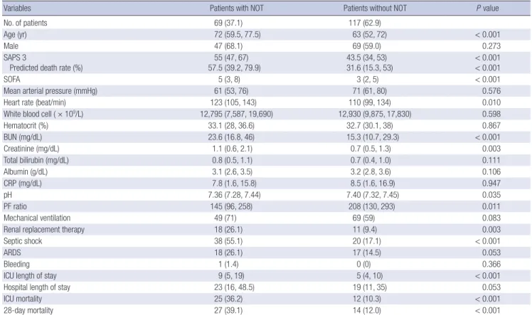
Comparison between patients with and without NOT Table 4 compares patients with and without NOT. Patients with NOT were older (72 vs 63 yr, P < 0.001) and had higher SAPS 3 (55 vs 43.5, P < 0.001) and SOFA scores (5 vs 3, P < 0.001) than patients without NOT. Patients with NOT had higher heart rates (123 vs 110 per min, P = 0.010) and higher creatinine levels (1.1 vs 0.7 mg/dL, P = 0.003) than patients without NOT. The length of ICU stay was significantly longer for patients with NOT (9 vs 5 days, P < 0.001). ICU mortality and 28-day mortality were high- er in patients with NOT than in patients without NOT (36.2% vs 10.3%, 39.1% vs 12%, respectively; P < 0.001).
The effects of NOT on 28-day mortality
Univariate and multivariate logistic regression analyses were performed to determine if there were any associations between risk factors and 28-day mortality in the 186 patients. The unad- justed odds ratio (OR) for NOT with regards to the 28-day mor- tality was 4.52 (95% CI, 2.18-9.36, P < 0.001) (Table 5). When ad- justed for age, gender, SAPS 3, and ICU LOS (model 1), NOT sig- Table 1. Baseline characteristics and outcomes of the all patients (n = 186)
Parameters No. (%) or Median (IQR)
Age (yr) 65.5 (55, 74)
Male 116 (62.4)
SAPS 3
Predicted death rate (%) 49 (37, 58)
43.8 (19.6, 64)
SOFA 4 (3, 5)
Medical history Hypertension Diabetes mellitus Solid cancer
Hematologic malignancy Neurologic disease Cardiovascular disease Chronic renal failure
47 (25.3) 34 (18.3) 67 (36) 11 (5.9) 11 (5.9) 18 (9.7) 13 (7) Primary reason for ICU admission
Respiratory Postoperative Septic shock Cardiovascular problem CRRT
Others
156 (83.9) 3 (1.6) 14 (7.5) 5 (2.7) 5 (2.7) 3 (1.6) Use of heparin
UFH LMWH Both
126 (67.7) 72 (38.7) 51 (27.4) 3 (1.6)
ICU length of stay 6 (4, 13)
Hospital length of stay 18 (8, 34)
ICU mortality 37 (20.3)
28-day mortality 41 (22.2)
Data are presented as number (percentage) or median (interquartile range). IQR, in- terquartile range; SAPS 3, simplified acute physiology score 3; SOFA, sequential or- gan failure assessment; ICU, intensive care unit; CRRT, continuous renal replacement therapy; UFH, unfractionated heparin; LMWH, low molecular weight heparin.
Table 3. Results of IgG-specific antibody to PF4/heparin Conditions of patients Exposure to
heparin No exposure to
heparin Total
No. of patients 126 (67.7) 60 (32.3) 186 (100%) New-onset thrombocytopenia 49 (38.9) 20 (33.3) 69 (37.1) Test of IgG-specific antibody to
PF4/heparin Performed Positive
43 (34.1) 3 (2.4)
20 (33.3) 0 (0)
63 (33.9) 3 (1.6) Data are presented as number (percentage).
Table 2. Causes of new-onset thrombocytopenia (n = 69)
Causes No. (%)
Sepsis with disseminated intravascular coagulation 46 (66.7) Drug induced thrombocytopenia
Penicillin/β-lactamase inhibitor Carbapenem
Cephalosporin Quinolone H2-blocker Anti-platelet agent
13 (18.8) 5 (38.5) 2 (15.4) 2 (15.4) 2 (15.4) 1 (7.7) 1 (7.7)
Heparin-induced thrombocytopenia 2 (2.9)
Liver disease 1 (1.4)
Unknown 7 (10.1)
Data are presented as number (percentage).
nificantly increased the odds ratio of 28-day mortality (4.70, 2.06- 10.70, P < 0.001). Even after adjusting for all variables with P values less than 0.25 in the univariate analysis and for age, gen- der, SAPS 3, and ICU LOS, NOT remained a significant risk fac- tor for mortality (model 2) (3.52, 1.32-9.38, P = 0.012) (Table 6).
DISCUSSION
This study confirms that NOT is common and an independent risk factor for mortality in Korean critically ill patients. Forty-three percent of patients had pre-existing thrombocytopenia upon admission and 37% of patients who were not thrombocytope- nic upon admission developed NOT in the ICU. The most com- mon causes of NOT were sepsis and drugs. HIT developed in Table 4. Comparison between the patients with and without new-onset thrombocytopenia
Variables Patients with NOT Patients without NOT P value
No. of patients 69 (37.1) 117 (62.9)
Age (yr) 72 (59.5, 77.5) 63 (52, 72) < 0.001
Male 47 (68.1) 69 (59.0) 0.273
SAPS 3
Predicted death rate (%) 55 (47, 67)
57.5 (39.2, 79.9) 43.5 (34, 53)
31.6 (15.3, 53) < 0.001
< 0.001
SOFA 5 (3, 8) 3 (2, 5) < 0.001
Mean arterial pressure (mmHg) 61 (53, 76) 71 (61, 80) 0.576
Heart rate (beat/min) 123 (105, 143) 110 (99, 134) 0.010
White blood cell ( × 109/L) 12,795 (7,587, 19,690) 12,930 (9,875, 17,830) 0.598
Hematocrit (%) 33.1 (28, 36.6) 32.7 (30.1, 38) 0.867
BUN (mg/dL) 23.6 (16.8, 46) 15.3 (10.7, 29.3) < 0.001
Creatinine (mg/dL) 1.1 (0.6, 2.1) 0.7 (0.5, 1.3) 0.003
Total bilirubin (mg/dL) 0.8 (0.5, 1.1) 0.7 (0.4, 1.0) 0.111
Albumin (g/dL) 3.1 (2.6, 3.5) 3.2 (2.8, 3.6) 0.106
CRP (mg/dL) 7.8 (1.6, 15.8) 8.5 (1.6, 16.9) 0.947
pH 7.36 (7.28, 7.44) 7.40 (7.32, 7.45) 0.035
PF ratio 145 (96, 258) 208 (130, 293) 0.011
Mechanical ventilation 49 (71) 69 (59) 0.083
Renal replacement therapy 18 (26.1) 11 (9.4) 0.003
Septic shock 38 (55.1) 20 (17.1) < 0.001
ARDS 18 (26.1) 17 (14.5) 0.053
Bleeding 1 (1.4) 0 (0) 0.366
ICU length of stay 9 (5, 19) 5 (4, 10) < 0.001
Hospital length of stay 23 (16, 48.5) 19 (11, 35) 0.053
ICU mortality 25 (36.2) 12 (10.3) < 0.001
28-day mortality 27 (39.1) 14 (12.0) < 0.001
Data are presented as number (percentage) or median (interquartile range). NOT, new-onset thrombocytopenia; SAPS 3, simplified acute physiology score 3; SOFA, sequential organ failure assessment; DIC, disseminated intravascular coagulation; ICU, intensive care unit; CRP, C-reactive protein; PF, PaO2/FiO2; ARDS, acute respiratory distress syn- drome.
Table 5. Univariate analysis of the effects of various clinical parameters on the odds ratio for 28-day mortality
Characteristics Odds ratio 95% confidence
intervals P value
Age 1.01 0.98, 1.03 0.649
Male 1.23 0.59, 2.54 0.581
SAPS 3 1.01 0.99, 1.03 0.067
SOFA 1.11 0.99, 1.23 0.063
Mean arterial pressure 1.00 0.98, 1.02 0.800
Heart rate 1.02 1.01, 1.03 0.009
White blood cell 1.00 1.00, 1.00 0.295
Hematocrit 1.02 0.96, 1.08 0.523
NOT 4.52 2.18, 9.36 < 0.001
BUN 1.00 0.94, 1.02 0.819
Creatinine 0.92 0.77, 1.11 0.387
Total bilirubin 1.00 0.97, 1.03 0.977
Albumin 0.89 0.51, 1.57 0.695
C-reactive protein 0.99 0.96, 1.02 0.629
pH 0.21 0.01, 3.66 0.282
PF ratio 1.00 1.00, 1.00 0.397
Mechanical ventilation 2.90 1.25, 6.72 0.013
Renal replacement therapy 2.57 1.10, 6.00 0.030
Septic shock 2.87 1.39, 5.91 0.004
ARDS 4.92 2.22, 10.88 < 0.001
ICU length of stay 1.01 0.99, 1.03 0.423
SAPS 3, simplified acute physiology score 3; SOFA, sequential organ failure assess- ment; PF, PaO2/FiO2; ARDS, acute respiratory distress syndrome; NOT, new onset throm- bocytopenia; ICU, intensive care unit.
Table 6. Relationship between new-onset thrombocytopenia and 28-day mortality
Adjustment OR (95% CI, P value)
New-onset thrombocytopenia
Unadjusted 4.52 (2.18 to 9.36, < 0.001)
Adjusted Model 1
Model 2 4.70 (2.06 to 10.70, < 0.001)
3.52 (1.32 to 9.38, 0.012)
Model 1 was adjusted for age, gender, SAPS 3, and ICU length of stay. Model 2 was adjusted for heart rate, mechanical ventilation, RRT, septic shock, ARDS, in addition to age, gender, SAPS 3, and ICU length of stay. OR, odds ratio; CI, confidence interval.
1.6% of the patients receiving heparin therapy, and thrombosis occurred in these two patients. NOT was associated with longer ICU LOS and higher mortality. Patients with NOT had 3.5-fold increased odds ratio of 28-day mortality compared to those with- out NOT even after adjusting for all relevant confounding vari- ables.
Wide ranges for the incidence (13.0%-44.1%) (3, 5-8) and prev- alence (8.3%-67.6%) of thrombocytopenia have been reported in the literature. However, most of these studies were conduct- ed in Europe or North America, and to our knowledge there has never been a study that examined patients specifically for NOT in Asia. A prospective study that was conducted in Singapore looked at the incidence of thrombocytopenia in 53 septic pa- tients (58%), however, did not report the incidence of thrombo- cytopenia in general medical ICU patients (4). Thus, this is the first prospective study of the incidence of NOT in Asian general medical ICU. The incidence of NOT in this study was similar to those reported for European and North American ICUs.
The causes of thrombocytopenia in critically ill patients are often complicatedly linked and physicians have difficulty to find the exact cause (5). In this study, sepsis with DIC was the main contributing factor for NOT. The mechanisms of sepsis-induced thrombocytopenia, although not fully understood, may include DIC (15), immune mediated platelet destruction, and hemo- phagocytic histiocytosis (16). Drug-induced thrombocytopenia was the second most common cause for NOT. A study conduct- ed in the USA reported that drug-induced thrombocytopenia, including cases caused by heparin, developed in 25% of acutely ill patients (17); this result was similar to our finding that 22% of thrombocytopenia cases were caused by drugs, including hep- arin. In a previous study, medications that were commonly as- sociated with drug-induced thrombocytopenia included glyco- protein IIb/IIIa inhibitors, antibiotics, anticonvulsants, and hep- arin (17).
HIT is a well-described complication of heparin therapy with an incidence of 1%-5% when UFH is used, but < 1% with LMWH (18). Although there have been isolated case reports of HIT in Korea as well as other Asian countries, the magnitude of this po- tentially life-threatening complication has never been systemi- cally studied in Asia. In this study, HIT occurred in Korean ICU patients at a similar frequency compared to Western ICU pa- tients.
In patients with suspected HIT, the two types of assays are used to detect HIT antibodies: functional and antigen assays (19). Serotonin release assay and heparin-induced platelet acti- vation assay are examples of functional assays (19). The strength of functional assays is their very high specificity (98%), if the ap- propriate controls are performed (20). However, though func- tional assays are technically demanded, they may involve deal- ing with radioactivity, require significant processing time, and can be performed only by a minority of experienced laborato-
ries (19). Antigen assays to detect HIT antibodies are relatively simple and has high sensitivity (up to 100%) (20), however, the specificity of antigen assays for detecting all types of antibodies is low (80%) (20-22). This is because some antibodies to PF4/
heparin complex, especially IgM and IgA, can bind to the anti- gen but is not functionally active. To increase specificity of the assay, IgG-specific assay was used in this study which is known to increase the specificity to nearly 96% (22). Even so, the possi- bility of antibody assays being false positive still exists. However, retrospective studies or even some prospective epidemiologic studies accept positivity to antigen assays alone as satisfying di- agnostic criteria for HIT. Also clinical risk stratification accord- ing to the 4T’s score for the 2 patients who showed reactivity to antigenic assays was high (6 points) with clinical thrombosis which suggest these patients had clinically significant HIT. The third patient who tested positive on antigen assay to HIT with high OD (3.48) did not meet our criteria because the patients had other explainable cause for NOT, namely sepsis with DIC and had low 4T’s score (3 points).
NOT was associated with an unfavorable prognosis. Many previous studies have reported an association between throm- bocytopenia and poor outcomes (2, 6, 23). Sprung et al. (23) re- ported that the presence of thrombocytopenia in patients with sepsis was associated with a relative risk of mortality of 1.7, and Vanderschueren et al. (6) noted that patients with thrombocy- topenia had significantly greater ICU mortality (33.8% vs 9.3%) and significantly longer ICU LOS (8 vs 5 days) compared to those without thrombocytopenia. Why is the patient who develops NOT more likely to die? First, thrombocytopenia is a marker of severe organ dysfunction, which is frequently seen in patients with greater disease severity. Second, many instances of throm- bocytopenia are associated with the underlying disease process- es that necessitated intensive care. For example, in our study, the most frequent cause of NOT was sepsis, which can frequent- ly run a fatal course. Third, thrombocytopenia is associated with hemostatic derangement and the complications associated with this condition, such as bleeding, transfusion, and thrombosis may adversely affect the patient’s prognosis.
This study has several limitations. First, this study was based on data from only one tertiary teaching hospital in Korea and the results may not be applicable to other hospitals in other re- gions and countries. Second, our conclusions were derived from medical ICUs and should be confirmed in other settings. Third, only antigen assays were used to detect antibodies to PF4/hep- arin complex which can result in false positive assays.
However, this study has several strengths. First, this study is the first prospective study designed to determine the incidence, causes, and outcomes of NOT in critically ill Korean patients.
Also for the first time, this study shows the possibility that HIT can be a significant problem in Korean ICU patients and pro- vides a rationale for the implementation of measures that sys-
temically look for and treat HIT, even in Asians.
In conclusion, NOT is common and is an independent risk factor for mortality in Korean medical ICU patients. Therefore, clinicians should make every effort to correct the cause of NOT to improve the outcomes of these patients.
REFERENCES
1. Bonfiglio MF, Traeger SM, Kier KL, Martin BR, Hulisz DT, Verbeck SR.
Thrombocytopenia in intensive care patients: a comprehensive analysis of risk factors in 314 patients. Ann Pharmacother 1995; 29: 835-42.
2. Crowther MA, Cook DJ, Meade MO, Griffith LE, Guyatt GH, Arnold DM, Rabbat CG, Geerts WH, Warkentin TE. Thrombocytopenia in medical- surgical critically ill patients: prevalence, incidence, and risk factors. J Crit Care 2005; 20: 348-53.
3. Strauss R, Wehler M, Mehler K, Kreutzer D, Koebnick C, Hahn EG.
Thrombocytopenia in patients in the medical intensive care unit: bleed- ing prevalence, transfusion requirements, and outcome. Crit Care Med 2002; 30: 1765-71.
4. Lee KH, Hui KP, Tan WC. Thrombocytopenia in sepsis: a predictor of mortality in the intensive care unit. Singapore Med J 1993; 34: 245-6.
5. Hui P, Cook DJ, Lim W, Fraser GA, Arnold DM. The frequency and clini- cal significance of thrombocytopenia complicating critical illness: a sys- tematic review. Chest 2011; 139: 271-8.
6. Vanderschueren S, De Weerdt A, Malbrain M, Vankersschaever D, Frans E, Wilmer A, Bobbaers H. Thrombocytopenia and prognosis in intensive care. Crit Care Med 2000; 28: 1871-6.
7. Akca S, Haji-Michael P, de Mendonca A, Suter P, Levi M, Vincent JL. Time course of platelet counts in critically ill patients. Crit Care Med 2002; 30:
753-6.
8. Shalansky SJ, Verma AK, Levine M, Spinelli JJ, Dodek PM. Risk markers for thrombocytopenia in critically ill patients: a prospective analysis. Phar- macotherapy 2002; 22: 803-13.
9. Drews RE, Weinberger SE. Thrombocytopenic disorders in critically ill patients. Am J Respir Crit Care Med 2000; 162: 347-51.
10. Fahey VA. Heparin-induced thrombocytopenia. J Vasc Nurs 1995; 13:
112-6.
11. Selleng K, Warkentin TE, Greinacher A. Heparin-induced thrombocyto-
penia in intensive care patients. Crit Care Med 2007; 35: 1165-76.
12. Linkins LA, Dans AL, Moores LK, Bona R, Davidson BL, Schulman S, Crowther M. Treatment and prevention of heparin-induced thrombocy- topenia: Antithrombotic Therapy and Prevention of Thrombosis, 9th ed:
American College of Chest Physicians Evidence-Based Clinical Practice Guidelines. Chest 2012; 141: e495S-530S.
13. Levi M. Disseminated intravascular coagulation. Crit Care Med 2007;
35: 2191-5.
14. Levy MM, Fink MP, Marshall JC, Abraham E, Angus D, Cook D, Cohen J, Opal SM, Vincent JL, Ramsay G. 2001 SCCM/ESICM/ACCP/ATS/SIS International Sepsis Definitions Conference. Intensive Care Med 2003;
29: 530-8.
15. Neame PB, Kelton JG, Walker IR, Stewart IO, Nossel HL, Hirsh J. Throm- bocytopenia in septicemia: the role of disseminated intravascular coag- ulation. Blood 1980; 56: 88-92.
16. Stephan F, Thioliere B, Verdy E, Tulliez M. Role of hemophagocytic his- tiocytosis in the etiology of thrombocytopenia in patients with sepsis syn- drome or septic shock. Clin Infect Dis 1997; 25: 1159-64.
17. Priziola JL, Smythe MA, Dager WE. Drug-induced thrombocytopenia in critically ill patients. Crit Care Med 2010; 38: S145-54.
18. Warkentin TE, Cook RJ, Marder VJ, Sheppard JA, Moore JC, Eriksson BI, Greinacher A, Kelton JG. Anti-platelet factor 4/heparin antibodies in orthopedic surgery patients receiving antithrombotic prophylaxis with fondaparinux or enoxaparin. Blood 2005; 106: 3791-6.
19. Sakr Y. Heparin-induced thrombocytopenia in the ICU: an overview.
Crit Care 2011; 15: 211-9.
20. Menajovsky LB. Heparin-induced thrombocytopenia: clinical manifes- tations and management strategies. Am J Med 2005; 118 Suppl 8A: 21S- 30S.
21. Greinacher A, Althaus K, Krauel K, Selleng S. Heparin-induced throm- bocytopenia. Hamostaseologie 2010; 30: 17-8, 20-8.
22. Morel-Kopp MC, Aboud M, Tan CW, Kulathilake C, Ward C. Heparin- induced thrombocytopenia: evaluation of IgG and IgGAM ELISA assays.
Int J Lab Hematol 2011; 33: 245-50.
23. Sprung CL, Peduzzi PN, Shatney CH, Schein RM, Wilson MF, Sheagren JN, Hinshaw LB. Impact of encephalopathy on mortality in the sepsis syn- drome. The Veterans Administration Systemic Sepsis Cooperative Study Group. Crit Care Med 1990; 18: 801-6.