Vitamin K Supplement Along with Vitamin D and Calcium Reduced Serum Concentration of Undercarboxylated
Osteocalcin While Increasing Bone Mineral Density in Korean Postmenopausal Women over Sixty-Years-Old
There are inconsistent findings on the effects of vitamin K on bone mineral density (BMD) and undercarboxylated osteocalcin (UcOC). The present intervention study evaluated the effect in subjects over 60-yr-old. The vitamin K group (vitamin K + vitamin D + calcium supplement; 15 mg of vitamin K2 [menatetrenone] three times daily, 400 IU of vitamin D once a day, and 315 mg of calcium twice daily) and the control group (vitamin D + calcium supplement) were randomly assigned. During the six months of treatment, seventy eight women participated (38 in the vitamin K group and 40 in the control group) and 45 women completed the study. The baseline characteristics of study participants did not differ between the vitamin K and the control groups. In a per protocol analysis after 6 months, L3 bone mineral density has increased statistically significantly in the vitamin K group compared to the control group (0.01 ± 0.03 g/cm2 vs -0.008 ± 0.04 g/cm2, P = 0.049). UcOC concentration was also significantly decreased in the vitamin K group (-1.6
± 1.6 ng/dL vs -0.4 ± 1.1 ng/dL, P = 0.008). In conclusion, addition of vitamin K to vitamin D and calcium supplements in the postmenopausal Korean women increase the L3 BMD and reduce the UcOC concentration.
Key Words: Vitamin K; Undercarboxylated osteocalcin; Bone Mineral Density; Korean Women
Sang Hyeon Je1, Nam-Seok Joo1, Beom-hee Choi2, Kwang-Min Kim1, Bom-Taeck Kim1, Sat-Byul Park1, Doo-Yeoun Cho1, Kyu-Nam Kim1 and Duck-Joo Lee1
1Department of Family Practice and Community Health, Ajou University School of Medicine, Suwon;
2Department of Family Medicine, CHA Biomedical Center, College of Medicine, CHA University, Seoul, Korea
Received: 16 January 2011 Accepted: 12 May 2011 Address for Correspondence:
Duck-Joo Lee, MD
Department of Family Practice and Community Health, Ajou University School of Medicine, 164 Worldcup-ro, Youngtong-gu, Suwon 443-721, Korea
Tel: +82.31-219-5309, Fax: +82.31-219-5218 E-mail: [email protected]
DOI: 10.3346/jkms.2011.26.8.1093 • J Korean Med Sci 2011; 26: 1093-1098 Musculoskeletal Disorders
INTRODUCTION
Health risk of osteoporosis is increasing in Korean women and there were many trials to reduce and prevent osteoporotic frac- ture risk (1, 2). Vitamin K plays a role in bone metabolism and may decrease the risk of fracture (3, 4). Vitamin K also acts syn- ergistically with Vitamin D on bone mineral density (BMD) and positively influences the balance of calcium, a key mineral in bone metabolism. As a cofactor for carboxylase activity, vitamin K facilitates the gamma-carboxylation of osteocalcin to increase the formation of bone (5). If the process of gamma-carboxylation is hindered by lack of vitamin K, the concentration of undercar- boxylated osteocalcin (UcOC), which has low affinity to hydroxy- apatite in bone, increases. An inverse relation between UcOC and BMD, and high serum UcOC levels in women in their 20 sec and 50 sec was recently reported (6). Since high UcOC level may make women in their 50 sec vulnerable to bone fracture, vitamin K supplementation may be worthwhile for women of this age group to improve BMD.
However, the effects of vitamin K supplementation are contro-
versial. Vitamin K supplementation provided protection against fractures, age related BMD decline, and osteoporosis in some studies (7-9), while other studies do not support a BMD benefit of vitamin K supplementation (10-14). Although studies exam- ining the effects of vitamin K supplementation on BMD or os- teoporosis have been conducted in many Asian countries (15- 18), the role of vitamin K in bone health in Korean postmeno- pausal women remains unclear.
The present intervention study was designed to test the effects of vitamin K supplementation with vitamin D and calcium for 6 months on BMD and UcOC among the advanced postmeno- pausal Korean women over sixty years old.
MATERIALS AND METHODS Subjects and study design
We enrolled the advanced postmenopausal women over 60-yr- old, who did not want to take anti-resorptive agent such as bis- phosphonate. Therefore, postmenopausal women over sixty years old living in Seoul, Korea, who visited the Out Patients Clin-
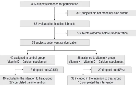
ic at Cha Hospital in Seoul from January-May 2010 were recruit- ed (n = 385) (Fig. 1). Of those screened, 103 women were exclud- ed for taking vitamin K, anti-lipid medications, hormone replace- ment therapy, or medications influencing bone metabolism as- pects such as bisphosphonate, calcitonin, steroids, phenytoin, carbamazepine, rifampicin, heparin, and warfarin. As well, 199 women with diabetes, hypertension, body mass index (BMI) > 30 kg/m2, or metabolic bone diseases were excluded. Five other subjects withdrew before randomization for personal reasons.
The remaining 78 women were randomly assigned to either the treatment group (vitamin K, n = 40) or a control group (n = 38).
For 6 months, the treatment group received 15 mg of vitamin K2
(menatetrenone) three times a day after every meal, combina- tion form containing 400 IU of active vitamin D3 once a day, and 315 mg of calcium carbonate twice daily. During the same peri- od, the control group received 400 IU of vitamin D and 315 mg of calcium twice daily. During the course of the study, 33 wom- en dropped out due to relocation or loss to follow-up. Forty five women completed the study.
Fasting blood (10 hr of fasting in the morning) was collected at baseline and at months 3 and 6. Total cholesterol, triglyceride and high density lipoprotein (HDL) cholesterol were measured by an enzymatic colorimetric assay using a model 7600 appara- tus (Hitachi, Tokyo, Japan). The BMD in the lumbar spine (L1- L4) and femur neck, and ward measurements were performed using a QDR 4500 apparatus (Hologic, Waltham, MA, USA) at baseline and 6 months. The serum UcOC was assayed by an en- zyme-linked immunosorbent assay using two monoclonal an- tibodies. Anti-osteocalcin (OC) antibody and a solid-phase anti- Glu 21, 24-OC antibody with recombinant human UcOC (Taka- ra Shuzo, Shiga, Japan) were used. The coefficients of variation (CVs) in the intra- and inter-assay were 7.3% and 9.7%, respec-
tively. CV was measured twice and the average was determined.
The study participants reported their medications to the princi- pal investigators at screening, and at months 3 and 6, and were interviewed about side effects at every visit. For assess study com- pliance, the remaining pill count was checked at all visits, and blood samples were collected at baseline and months 3 and 6.
Statistical analysis
An independent t test was used to compare the baseline demo- graphics and any changes that occurred after 6 months of inter- vention between the vitamin K group and the control group. To compare any changes in each group after 6 months, the paired t test was used. An intention to treat (ITT) analysis was conduct- ed to obtain standardized outcome variables. In addition, per- protocol (PP) analysis of the 45 subjects was also conducted. A P value < 0.05 was statistically significant. These analyses were done using SPSS version 18.0 (SPSS, Chicago, IL, USA).
Ethics statements
All participants provided written informed consent and this study was reviewed and approved by the institutional review board of Cha Hospital (EKI-GLA-06-32).
RESULTS
A total of 385 postmenopausal women over sixty were screened and 78 women were enrolled in the study (Fig. 1). The main rea- sons for ineligibility were the used of restricted medications and occurrence of diseases that could affect bone metabolism. In the ITT analysis comparing the characteristics of the vitamin K group with the control group (Table 1), the age at menopause in the vitamin K group (49.6 ± 2.7 yr) was significantly different
13 dropped out (32.5%) 20 dropped out (53%)
40 included in the intention to treat group 27 completed the intervention
38 included in the intention to treat group 18 completed the intervention
302 subjects did not meet inclusion criteria
5 subjects withdrew before randomization
Fig. 1. Screening, randomization and follow up for study subjects.
40 assigned to control group Vitamin D + Calcium supplement
38 assigned to vitamin K group Vitamin K + Vitamin D + Calcium supplement 385 subjects screened for participation
83 evaluated for baseline lab tests
78 subjects underwent randomization
from the age at menopause in the control group (50.8 ± 1.7 yr, P
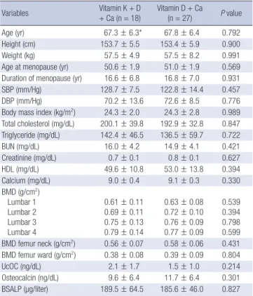
= 0.03). Other characteristics did not differ in the treatment group in both ITT and PP analyses. In the PP analysis (Table 2), char- acteristics of women in the vitamin K group were not different from those in the control group. The mean (± SD) age, age at menopause, and duration of menopause were 67.3 ± 6.3, 50.6 ± 1.9, and 16.6 ± 6.8 yr, respectively, in the vitamin K group and 67.8 ± 6.4, 51.0 ± 1.9, and 16.8 ± 7.0 yr, respectively, in the con- trol group. BMDs in lumbar and femur were similar in both the vitamin K and the control groups. In addition, the UcOC con- centration in the vitamin K group (2.1 ± 1.7 ng/dL) was not sig- nificantly different from that in the control group (1.5 ± 1.0 ng/
dL, P = 0.214). The mean (± SD) osteocalcin level was 9.6 ± 6.4 ng/dL in the vitamin K group and 11.7 ± 6.4 ng/dL in the con- trol group. There were no significant differences in blood urea nitrogen, creatinine, calcium and serum bone-specific alkaline phosphatase (BSALP) between the vitamin K and the control groups. The BMD in L1-L4 and femur neck, and in the ward in the vitamin K group were not different from those in the control group. The serum UcOC level in the vitamin K group (2.1 ± 1.7 ng/dL) was higher than that in the control group (1.5 ± 1.0 ng/
dL), but showed no significant difference (P = 0.21). Osteocalcin level was nonsignificantly lower in the vitamin K group (9.6 ±
6.4 ng/dL) than in the control group (11.7 ± 6.4 ng/dL, P = 0.29).
The BSALP level was 189.5 ± 64.5 μg/L in the vitamin K group and 185.6 ± 46.0 μg/L in the control group (P = 0.827). No signif- icant differences in BMD and biomarker values between the vi- tamin K and the control groups indicated that the randomiza- tion worked well. The ITT analysis showed similar results.
After 6 months of treatment, the L3 BMD in the vitamin K group compared to the control group increased statistically sig- nificantly (0.01 ± 0.03 g/cm2 vs -0.008 ± 0.04 g/cm2, P = 0.049).
Compared to BMD at baseline, BMD in femur at month 6 was significantly increased in both the vitamin K and the control groups. However, after 6 months of treatment, BMD in the vita- min K group was not statistically different from BMD in the con- trol group. In addition, compared to the baseline, the vitamin K group significantly decreased UcOC concentration (-1.6 ± 1.6 ng/dL, P < 0.01), whereas the UcOC level in the control group did not change (-0.4 ± 1.1 ng/dL). The change in UcOC concen- tration in the vitamin K group, but not in the control group, con- firmed the compliance of the participants in the vitamin K group with the study protocol. Osteocalcin was also non-significantly increased in the vitamin K group (1.6 ± 5.8 ng/dL), but not in the control group (-1.1 ± 6.0 ng/dL). Triglyceride level decreased in the vitamin K group (-10.0 ± 59.1 ng/dL) (Table 3). Three people Table 1. Characteristics of study participants at baseline by treatment group: ITT
analysis
Variables Vitamin K + D
+ Ca (n = 38)
Vitamin D + Ca
(n = 40) P value
Age (yr) 68.1 ± 6.3 67.6 ± 6.2 0.750
Height (cm) 153.1 ± 4.9 153.2 ± 5.5 0.956
Weight (kg) 56.0 ± 6.3 57.7 ± 7.6 0.292
Age at menopause (yr) 49.6 ± 2.7 50.8 ± 1.7 0.028 Duration of menopause (yr) 18.4 ± 7.2 16.8 ± 6.8 0.312 SBP (mm/Hg) 128.8 ± 27.0 119.2 ± 17.8 0.317 DBP (mm/Hg) 75.0 ± 19.5 70.2 ± 10.5 0.453 Body mass index (kg/m2) 23.8 ± 2.5 24.5 ± 2.9 0.278 Total cholesterol (mg/dL) 198.4 ± 36.1 204.0 ± 36.6 0.501
BUN (mg/dL) 15.3 ± 4.0 15.3 ± 3.9 0.993
Creatinine (mg/dL) 0.8 ± 0.1 0.8 ± 0.1 0.639 HDL (mg/dL) 50.8 ± 10.3 52.9 ± 12.7 0.442 Calcium (mg/dL) 9.0 ± 0.4 9.1 ± 0.2 0.449 BMD (g/cm2)
Lumbar 1 Lumbar 2 Lumbar 3 Lumbar 4
0.63 ± 0.10 0.70 ± 0.10 0.76 ± 0.11 0.82 ± 0.13
0.62 ± 0.07 0.72 ± 0.10 0.77 ± 0.10 0.78 ± 0.10
0.994 0.333 0.897 0.190 BMD femur neck (g/cm2) 0.57 ± 0.07 0.58 ± 0.07 0.473 BMD femur ward (g/cm2) 0.39 ± 0.12 0.39 ± 0.09 0.810
UcOC (ng/dL) 2.2 ± 1.8 1.6 ± 1.0 0.094
Osteocalcin (ng/dL) 10.9 ± 5.4 12.0 ± 6.3 0.484 BSALP (µg/liter) 197.2 ± 54.4 184.2 ± 47.6 0.269 All values are mean ± standard deviation. P values are from independent t test. D, vitamin D; Ca, calcium supplement; SBP, systolic blood pressure; DBP, diastolic blood pressure; BUN, blood urea nitrogen; HDL, high-density lipoprotein; BMD, bone miner- al density; UcOC, undercarboxylated osteocalcin; BSALP, serum bone-specific alka- line phosphatase.
Table 2. Characteristics of study participants at baseline by treatment group: PP anal- ysis
Variables Vitamin K + D
+ Ca (n = 18)
Vitamin D + Ca
(n = 27) P value
Age (yr) 67.3 ± 6.3* 67.8 ± 6.4 0.792
Height (cm) 153.7 ± 5.5 153.4 ± 5.9 0.900
Weight (kg) 57.5 ± 4.9 57.5 ± 8.2 0.991
Age at menopause (yr) 50.6 ± 1.9 51.0 ± 1.9 0.569 Duration of menopause (yr) 16.6 ± 6.8 16.8 ± 7.0 0.931
SBP (mm/Hg) 128.7 ± 7.5 122.8 ± 14.4 0.457
DBP (mm/Hg) 70.2 ± 13.6 72.6 ± 8.5 0.776
Body mass index (kg/m2) 24.3 ± 2.0 24.3 ± 2.8 0.989 Total cholesterol (mg/dL) 200.1 ± 39.8 192.9 ± 32.8 0.847 Triglyceride (mg/dL) 142.4 ± 46.5 136.5 ± 59.7 0.722
BUN (mg/dL) 16.0 ± 4.2 14.9 ± 4.1 0.421
Creatinine (mg/dL) 0.7 ± 0.1 0.8 ± 0.1 0.627 HDL (mg/dL) 49.6 ± 10.8 53.0 ± 13.8 0.394 Calcium (mg/dL) 9.0 ± 0.4 9.1 ± 0.3 0.330 BMD (g/cm2)
Lumbar 1 Lumbar 2 Lumbar 3 Lumbar 4
0.61 ± 0.11 0.69 ± 0.11 0.75 ± 0.13 0.79 ± 0.14
0.63 ± 0.08 0.72 ± 0.10 0.76 ± 0.09 0.77 ± 0.09
0.539 0.394 0.798 0.599 BMD femur neck (g/cm2) 0.56 ± 0.07 0.58 ± 0.06 0.431 BMD femur ward (g/cm2) 0.38 ± 0.08 0.39 ± 0.09 0.804
UcOC (ng/dL) 2.1 ± 1.7 1.5 ± 1.0 0.214
Osteocalcin (ng/dL) 9.6 ± 6.4 11.7 ± 6.4 0.301 BSALP (µg/liter) 189.5 ± 64.5 185.6 ± 46.0 0.827 All values are mean ± standard deviation. P values were from independent t test. D, vitamin D; Ca, calcium supplement; SBP, systolic blood pressure; DBP, diastolic blood pressure; BUN, blood urea nitrogen; HDL, high-density lipoprotein; BMD, bone mineral density; UcOC, undercarboxylated osteocalcin; BSALP, serum bone-specific alkaline phosphatase.
complained of nausea sensation twice after taking vitamin K.
No other participants including people in the control group re- ported adverse events during the study period.
In summary, after 6 months treatment of vitamin K to the post- menopausal women over sixty, the L3 BMD increased signifi- cantly compared to the control group. In addition, the changes in some biochemical markers differed by treatment. Compared to the control group, the vitamin K group significantly decreased serum UcOC concentration (P = 0.008). Osteocalcin level was higher in the vitamin K group than in the control group, but the difference was not significant (P = 0.14).
DISCUSSION
In this study, supplementation with vitamins K and D, and with calcium for 6 months of treatment significantly improved the L3 BMD compared to supplementation with vitamin D and calci- um in postmenopausal Korean women over sixty years old. In addition, femur BMD was increased in the vitamin K group (vi- tamin K + vitamin D + calcium), but a similar increase was ob- served in the control group (vitamin D + calcium supplementa- tion). The UcOC concentration significantly decreased in the vitamin K group compared to the control group (P < 0.01), and the osteocalcin level increased nonsignificantly in the vitamin K group (P = 0.14). The triglyceride level decreased in the vita- min K group, but was not statistically different from the control group. Similar results were observed in the ITT analysis.
When vitamin K concentration in the body is low, insufficient amount of osteocalcin completes γ-carboxylation; consequent- Table 3. Changes in BMD and biochemical markers from the baseline to 6 months
Variables Vitamin K + D
+ Ca (n = 18) Vitamin D + Ca
(n = 27) P value SBP (mm/Hg) 0.5 ± 3.1 1.2 ± 6.1 0.832 DBP (mm/Hg) 11.0 ± 13.1 5.6 ± 4.6 0.480 BMD (g/cm2)
Lumbar 1 Lumbar 2 Lumbar 3 Lumbar 4
-0.0009 ± 0.04 -0.01 ± 0.04 0.01 ± 0.03 0.007 ± 0.05
0.009 ± 0.04 -0.0048 ± 0.03 -0.008 ± 0.04 0.005 ± 0.04
0.377 0.618 0.049 0.888 BMD femur neck (g/cm2) 0.01 ± 0.031 0.01 ± 0.027 0.890 BMD femur ward (g/cm2) 0.06 ± 0.10 0.03 ± 0.07 0.269 Total cholesterol (mg/dL) -6.1 ± 42.8 10.6 ± 29.2 0.169 Triglyceride (mg/dL) -10.0 ± 59.1 10.5 ± 78.7 0.335 UcOC (ng/dL) -1.6 ± 1.6 -0.4 ± 1.1 0.008 Osteocalcin (ng/dL) 1.6 ± 5.8 -1.1 ± 6.0 0.136 BSALP (µg/liter) -2.1 ± 34.3 7.6 ± 41.0 0.390 BUN (mg/dL) -0.5 ± 3.6 0.0 ± 4.1 0.618 Creatinine (mg/dL) 0.02 ± 0.09 0.007 ± 0.08 0.566 HDL (mg/dL) 2.0 ± 9.4 3.0 ± 8.1 0.713 Calcium (mg/dL) 0.2 ± 0.3 0.1 ± 0.3 0.232 All values are mean ± standard deviation. P values from independent t test. D, vita- min D; Ca, calcium supplement; SBP, systolic blood pressure; DBP, diastolic blood pressure; BMD, bone mineral density; UcOC, undercarboxylated osteocalcin; BSALP, serum bone-specific alkaline phosphatase; BUN, blood urea nitrogen; HDL, high-den- sity lipoprotein.
ly, UcOC concentration increases and the affinity to calcium to bone matrix decreases. Gla-residues of osteocalcin combine with calcium and require vitamin K to activate the reaction site.
Therefore, measuring UcOC concentration is a more accurate method of monitoring vitamin K than prothrombin in the liver (19).
In a previous cross-sectional study in postmenopausal wom- en, elevated serum UcOC and low spinal BMD were observed in the low plasma phylloquinone (vitamin K1) group (20). In an- other study, healthy elderly Scottish women who took vitamin K1 with vitamin D and calcium showed a sustained increase in BMD at the site of the ultradistal radius in comparison with wom- en taking calcium and vitamin D (21). A combined vitamin K2
therapy with vitamin D3 for postmenopausal Japanese women produced low serum UcOC and improved BMD values (22). In an examination of the effects of vitamin K2 (45 mg/day) supple- mentation with calcium (1,500 mg/day) on lumbar BMD in post- menopausal women in Indonesia, a meaningful increase in BMD and a decrease in UcOC level were reported (18). On the other hand, a study reported no vitamin K effect on BMD in North American women, prompting the suggestion that wom- en receiving calcium and vitamin D supplements do not need additional vitamin K supplementation to prevent osteoporosis (23). Another recent study of vitamin K1 supplementation for 2-4 yr in Canadian women also reported no benefits on age-re- lated decline in BMD (12). Our study showed that compared to the control group that took vitamin D and calcium, additional vitamin K supplementation to vitamin D and calcium improved L3 BMD in Korean postmenopausal women over sixty. Our pre- vious cross-sectional study involving Korean women reported an inverse association between UcOC and BMD (6). This dichot- omy may partially be due to an ethnic difference and subjects’
age in the bone metabolism or response to vitamin K or UcOC.
A study of inflammatory bowel disease patients in Japanese women reported a decrease in the concentrations of vitamin K and D, and a resulting decrease in BMD (24). The foregoing re- sults imply that ethnic-related diversity in gastrointestinal ab- sorption may explain the different response to vitamin K sup- plement and its subsequent effects on BMD (25).
Several studies reported that obesity has been related to the level of vitamin K in adipose tissue, reflecting it’s fat-soluble na- ture. In one study, obese patients displayed a lower UcOC con- centration and their UcOC/osteocalcin ratio was negatively re- lated with body mass index (26). Another study reported that people with a high serum triglyceride level had high BMD in skeletal muscles (27). On the other hand, adult obesity has been inversely related to circulating indicators of serum vitamin K (28).
Limitations of the study were the small number of participants, relatively high dropout rate, and lack of a placebo group (i.e., no supplementation). The dosages of vitamin K, vitamin D and cal- cium and the duration of treatment may have been insufficient
to induce responses in related biochemical markers and total BMD. Moreover, we did not measure and compare the dietary vitamin K intake. Also, bone quality was not measured, which might be only a minor limitation. A previous study (6) suggest- ed that UcOC concentration is inversely associated with age, with higher UcOC concentrations in younger women. This sug- gests that the effect of the vitamin K on osteocalcin may also be age-related. Given the advanced age of our study subjects (mean age 68 yr) and relatively short treatment period, the study may have not been able to discern an increase all part of BMD. How- ever, it is noteworthy that changes in UcOC concentration among older women have been reported, with UcOC concentration in women in their 50 sec being higher than that in women in their 60 sec and 70 sec (6). Therefore, vitamin K may be more effec- tive in preventing bone loss or improving BMD in women aged 50-59 yr, especially in perimenopausal women or women with few years of menopause. A study testing the influence of vitamin K supplementation to perimenopausal women or women with few years of menopause for more than 6 months is needed. In addition, inconsistent findings from different countries suggest that a comparison of Korean data with data from other coun- tries is warranted. A study design involving a longer duration of vitamin K supplementation and with postmenopausal women having a short duration of menopause may prove valuable. Fi- nally, physical activity was not measured.
In conclusion, in this randomized study, supplementation with vitamin K, D and calcium for 6 months improves L3 BMD and reduces UcOC concentration in Korean postmenopausal women over sixty.
REFERENCES
1. Rowe SM, Song EK, Kim JS, Lee JY, Park YB, Bae BH, Hur CI. Rising in- cidence of hip fracture in Gwangju City and Chonnam Province, Korea.
J Korean Med Sci 2005; 20: 655-8.
2. Lee DY, Lim SJ, Moon YW, Min YK, Choi D, Yoon BK, Park YS. Determi- nation of an applicable FRAX model in Korean women. J Korean Med Sci 2010; 25: 1657-60.
3. Forli L, Bollerslev J, Simonsen S, Isaksen GA, Kvamsdal KE, Godang K, Gadeholt G, Pripp AH, Bjortuft O. Dietary vitamin K2 supplement im- proves bone status after lung and heart transplantation. Transplanta- tion 2010; 89: 458-64.
4. Reid DM, Macdonald HM. Nutrition and bone: is there more to it than just calcium and vitamin D? QJM 2001; 94: 53-6.
5. Furie B, Bouchard BA, Furie BC. Vitamin K-dependent biosynthesis of gamma-carboxyglutamic acid. Blood 1999; 93: 1798-808.
6. Kim SM, Kim KM, Kim BT, Joo NS, Kim KN, Lee DJ. Correlation of un- dercarboxylated osteocalcin (ucOC) concentration and bone density with age in healthy Korean women. J Korean Med Sci 2010; 25: 1171-5.
7. Booth SL, Broe KE, Peterson JW, Cheng DM, Dawson-Hughes B, Gund- berg CM, Cupples LA, Wilson PW, Kiel DP. Associations between vita- min K biochemical measures and bone mineral density in men and wom-
en. J Clin Endocrinol Metab 2004; 89: 4904-9.
8. Stevenson M, Lloyd-Jones M, Papaioannou D. Vitamin K to prevent frac- tures in older women: systematic review and economic evaluation. Health Technol Assess 2009; 13: iii-xi, 1-134.
9. Troy LM, Jacques PF, Hannan MT, Kiel DP, Lichtenstein AH, Kennedy ET, Booth SL. Dihydrophylloquinone intake is associated with low bone mineral density in men and women. Am J Clin Nutr 2007; 86: 504-8.
10. Emaus N, Gjesdal CG, Almås B, Christensen M, Grimsgaard AS, Bern- tsen GK, Salomonsen L, Fønnebø V. Vitamin K2 supplementation does not influence bone loss in early menopausal women: a randomised dou- ble-blind placebo-controlled trial. Osteoporos Int 2010; 21: 1731-40.
11. Volpe SL, Leung MM, Giordano H. Vitamin K supplementation does not significantly impact bone mineral density and biochemical markers of bone in pre-and perimenopausal women. Nutr Res 2008; 28: 577-82.
12. Cheung AM, Tile L, Lee Y, Tomlinson G, Hawker G, Scher J, Hu H, Vieth R, Thompson L, Jamal S, Josse R. Vitamin K supplementation in post- menopausal women with osteopenia (ECKO trial): a randomized con- trolled trial. PLoS Med 2008; 5: e196.
13. Booth SL, Dallal G, Shea MK, Gundberg C, Peterson JW, Dawson-Hughes B. Effect of vitamin K supplementation on bone loss in elderly men and women. J Clin Endocrinol Metab 2008; 93: 1217-23.
14. Knapen MH, Schurgers LJ, Vermeer C. Vitamin K2 supplementation im- proves hip bone geometry and bone strength indices in postmenopausal women. Osteoporos Int 2007; 18: 963-72.
15. Arunakul M, Niempoog S, Arunakul P, Bunyaratavej N. Level of under- carboxylated osteocalcin in hip fracture Thai female patients. J Med As- soc Thai 2009; 92(Suppl 5): S7-11.
16. Booth SL, Lichtenstein AH, O’Brien-Morse M, McKeown NM, Wood RJ, Saltzman E, Gundberg CM. Effects of a hydrogenated form of vitamin K on bone formation and resorption. Am J Clin Nutr 2001; 74: 783-90.
17. Tsugawa N, Shiraki M, Suhara Y, Kamao M, Ozaki R, Tanaka K, Okano T.
Low plasma phylloquinone concentration is associated with high inci- dence of vertebral fracture in Japanese women. J Bone Miner Metab 2008;
26: 79-85.
18. Purwosunu Y, Muharram, Rachman IA, Reksoprodjo S, Sekizawa A. Vi- tamin K2 treatment for postmenopausal osteoporosis in Indonesia. J Ob- stet Gynaecol Res 2006; 32: 230-4.
19. Furie B, Furie BC. Molecuar basis of vitamin K-dependent gamma-car- boxylation. Blood 1990; 75: 1753-62.
20. Hodges SJ, Akesson K, Vergnaud P, Obrant K, Delmas PD. Circulating levels of vitamins K1 and K2 are decreased in elderly women with hip fracture. J Bone Miner Res 1993; 8: 1241-5.
21. Bolton-Smith C, McMurdo ME, Paterson CR, Mole PA, Harvey JM, Fen- ton ST, Prynne CJ, Mishra GD, Shearer MJ. Two-year randomized con- trolled trial of vitamin K1 (phylloquinone) and vitamin D3 plus calcium on the bone health of older women. J Bone Miner Res 2007; 22: 509-19.
22. Yasui T, Miyatani Y, Tomita J, Yamada M, Uemura H, Miura M, Irahara M. Effect of vitamin K2 treatment on carboxylation of osteocalcin in ear- ly postmenopausal women. Gynecol Endocrinol 2006; 22: 455-9.
23. Binkley N, Harke J, Krueger D, Engelke J, Vallarta-Ast N, Gemar D, Che- covich M, Chappell R, Suttie J. Vitamin K treatment reduces undercar- boxylated osteocalcin but does not alter bone turnover, density, or geom- etry in healthy postmenopausal North American women. J Bone Miner Res 2009; 24: 983-91.
24. Kuwabara A, Tanaka K, Tsugawa N, Nakase H, Tsuji H, Shide K, Kamao
AUTHOR SUMMARY
Vitamin K Supplement Along with Vitamin D and Calcium Reduced Serum Concentration of Undercarboxylated Osteocalcin While Increasing Bone Mineral Density in Korean Postmenopausal Women over Sixty-Years-Old
Sang Hyeon Je, Nam-Seok Joo, Beom-hee Choi, Kwang-Min Kim, Bom-Taeck Kim, Sat-Byul Park, Doo-Yeoun Cho, Kyu-nam-Kim and Duck-Joo Lee
This study is to evaluate effects of vitamin K supplementation on bone mineral density (VMD) in postmenopausal Korean women.
78 postmenopausal women over sixty years old were assigned to either control (Calcium & Vitamin D, n = 40) or vitamin K-supplemented group (Calcium, Vitamin D & Vitamin K, n = 38). After 6 months of treatment, the vitamin K group showed decrease of undercarboxylated osteocalcin and increase of BMD. The Intention to Treat (ITT) analysis showed the same result. Such result is contrary to the previous study in North American postmenopausal women.
M, Chiba T, Inagaki N, Okano T, Kido S. High prevalence of vitamin K and D deficiency and decreased BMD in inflammatory bowel disease.
Osteoporos Int 2009; 20: 935-42.
25. Menzies IS, Zuckerman MJ, Nukajam WS, Somasundaram SG, Murphy B, Jenkins AP, Crane RS, Gregory GG. Geography of intestinal permea- bility and absorption. Gut 1999; 44: 483-9.
26. Foresta C, Strapazzon G, De Toni L, Gianesello L, Calcagno A, Pilon C, Plebani M, Vettor R. Evidence for osteocalcin production by adipose tis- sue and its role in human metabolism. J Clin Endocrinol Metab 2010;
95: 3502-6.
27. Brownbill RA, llich JZ. Lipid profile and bone paradox: higher serum lip- ids are associated with higher bone mineral density in postmenopausal women. J Womens Health (Larchmt) 2006; 15: 261-70.
28. Shea MK, Booth SL, Gundberg CM, Peterson JW, Waddell C, Dawson- Hughes B, Saltzman E. Adulthood obesity is positively associated with adipose tissue concentrations of vitamin K and inversely associated with circulating indicators of vitamin K status in men and women. J Nutr 2010;
140: 1029-34.