INTRODUCTION
Rhabdoid tumor was originally described in 1978 by Beck- with et al. (1) as a highly malignant pediatric tumor of the kidney, and Rorke et al. (2) named primary central nervous system malignant rhabdoid tumor as ‘atypical teratoid/rhab- doid tumor (ATT/RhT)’. These tumors resemble rhabdomy- osarcomas, but their immunohistochemical and ultrastruc- tural features allow a clear distinction between the two types of tumors. Recently, some authors have reported a small num- ber of cases of extrarenal rhabdoid tumors. Primary central nervous system (CNS) malignant rhabdoid tumors are rare.
Since 1985, 85 cases of primary CNS malignant rhabdoid tumor have been documented (2-6). Intracranial ATT/RhT is a disease of infancy and childhood, usually occurring dur- ing the first decade of life (2). The most common biological- ly malignant CNS tumor in this period is a primitive neu- roectodermal tumor-medulloblastoma (PNET-MB). ATT/
RhT has been frequently misdiagnosed as a PNET-MB be- cause 70% of ATT/RhTs contain histological fields indistin- guishable from classic PNET-MB. However, microscopic features in routine and special studies allow the differential diagnosis of PNET-MB and ATT/RhT (1-6). We report a case of a primary CNS ATT/RhT arising from the posterior cranial fossa in a child. The clinical course of the patient is described with a special emphasis on the differential diagno-
sis between ATT/RhT and PNET-MB.
CASE REPORT
A 16-month-old baby boy had been suffering from vom- iting and progressive gait disturbance since the age of 14 months. He was irritable, drowsy, and showed bilateral pa- pilledema and bilateral Babinski’s sign on admission.
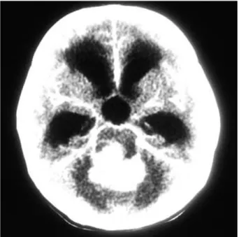
Computerized tomography (CT) showed a 4×4 cm-sized tumor with calcification in the posterior cranial fossa. The mass revealed heterogeneous density on the pre-contrast CT and heterogeneous enhancement with contrast infusion (Fig.
1). Subsequent magnetic resonance imaging (MRI) demon- strated a mass occupying the fourth ventricle and compress- ing the right posterior aspect of the brainstem (Fig. 2).
With a preoperative diagnosis of PNET-MB, surgery was performed via a median suboccipital craniotomy, and the ce- rebellar vermian incision. The mass, which originated from the fourth ventricle invading both cerebellar hemisphere and brainstem, was grayish, friable and highly vascular. Subtotal removal of the tumor was performed leaving a small portion of the tumor which was tightly adhered to the ventricular floor.
Histopathological findings depicted the aggregation of large, pale, and polygonal cells with distinct cell borders, ve-
You-Nam Chung, Kyu-Chang Wang, Sang-Hoon Shin, Narae Kim*, Je G. Chi*, Kyung-Soo Min�, Byung-Kyu Cho
Department of Neurosurgery, Seoul National University College of Medicine & Neurological Research Institute, SNUMRC, Seoul; Department of Pathology*, Seoul National University College of Medicine, Seoul; Department of Neurosurgery�, Chungbuk National University College of Medicine, Cheongju, Korea
Address for correspondence Kyu-Chang Wang, M.D.
Department of Neurosurgery, Seoul National University College of Medicine, 28 Yongon-dong, Chongno-gu, Seoul 110-744, Korea
Tel : +82.2-760-3489, Fax : +82.2-747-5130 E-mail : [email protected]
723 J Korean Med Sci 2002; 17: 723-6
ISSN 1011-8934
Copyright � The Korean Academy of Medical Sciences
Primary Intracranial Atypical Teratoid/Rhabdoid Tumor in a Child
: A Case Report
Rhabdoid tumors of the central nervous system are rare malignancies. Primary central nervous system atypical teratoid/rhabdoid tumors (ATT/RhTs) mostly occur during early childhood and are almost invariably fatal. These tumors show similar histological and radiological features to primitive neuroectodermal tumor-medul- loblastoma (PNET-MB) but have different biological behaviors. We report a case of primary intracranial ATT/RhT in the posterior cranial fossa of a child. Preoper- ative radiological diagnosis was PNET-MB, but pathological diagnosis is ATT/
RhT. The case involved a 16-month-old baby boy who presented with severe headache, vomiting, and gait disturbance. He was treated by surgical resection, chemotherapy, and radiotherapy. Despite aggressive therapy, he died 19 months after diagnosis. Clinical, radiological, and histopathological features of primary intracranial ATT/RhT are discussed with a special emphasis on the differential diagnosis from PNET-MB.
Key Words : Atypical Teratoid/Rhabdoid Tumor; Primitive Neuroectodermal Tumor; Medulloblastoma;
Child
Received : 22 May 2001 Accepted : 28 September 2001
724 Y.N. Chung, K.C. Wang, S.H. Shin, et al.
sicular nuclei, occasional single prominent nucleolus, and moderate amounts of eosinophilic or pale cytoplasm (Fig. 3A).
In addition to these rhabdoid cells, some cells with intracy- toplasmic hyaline-like eosinophilic material were found. The material was displacing the nuclei to the eccentric side, and creating the classic ‘rhabdoid’ appearance, as seen in the rhab- doid tumor of the kidney. Portions consisting of small undif-
ferentiated cells with indistinct cell borders resembling me- dulloblastoma were also found. Immunohistochemical study revealed both large and small cells which were focally posi- tive for cytokeratin (Fig. 3B), epithelial membrane antigen (EMA, Fig. 3C), synaptophysin, and vimentin (Fig. 3D).
They were totally negative for glial fibrillary acidic protein (GFAP). These histologic and immunohistochemical find- ings were compatible with a diagnosis of ATT/RhT.
After surgical removal of the tumor, the patient received four cycles of chemotherapy according to the eight-drugs-in- one-day protocol. A small enhancing nodule was detected on the left cerebellum on brain MRI after the second chemother- apy. Subsequent spinal MRI showed linearly enhancing le- sions along the surface of the spinal cord, suggesting an in- traspinal dissemination. Radiotherapy was given with a total intracranial dose of 4,860 cGy in 27 fractions and a total spinal dose of 2,400 cGy in 16 fractions. Though he received two additional cycles of chemotherapy, the tumor volume was not reduced. He died 19 months after the diagnosis. Autopsy was not performed.
DISCUSSION
ATT/RhT is a tumor of infancy and childhood, and very rare in adults (5, 6, 9). The mean age of the patients is 2.9 yr and three-quarters of them are 3 yr or younger at the time of diagnosis with a male predominance (10). Childhood PNET- MB, tends to appear between 3 and 5 yr of age. Eighty per- cent of classic PNET-MBs arise in the cerebellum, but the ATT/RhTs develop on that site in only slightly over half (3-
Fig. 1.The computerized tomography showed a 4×4 cm-sized tumor in the posterior cranial fossa with calcification and strong enhancement.
Fig. 2.Magnetic resonance imaging demonstrated a mass occupying the fourth ventricle compressing the right posterior aspect of the brainstem. axial image (A), sagittal image (B).
A B
6, 10). The cerebellopontine angle appears to be a common location for ATT/RhT; 15% in series of Rorke et al. (6). ATT/
RhT is a highly malignant neoplasm involving leptomeningeal dissemination in 10-30% of patients (3-6, 10).
Clinical presentation depends on the age of onset and the location of the tumor. Children younger than 3 yr of age usu- ally present with nonspecific symptoms and signs, such as vomiting, lethargy, irritability, loss of weight, macrocephaly, and failure to thrive. Older patients commonly present with increased intracranial pressure or localizing signs. Cranial nerve palsies, headache, and hemiplegia are common (2-4, 6).
Many authors have reported that the clinical symptoms and signs, and the radiological appearance of ATT/RhT are similar to those of PNET-MB, which frequently misleads ATT/RhT to a preoperative diagnosis of PNET/MB. The characteristic findings of ATT/RhT are heterogeneous den- sity on pre-contrast CT, and inhomogeneous enhancement.
Calcification, cyst formation, and hemorrhage are frequent- ly associated. On MRI, ATT/RhT usually shows a decreased signal intensity on T2-weighted images, an isosignal inten- sity on proton image, and inhomogeneous enhancement with gadolinium. Differential diagnosis between ATT/RhT and
Primary Intracranial Atypical Teratoid/Rhabdoid Tumor in a Child 725
Fig. 3.Photomicrography of the tumor discloses aggregations of large, pale, polygonal cells with vesicular nuclei, occasional single prominent nucleolus, and moderate amount of eosinophilic cytoplasm (H&E, A). Immunohistochemical stains for cytokeratin (B), epithe- lial membrane antigen (C), and vimentin (D) reveal focal positive reactions (A-D, original magnification×200).
A B
C D
PNET-MB is difficult by radiological findings, but is easier by histopathological findings (6). The malignant rhabdoid tumors are mainly composed of rhabdoid cells, but they also contain areas of primitive neuroectodermal cells with both epithelial and mesenchymal components, which explains why these tumors have been misdiagnosed as PNET-MB in the past. This variety of histological components in the malig- nant rhabdoid tumor suggests that this tumor is a special type of teratoma, and this motivated Rorke et al. to propose the name ‘atypical teratoid/rhabdoid tumor’ for the malignant form of rhabdoid tumor (5). Light microscopic examination of ATT/RhT reveals a diffuse growth pattern of predominant- ly polygonal cells arranged in a focally trabecular or alveolar fashion, cells with vesicular nuclei and prominent nucleoli, and scattered cells with globular hyaline cytoplasmic inclu- sions in the vicinity of the nuclei. Electron microscopy shows whorls of filaments in the cytoplasm, which can be classified as intermediate filaments and represent vimentin (1, 2, 5, 6).
Results of the immunohistochemical studies show positivi- ties for three antibodies whose epitopes are almost always ex- pressed: EMA, vimentin, and smooth-muscle actin (SMA) (3, 4, 6, 7, 10). These positivities are not found in PNET- MBs: in particular, EMA is always negative in PNET-MBs (10). Results for GFAP, germ cell tumor markers, desmin, neurofilament, and myoglobulin are negative (2, 4-6, 10).
Some authors have described deletion or monosomy of chro- mosome 22 as the most common abnormality in the CNS ATT/RhT (5, 6, 8). Differential diagnosis must include PN ET-MB, ependymoma, choroid plexus papilloma, and tera- toma.
There has hitherto been no satisfactory treatment for ATT/
RhT. The majority of patients die within the first year of local tumor relapse or of leptomeningeal dissemination (2, 4, 5).
ATT/RhT is frequently disseminated along the neuraxis at the time of disease relapse. The purpose of surgery is to make a diagnosis and to reduce the tumor burden. Children with ATT/RhT rarely respond to treatment despite the use of ag- gressive chemotherapy and/or radiotherapy (6).
The median time to tumor progression is 4.5 months, and the mean survival duration is 6 months (6). Survival is poor and unrelated to the age of the patient at the time of diagno- sis, the extent of resection, or the type of adjuvant postoper- ative therapy.
In summary, we reported a case of a 16-month-old male
who presented with vomiting, lethargy, and gait disturbance.
Preoperative CT and MRI seemed to indicate PNET-MB.
Total tumor removal was not possible owing to the invasion of the brainstem. The pathological findings of the resected tissue were consistent with ATT/RhT. CNS ATT/RhT is very rare, and its prognosis is invariably poor according to the pre- vious reports. Considerations on ATT/RhT in the differen- tial diagnosis of posterior cranial fossa tumors may help to avoid misdiagnosis and erroneous prognostication.
REFERENCES
1. Beckwith JB, Palmer NF. Histopathology and prognosis of Wilms’
tumor. Results from the First National Wilms’ Tumor Study. Cancer 1978; 41: 1937-48.
2. Rorke LB, Packer R, Biegel J. Central nervous system atypical ter- atoid/rhabdoid tumors of infancy and childhood. J Neurooncol 1995; 24: 21-8.
3. Martinez-Lage JF, Nieto A, Sola J, Domingo R, Costa TR, Poza M.
Primary malignant rhabdoid tumor of the cerebellum. Child’ Nerv Syst 1997; 13: 418-21.
4. Munoz A, Carrasco A, Munoz MJ, Esparza J. Cranial rhabdoid tumor with marginal tumor cystic component and extraaxial exten- sion. Am J Neuroradiol 1995; 16: 1727-8.
5. Caldemeyer KS, Smith RR, Azzarelli B, Boaz JC. Primary central nervous system malignant rhabdoid tumor: CT and MR appearance simulates a primitive neuroectodermal tumor. Pediatr Neurosurg 1994; 21: 232-6.
6. Rorke LB, Packer RJ, Biegel JA. Central nervous system atypical teratoid/rhabdoid tumors of the infancy and childhood: definition of an entity. J Neurosurg 1996; 85: 56-65.
7. Bonnin JM, Rubinstein LJ, Palmer NF, Beckwith JB. The associa- tion of embryonal tumors originating in the kidney and in the brain:
a report of seven cases. Cancer 1984; 54: 2137-46.
8. Biegel JA, Rorke LB, Packer RJ, Emanuel BS. Monosomy 22 in rhabdoid or atypical teratoid tumors of the brain. J Neurosurg 1990: 73: 710-4.
9. Horn M, Schlote W, Lerch KD, Steudel WI, Harms D, Thomas E.
Malignant rhabdoid tumor: primary intracranial manifestation in an adult. Acta Neuropathol (Berl) 1992; 83: 445-8.
10. Agranovich AL, Ang LC, Griebel RW, Kobrisky NL, Lowry N, Tchang SP. Malignant rhabdoid tumor of the central nervous system with subarachnoid dissemination. Surg Neurol 1992; 37: 410-4.
726 Y.N. Chung, K.C. Wang, S.H. Shin, et al.