INTRODUCTION
Endocarditis related to pacemaker lead infection is a rare, but serious condition in permanent venous tracing. Staphy- lococci are involved in the majority of these infections (1-4) and Achromobacter xylosoxidans is very rare cause of organism.
Although some studies reported that medical extraction of even large vegetation appeared to be safe (5), usually the infect- ed endocardial system must be entirely removed and appro- priate antibiotic therapy pursued for 6 weeks (1, 6).
CASE REPORT
A 35-yr-old patient visited our hospital due to a unremitting high fever and chills for 10 days. He had undergone a patch closure of the ventricular septal defect (VSD) 18 yr before.
One year later, a VVI pacemaker was implanted via the right subclavian vein because of complete heart block. Nine years after that, a new VVI pacemaker with another right ventricu- lar electrode was inserted controlaterally and the old pacing lead was abandoned. Several months before admission, he had the scaling and root planning at local dental clinic. He did not have any intravenous treatment history.
On admission, vital signs were blood pressure 120/75 mmHg, body temperature 39℃, and pulse rate 75/min.
Physical examination revealed systolic ejection murmur in the
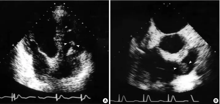
left second intercostal area. The results of a complete blood count were normocytic, normochromic anemia, and leuko- cytosis (15,400/ L). An elevated erythrocyte sedimentation rate (ESR) (135 mm/hr) and positive C-reactive protein (CRP) (13.5 mg/dL) were demonstrated. Chest radiography revealed a mild cardiomegaly without active lesions in the lung paren- chyma. Blood cultures were positive for Achromobacter xylosoxi- dans (colorless growth on MacConkey agar, oxidase positive, indole negative, saccharolytic, motile, rod-shaped nonfermen- ters analyzed by Vitec) 5 times. Susceptibility study showed that the strain was susceptible to ceftazidime, piperacillin, cefoperazone, imipenem, trimethoprim-sulfamethoxazole, and was resistant to aminoglycosides. Trans-thoracic echocar- diogram (TTE) (Fig. 1A) and trans-esophageal echocardio- gram (TEE) (Fig. 1B) identified the pacemaker lead in the right ventricle (RV) attaching hyperechoic materials and also a fluttering round hyperechoic mass with a stalk in the RV outflow tract (RVOT). No shunt flow was detected through the previous patch closure site in TTE and TEE. The function of pacemaker was normal on pacemaker analysis and 24-hr Holter monitoring.
Despite sensitive antibiotics of ceftazidime and piperacillin for 3 weeks, patient’s status was not stabilized. Therefore, the patient underwent a cardiac surgery through median sternoto- my under extracorporeal circulation. Intraoperatively, yellow- brown friable materials attached to the older electrode (Fig.
2) and the septal wall of RVOT were confirmed. Removal Youngkeun Ahn, Nam Ho Kim*,
Dong Hyeon Shin, Ok Young Park, Won Kim, Myung Ho Jeong, Jeong Gwan Cho, Jong Chun Park, Jung Chaee Kang
Divisions of Cardiology and Infectious Disease, Chonnam National University Hospital, Gwangju;
*Division of Cardiology, Wonkwang University Hospital, Iksan, Korea
Received : 4 February 2003 Accepted : 22 April 2003
Address for correspondence Jeong Gwan Cho, M.D.
Division of Cardiology, Chonnam National University Hospital, 8 Hak-dong, Dong-gu, Gwangju 501-757, Korea
Tel : +82.62-220-6242, Fax : +82.62-225-8578 E-mail : [email protected]
291 J Korean Med Sci 2004; 19: 291-3
ISSN 1011-8934
Copyright � The Korean Academy of Medical Sciences
Pacemaker Lead Endocarditis Caused by Achromobacter xylosoxidans
We report the case of a 35-yr-old patient who presented with high fever and chills.
He had undergone a patch closure of the ventricular septal defect 18 yr before.
One year later, a VVI pacemaker was implanted via the right subclavian vein because of complete heart block. Nine years after that, a new VVI pacemaker with another right ventricular electrode was inserted controlaterally and the old pacing lead was abandoned. Trans-thoracic and trans-esophageal echocardiogram iden- tified the pacemaker lead in the right ventricle (RV) attaching hyperechoic materi- als and also a fluttering round hyperechoic mass with a stalk in the RV outflow tract. Cultures in blood and pus from pacemaker lead grew Achromobacter xylosoxi- dans. A diagnosis of pacemaker lead endocarditis due to Achromobacter xylosoxi- dans was made. In this regards, the best treatment is an immediate removal of the entire pacing system and antimicrobial therapy.
Key Words :Endocarditis; Pacemaker, Artificial; Achromobacter xylosoxidans
292 Y. Ahn, N.H. Kim, D.H. Shin, et al.
of infected wires, infected Dacron patch, and vegetations in the RVOT, and curretage of infected RV wall were performed.
Cultures for Achromobacter xylosoxidans were positive in the pus attached to the pacemaker lead. With sensitive antibi- otics treatment, the patient was doing well 6 weeks postop- eratively without any signs for infection. Epicardial pacing was maintained. There were no adverse changes in the nor- malized CRP and ESR. Thus, a transvenous permanent pace- maker implantation was performed after confirming the paten- cy of the left subclavian vein with venography.
A follow-up echocardiogram 1 yr later demonstrated no detectable vegetations. The patients has been doing well with- out recurrence of infection for 2 yr.
DISCUSSION
Infection of the pacemaker pouch and lead may occur in 1% to 7% of patients with a permanent pacemaker (3). The microorganism most responsible for a late pacemaker lead infection is Staphylococcus epidermidis (4). Achromobacter xylosoxi- dans is a nonfermentative Gram-negative bacilli. This organ- ism is opportunistic and usually affects severely immuno- compromised patients such as those with neutropenia and those with a malignant or cardiovascular disease (7, 8). To the best of our knowledge, however, the present case is unique in that there has been no report such as ours. A case report of aortic valve and VSD Dacron patch infective endocarditis due to Achromobacter xylosoxidans 15 yr after a VSD patch clo- sure appeared on Medline search (9). He did not have any definite cause of pacemaker lead endocarditis. Several months before admission, he had the scaling and root planning at local dental clinic.
The diagnosis is difficult by using conventional imaging methods such as transthoracic echocardiogram. TEE can facil- itate the diagnosis of pacemaker lead endocarditis, however, it is sometimes not diagnostic (10, 11). In our case, the hyper- echoic materials clearly appeared in TTE and TEE.
Some studies reported successful treatment with the use of antibiotics alone in patients with a pacemaker lead endocardi- tis, however, the main management modality is cardiac surgery (12). Patients are recommended to be treated with prolonged antibiotic regimens before and after electrode removal (3). The electrode removal can be achieved by surgery or traction. Per- cutaneous traction is not simple and associated with compli- cations, such as a tear of the tricuspid valve. Some patients
Fig. 1.(A) Transthoracic echocardiogram, apical four-chamber view. A pacemaker lead with attached hyperechoic materials is seen in the right ventricle. (B) Transesophageal echocardiogram. A hyperechoic mass with a stalk is freely movable in the right ventricular out- flow tract.
A B
Fig. 2.The removed electrode. It was destroyed and attached with yellow-brown friable materials.
Pacemaker Lead Endocarditis 293
need a new permanent pacemaker (3).
REFERENCES
1. Klug D, Lacroix D, Savoye C, Goullard L, Grandmougin D, Hen- nequin JL, Kacet S, Lekieffre J. Systemic infection related to endo- carditis on pacemaker leads: clinical presentation and manage- ment. Circulation 1997; 95: 2098-107.
2. Banos R, Gomez J, Sanchez B, de la Morena G, Simarro E, Garcia del Real F. Pacemaker lead endocarditis: analysis of 11 cases. Enferm Infecc Microbiol Clin 2000; 18: 267-70.
3. Cacoub P, Leprince P, Nataf P, Hausfater P, Dorent R, Wechsler B, Bors V, Pavie A, Piette JC, Gandjbakhch I. Pacemaker infective endo- carditis. Am J Cardiol 1998; 82: 480-4.
4. Voet JG, Vandekerckhove YR, Muyldermans LL, Missault LH, Ma- tthys LJ. Pacemaker lead infection: report of three cases and review of the literature. Heart 1999; 81: 88-91.
5. Victor F, De Place C, Camus C, Le Breton H, Leclercq C, Pavin D, Mabo P, Daubert C. Pacemaker lead infection: echocardiographic features, management, and outcome. Heart 1999; 81: 82-7.
6. Miralles A, Moncada V, Chevez H, Rodriguez R, Granados J, Castells
E. Pacemaker endocarditis: approach for lead extraction in endocardi- tis with large vegetations. Ann Thorac Surg 2001; 72: 2130-2.
7. Jacobs JA, Stobberingh EE, Schouten HC. Fatal infection due to Alcali- genes xylosoxidans subsp. Xylosoxidans in a neutropenic host. Clin Microbiol Newslett 1992; 14: 182-4.
8. Duggan JM, Goldstein SJ, Chenoweth CE, Kauffman CA, Bradley SF. Achromobacter xylosoxidans bacteremia: report of four cases and review of the literature. Clin Infect Dis 1996; 23: 569-76.
9. Sasaki H, Kawai H, Sawamura T, Takiya H. A case report of aortic valve and VSD Dacron patch infective endocarditis after VSD patch closure 15 years ago. Nippon Kyobu Geka Gakkai Zasshi 1993; 41:
1373-7.
10. Dalal A, Asirvatham SJ, Chandrasekaran K, Seward JB, Tajik AJ.
Intracardiac echocardiography in the detection of pacemaker lead endocarditis. J Am Soc Echocardiogr 2002; 15: 1027-8.
11. Tan HH, Ling LH, Ng WL, Cheng A. Diagnosis of pacemaker lead infection using transoesophageal echocardiography: a case report.
Ann Acad Med Singapore 2000; 29: 97-100.
12. Ku GW, Kang SK, Won TH, Kim SW, Yu JH, Na MH, Lim SP, Lee Y. Endocarditis with intracardiac migration of transvenous perma- nent pacing lead: 1 case report. Korean J Thorac Cardiovasc Surg 2002; 35: 831-4.