© 2013 Korean Breast Cancer Society. All rights reserved. http://ejbc.kr | pISSN 1738-6756
INTRODUCTION
Axillary lymph node (ALN) status is an important prognos- tic factor in breast cancer [1]. Accurate lymph node staging and adequate locoregional control can be achieved by axillary lymph node dissection (ALND), which, however, is often fol- lowed by significant morbidities including lymphedema and nerve injury [2]. Sentinel lymph node biopsy (SLNB) has been suggested as an alternative method, associated with fewer complications. Over the years, the accuracy of SLNB has been confirmed in several studies, and SLNB has now become a
standard surgical procedure for axillary staging in clinically node-negative primary breast cancer [3].
However, the effectiveness of SLNB after neoadjuvant che- motherapy (NAC) is less clear. Conflicting results on the ac- curacy of SLNB have been reported and ALND remains the standard of care for nodal staging and evaluation of local con- trol after NAC [4-8]. The possibility of high false negative rates is a major concern in implementing SLNB in patients who re- ceive NAC. The reported rate of sentinel node identification failure is another matter of contention [9,10].
In the present study, we evaluated the reliability of SLNB in predicting axillary lymph node status in breast cancer patients after NAC by assessing its identification and false negative rates. We also examined the accuracy of intraoperative frozen section examination of sentinel lymph nodes (SLNs) after NAC.
METHODS
From January 2008 to December 2011, 350 pathologically proven breast cancer patients underwent NAC and subse- quent definitive surgery at Seoul National University Hospital.
Among these patients, 281 underwent SLNB for axillary stag-
Reliability of Sentinel Lymph Node Biopsy after Neoadjuvant Chemotherapy in Breast Cancer Patients
Ahram Han1,*, Hyeong-Gon Moon1,*, Jisun Kim1, Soo Kyung Ahn1, In Ae Park2, Wonshik Han1,3, Dong-Young Noh1,3
Departments of 1Surgery and 2Pathology, Seoul National University Hospital, Seoul; 3Cancer Research Institute, Seoul National University College of Medicine, Seoul, Korea
ORIGINAL ARTICLE
Purpose: Sentinel lymph node biopsy (SLNB) is an accurate and effective means of axillary nodal staging in early breast cancer.
However its indication after neoadjuvant chemotherapy (NAC) is under constant debate. The present study evaluates the reliabili- ty of SLNB in assessing axillary nodal status after NAC. Methods:
Data from 281 patients who had received NAC and subsequent SLNB were reviewed. The identification and false negative rates of SLNB were determined and the clinicopathologic factors as- sociated with false negative results were investigated using uni- variate analysis. Results: The identification rate of SLNB after NAC was 93.6% and the false negative rate was 10.4%. Hor- mone receptor status, especially progesterone receptor positivi- ty, was significantly associated with false negative results. The
accuracy of intraoperative frozen section examination of sentinel lymph nodes was 91.2%. Conclusion: The identification rate of SLNB and the accuracy of intraoperative frozen section exami- nation after NAC are comparable to the results without NAC in patients with early breast cancer. However considering the high false negative rates, general application of SLNB after NAC should be avoided. Patients with progesterone-positive tumors and non-triple-negative breast cancers may be a select group of patients in whom SLNB can be employed safely after NAC, but further studies are necessary.
Key Words: Breast neoplasms, Neoadjuvant therapy, Sentinel lymph node biopsy
Correspondence to: Dong-Young Noh
Department of Surgery, Seoul National University Hospital, 101 Daehak-ro, Jongno-gu, Seoul 110-744, Korea
Tel: +82-2-2072-2921, Fax: +82-2-3673-4250 E-mail: [email protected]
*These authors contributed equally to this work.
This work was supported by the Basic Science Research Program through the National Research Foundation of Korea (NRF) funded by the Ministry of Education, Science and Technology (2012R1A1A2005929) and by a grant from the National R&D Program for Cancer Control, Ministry for Health and Welfare, Republic of Korea (A1220200).
Received: June 27, 2013 Accepted: October 24, 2013
Cancer
ing during surgery and were included in the final analysis. The reliability of SLNB after NAC was examined by evaluating the sentinel node identification rate and false negative rate. During the study period, subsequent axillary dissection after SLNB was performed at the discretion of the responsible surgeon, because of a lack of safety data on SLNB in patients who re- ceive NAC. Thus, axillary dissection was frequently performed even in SLN-negative patients. The false negative rate of SLNB in this study was evaluated only in patients who underwent subsequent ALND. This study was reviewed and approved by the Institutional Review Board of Seoul National University Hospital (IRB number: H-1309-098-522).
For breast cancer diagnosis and staging, core needle biopsy and multiple imaging studies were performed. Initial imaging studies included breast and axilla sonography, mammography, chest computed tomography, breast magnetic resonance im- aging (MRI), and bone scanning. Pathologic examination of biopsied tissue included immunohistochemistry (IHC) for es- trogen receptor (ER), progesterone receptor (PR), c-erbB-2, p53, Bcl-2, and Ki-67. Formalin-fixed, paraffin-embedded tis- sue blocks were serially sectioned at 4-µm thickness and slides were subjected to our previously described IHC method [11].
Briefly, after deparaffinization in xylene and dehydration in a graded alcohol series, sections were treated to enhance anti- gen retrieval. The following mouse monoclonal antibodies were used as primary antibodies: ER (1:50; Dako Co., Carpin- teria, USA), PR (1:50; Dako Co.), c-erbB-2 (1:200; Novocastra Laboratories Ltd., Newcastle, UK), p53 (1:1,200; Dako Co.), Bcl-2 (1:50; Dako Co.), and Ki-67 (1:800; Dako Co.). The an- tigen-antibody complex was detected using the labeled strep- tavidin-biotin method, using anti-mouse antibody and strep- tavidin horseradish peroxidase (Zymed Laboratories Inc., San Francisco, USA). Tumors were considered ER and PR positive if 10% or more nuclei were positively stained in 10 high-power fields. Human epidermal growth factor receptor 2 (HER2) overexpression was defined as a c-erbB-2 membrane staining score of 3+ (uniform, strong membranous staining in more than 30% of cancer cells) or a positive result on fluorescence in situ hybridization.
Neoadjuvant chemotherapy
Patients received 3 to 12 cycles of NAC before surgery. NAC regimens were mainly anthracycline- and/or taxane-based.
Most patients received 3 to 8 cycles of chemotherapy unless their tumors were inoperable. Clinical response was deter- mined on the basis of physical and radiologic examinations [12]. Complete clinical response (cCR) of the primary tumor was defined according to the Response Evaluation Criteria in Solid Tumors (RECIST) guidelines. In most cases, MRI mea-
surements were used to assess tumor regression. For 35 cases for which MRI measurements were not available, sonographic size estimates were used instead. ALN status was evaluated before and after NAC with high-resolution ultrasonography performed by experienced radiologists, and was categorized according to the maximum thickness of the cortex and the ap- pearance of the fatty hilum [13]. Pathologic complete response (pCR) of the primary tumor was defined as the absence of in- vasive cancer cells in the breast and axilla; a residual in situ le- sion in the breast was permitted. When referring to the res- ponse in the breast separately, we designated this as “TpCR.”
Sentinel lymph node biopsy
SLNs were detected using a blue dye and/or a radioisotope technique. Subareolar intradermal injection of 0.8% indigo carmine (1 cc) dye in four areas around the areola was per- formed immediately before the surgery. For the radioisotope technique, Tc-99m antimony sulfur colloid (0.4 mCi) was in- tradermally injected 1 to 6 hours prior to surgery, in the quad- rant where the tumor was located. Lymphoscintigraphic im- ages were obtained approximately 40 minutes after injection, and SLNs were intraoperatively detected using a gamma probe (NEO2000; Neoprobe Co., Dublin, USA). SLNs were identified as any blue-stained nodes or any nodes with radio- active counts of 10% or greater than the count of the most ra- dioactive node. SLNs and grossly enlarged non-SLNs suspi- cious for metastasis were harvested and were, in most cases, bisected and examined intraoperatively by hematoxylin and eosin staining of frozen sections. Postoperatively, SLNs were formalin-fixed, paraffin-embedded, and sectioned in 4 µm thickness for pathologic examination.
Definitions and statistical analysis
The identification rate was defined as the proportion of pa- tients with successful detection of SLNs among the total num- ber of patients who underwent SLNB (identification rate=
number of patients in whom SLNs were detected/number of patients in whom SLNB was attempted). The false negative rate was defined as the number of patients with confirmed ALN metastasis but with negative SLN divided by the total number of patients with positive nodes (false negative rate=
SLN-negative patients with ALN metastasis∕total number of ALN-positive patients).
The chi-square test or Fisher exact test was used to assess the association between the false negative rate and various clinicopathologic factors. All statistical analyses were per- formed using SPSS Statistics version 18.0 software (SPSS Inc., Chicago, USA), and p<0.05 was considered statistically sig- nificant.
RESULTS
Patient demographic and tumor characteristics
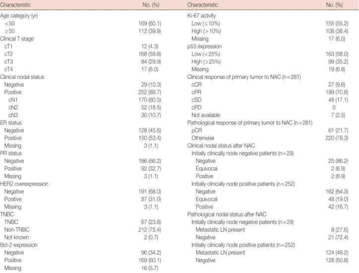
Clinicopathologic characteristics at the time of initial diag- nosis are illustrated in Table 1. The median age of patients was 46 years (range, 24-73 years). Among the 281 patients studied, 204 underwent breast-conservation surgery (72.6%). Most pa- tients had cT2 (n=168, 59.8%) or cT3 (n=84, 29.9%) tumors, with a mean tumor size of 4.8 cm (range, 1.0-13.5 cm). Two hundred fifty-two patients (89.7%) had clinically positive lymph nodes at diagnosis. In 85 patients, the presence of lymph node metastasis was confirmed by needle biopsy. Among 281 patients, 150 (53.4%) were ER positive, 92 (32.7%) were PR positive, and 87 (31.0%) showed HER2 overexpression. Sixty- seven patients (23.8%) had triple negative (ER negative, PR negative, HER2 negative) breast cancer.
Neoadjuvant chemotherapy and tumor response after treatment
All patient received NAC prior to definitive surgery. Twen- ty-seven patients (9.6%) presented no evidence of residual tu- mor on physical and radiologic examination after NAC. The incidence of pCR was higher than that of cCR with a rate of 21.7% (61 patients). Regarding nodal status, among 252 clini- cally node positive patients at diagnosis, 162 (64.3%) became clinically node negative after chemotherapy. According to the final pathologic results, NAC resulted in complete nodal ster- ilization in 49.2% (125) of initially clinically node-positive breast cancer patients.
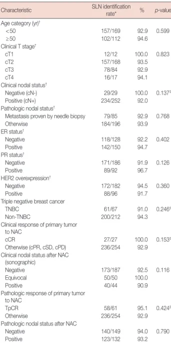
SLN identification rate and related factors
After NAC, the overall SLN identification rate was 93.6%
(263/281). SLN identification rate was 93.0% (186/200) in 200
Table 1. Patient and tumor characteristics
Characteristic No. (%) Characteristic No. (%)
Age category (yr) Ki-67 activity
<50 169 (60.1) Low (≤10%) 155 (55.2)
≥50 112 (39.9) High (>10%) 108 (38.4)
Clinical T stage Missing 17 (6.0)
cT1 12 (4.3) p53 expression
cT2 168 (59.8) Low (<25%) 163 (58.0)
cT3 84 (29.9) High (≥25%) 99 (35.2)
cT4 17 (6.0) Missing 19 (6.8)
Clinical nodal status Clinical response of primary tumor to NAC (n=281)
Negative 29 (10.3) cCR 27 (9.6)
Positive 252 (89.7) cPR 199 (70.8)
cN1 170 (60.5) cSD 48 (17.1)
cN2 52 (18.5) cPD 0
cN3 30 (10.7) Not available 7 (2.5)
ER status Pathological response of primary tumor to NAC (n=281)
Negative 128 (45.6) pCR 61 (21.7)
Positive 150 (53.4) Otherwise 220 (78.3)
Missing 3 (1.1) Clinical nodal status after NAC
PR status Initially clinically node negative patients (n=29)
Negative 186 (66.2) Negative 25 (86.2)
Positive 92 (32.7) Equivocal 2 (6.9)
Missing 3 (1.1) Positive 2 (6.9)
HER2 overexpression Initially clinically node positive patients (n=252)
Negative 191 (68.0) Negative 162 (64.3)
Positive 87 (31.0) Equivocal 48 (19.0)
Missing 3 (1.1) Positive 42 (16.7)
TNBC Pathological nodal status after NAC
TNBC 67 (23.8) Initially clinically node negative patients (n=29)
Non-TNBC 212 (75.4) Metastatic LN present 8 (27.6)
Not known 2 (0.7) Negative 21 (72.4)
Bcl-2 expression Initially clinically node positive patients (n=252)
Negative 96 (34.2) Metastatic LN present 124 (49.2)
Positive 169 (60.1) Negative 128 (50.8)
Missing 16 (5.7)
ER=estrogen receptor; PR=progesterone receptor; HER2=human epidermal growth factor receptor 2; TNBC=triple negative breast cancer; NAC=neoadjuvant chemotherapy; cCR =clinical complete response; cPR =clinical partial response; cSD =clinical stable disease; cPD =clinical progressive disease;
pCR=pathological complete response; LN=lymph node.
patients who underwent intraoperative lymphatic mapping with blue dye alone. When both blue dye and radioactive col- loid were used, SLNs were successfully detected in 96.2%
(76/79) of cases. In two patients in whom only radioactive col- loid was used, SLNs were identified in one patient (50%). Ac- cording to univariate analysis, no clinicopathologic factors in- cluding age at diagnosis, clinical T stage, initial nodal status, hormonal receptor status, and degree of clinical/pathological response to NAC was significantly related to the SLN identifi- cation rate (Table 2).
For 18 patients in whom SLNs were not identified, subse- quent axillary dissection revealed the presence of metastatic cancer cells in nine cases (50%).
False negative rate of SLNB and related factors
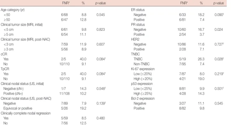
Among 263 patients with identified SLNs, 202 (76.8%) un- derwent subsequent ALND (Figure 1). No further ALND was performed on the other 61 patients who had negative frozen section results. The false negative rate of SLNB was deter- mined in the 202 patients who underwent SLNB followed by ALND (Table 3). According to patients’ final pathology re- ports, SLNB results accurately predicted ALN status in 190 of 202 patients. However, in 12 patients with residual cancer cells after NAC, SLNB failed to identify the metastatic nodes, re- sulting in a false negative rate of 10.4%.
The false negative rate was significantly higher in patients with PR-negative tumors than in those with PR-positive tu- mors (16.7% vs. 3.7%, p=0.024) (Table 4). Although ER status and HER2 expression were not significant factors affecting the false negative rate, triple-negative breast cancer (TNBC) pa- tients showed a significantly higher false negative rate than non-TNBC patients (26.3% vs. 7.4%, p=0.028). No other in- vestigated factors including age, response to NAC, and nodal status were positively associated with the false negative rate, as shown in Table 4. Multivariate logistic regression analysis showed that TNBC was an independent predictor of false negative SLNB in patients who received NAC (hazard ratio [HR], 0.155; p=0.045).
Table 2. Sentinel lymph node identification rate according to clinico- pathologic characteristics
Characteristic SLN identification
rate* % p-value
Age category (yr)†
<50 157/169 92.9 0.599
≥50 102/112 94.6
Clinical T stage†
cT1 12/12 100.0 0.823
cT2 157/168 93.5
cT3 78/84 92.9
cT4 16/17 94.1
Clinical nodal status†
Negative (cN-) 29/29 100.0 0.137‡
Positive (cN+) 234/252 92.0
Pathologic nodal status†
Metastasis proven by needle biopsy 79/85 92.9 0.768
Otherwise 184/196 93.9
ER status†
Negative 118/128 92.2 0.402
Positive 142/150 94.7
PR status†
Negative 171/186 91.9 0.126
Positive 89/92 96.7
HER2 overexpression†
Negative 172/182 94.5 0.360
Positive 88/96 91.7
Triple negative breast cancer
TNBC 61/67 91.0 0.246‡
Non-TNBC 200/212 94.3
Clinical response of primary tumor to NAC
cCR 27/27 100.0 0.153‡
Otherwise (cPR, cSD, cPD) 236/254 92.9 Clinical nodal status after NAC
(sonographic)
Negative 173/187 92.5 0.116
Equivocal 50/50 100.0
Positive 40/44 90.9
Pathologic response of primary tumor to NAC
TpCR 58/61 95.1 0.424‡
Otherwise 236/254 92.9
Pathologic nodal status after NAC
Negative 140/149 94.0 0.790
Positive 123/132 93.2
SLN=sentinel lymph node; cN-=clinically node negative; cN+=clinically node positive; ER=estrogen receptor; PR=progesterone receptor; HER2=human epidermal growth factor receptor 2; TNBC=triple negative breast cancer;
NAC=neoadjuvant chemotherapy; cCR=clinical complete response; cPR=
clinical partial response; cSD=clinical stable disease; cPD=progressive dis- ease; TpCR=pathological complete response of primary tumor.
*SLN identification rate=number of patients in whom SLNs were detected/
number of patients in whom sentinel lymph node biopsy was attempted; †Clini- copathologic variables at the time of initial diagnosis; ‡p-values from Fisher ex- act test.
Table 3. Axillary lymph node status after neoadjuvant chemotherapy in patients who underwent sentinel lymph node biopsy followed by axillary lymph node dissection (n=202)
Axillary LN
Positive Negative
SLN
Positive 103 0
Negative 12* 87
Total 115 87
LN=lymph node; SLN=sentinel lymph node.
*False negative rate=SLN-negative patients with axillary lymph node (ALN) metastasis∕total number of ALN-positive patients =12/(103+12) =12/115 (10.4%).
attemptedSLN
Total
(n=281) SLN
not identified (n=18)
ALN (+) (n=9)
SLN (+) (n=18)
SLN (+) (n=71)
SLN (+) (n=20)*
ALN (-) (n=9)
SLN (-), NSLN (-) (n=19)
SLN (-), NSLN (-) (n=121) SLN (-), NSLN (+) (n=14)† SLN (-), NSLN (+) (n=0)
SLN identified (n=263)
identified?SLN SLN Frozen Bx.
Frozen Bx.
not done (n=37)
All underwent ALND (n=18)
All underwent ALND (n=108)
ALND (n=14), SLNB (n=6) ALND (n=68), SLNB (n=53) ALND (n=12), SLNB (n=2) SLN frozen Bx.
positive (n=71)
SLN frozen Bx.
negative (n=155)
Permanent Bx.
Figure 1. Description of the study population by treatment/procedure performed.
SLN=sentinel lymph node; Bx=biopsy; ALN=axillary lymph node; ALND=axillary lymph node dissection; SLNB=sentinel lymph node biopsy. *Frozen biopsy and permanent biopsy discordant cases; †False negative cases (based on permanent biopsy results).
Table 4. False negative rate of sentinel lymph node biopsy according to clinicopathologic characteristics
FNR* % p-value FNR* % p-value
Age category (yr) ER status
<50 6/68 8.8 0.545 Negative 6/33 18.2 0.090†
≥50 6/47 12.8 Positive 6/81 7.4
Clinical tumor size (MRI, initial) PR status
<5 cm 6/61 9.8 0.823 Negative 10/60 16.7 0.024
≥5 cm 6/54 11.1 Positive 2/54 3.7
Clinical tumor size (MRI, post-NAC) HER2
<3 cm 7/59 11.9 0.607 Negative 10/86 11.6 0.727†
≥3 cm 5/56 8.9 Positive 2/28 7.1
cCR TNBC
Yes 2/5 40.0 0.084† TNBC 5/19 26.3 0.028†
No 10/110 9.1 Non-TNBC 7/95 7.4
TpCR Ki-67 expression
Yes 2/5 40.0 0.084† Low (≤20%) 7/87 8.0 0.219†
No 10/110 9.1 High (>20%) 4/21 19.0
Clinical nodal status (US, initial) p53 expression
Negative (cN-) 1/7 14.3 0.548† Low (<25%) 8/81 9.9 0.501†
Positive (cN+) 11/108 10.2 High (≥25%) 4/28 14.3
Clinical nodal status (US, post-NAC) Bcl-2 expression
Negative 7/89 7.9 0.139† Negative 3/27 11.1 0.545
Equivocal or positive 5/26 19.2 Positive 8/82 9.8
Clinically complete nodal regression
Yes 5/59 8.5 0.480
No 7/56 12.5
FNR=false negative rate; MRI=magnetic resonance imaging; NAC=neoadjuvant chemotherapy; cCR=clinical complete response; TpCR=pathological complete re- sponse of primary tumor (regardless of axillary status); US=ultrasonography; cN-=clinically node negative; cN+=clinically node positive; ER=estrogen receptor;
PR=progesterone receptor; HER2=human epidermal growth factor receptor 2; TNBC=triple negative breast cancer.
*FNR=sentinel lymph node-negative patients with axillary lymph node (ALN) metastasis∕ total number of ALN-positive patients; †p-values from Fisher exact test.
Discordance between intraoperative frozen section results and final permanent section results of SLNB
The final pathologic results of SLN status were not always in accordance with intraoperative frozen section findings.
Among 226 patients receiving intraoperative SLN examina- tion, frozen sections indicated no tumor cells in 155 patients (Figure 1). However, the final pathologic examination of the SLNs showed metastatic cells in 20 patients with negative fro- zen section results, yielding an accuracy of 91.2%, a sensitivity of 78%, and a specificity of 100% for the frozen section study.
DISCUSSION
The accuracy of SLNB after NAC remains controversial for several reasons. First, lymphatic fibrosis or tumor debris oc- curring after NAC may result in changes in the lymphatic drainage pattern, leading to a decrease in SLNB accuracy. Al- though direct comparison of the lymphatic drainage pattern before and after NAC has not been reported, Brown et al. [14]
recently demonstrated histologic changes including fibrosis and obliteration of lymph node architecture after NAC. They performed SLNB and subsequent axillary dissection after NAC in patients with pathologically proven positive ALNs and demonstrated that SLNs from patients with sterilized ALNs generally exhibited the histologic changes mentioned
above, indicative of treatment effect. Another possible expla- nation for the increased false negative rate is the nonsequen- tial therapeutic effect of NAC on lymph nodes, which indi- cates SLNs may be sterilized before non-SLNs.
In the present study, we aimed to assess the reliability of SLNB after NAC by evaluating its identification rate and false negative rate.
The overall identification rate achieved in our study was 93.6%, similar to the pooled value from four previously pub- lished meta-analyses (Table 5) [7,8,15-26]. However, identifi- cation rates from recent independent reports show significant variation, ranging from 75.7% to 98.7%, which is often ex- plained by study population heterogeneity. Clinicopathologic factors such as initial clinical nodal status [17,22], residual tu- mor size [19], degree of tumor response to NAC [21], age, ER status, proliferation index, and lymphovascular invasion [22]
have been suggested to affect identification rates. However, no single clinicopathologic factor has consistently been shown to affect the identification rate of SLNB after NAC. In our study, none of the above factors were significantly associated with the identification rate. SLNs were successfully detected in all initially node-negative patients, thus resulting in an identifica- tion rate of 100%, but this result did not reach statistical sig- nificance (p=0.137). The insignificance may be due to the small study population size as suggested by the fact that the Table 5. Meta-analysis and recent reports on sentinel lymph node biopsy after neoadjuvant chemotherapy
Year No. Study design/population IR Associated factors FNR Associated factors
Xing et al. [15] 2006 1,273 Meta-analysis 90 12
van Deurzen et al. [7] 2009 2,148 Meta-analysis 90.9 10.5
Kelly et al. [16] 2009 1,799 Meta-analysis 89.6 8.4
Tan et al. [8] 2011 449 Meta-analysis, clinically node negative after NAC 94.3 9.4 Classe et al. [17] 2009 195 Prospective, multicenter/Operable,
noninflammatory, unifocal, large, N0 or N1
90.1 Initial cN0 11.5 -
Schwartz et al. [18] 2010 79 Retrospective/T0-T4, N0-N2 98.7 - 1/23 -
Ozmen et al. [19] 2010 77 Retrospective/Stage IIB, IIIA, IIIB, clinically node
negative after NAC, NAC=FAC or AC/docetaxel 92 Residual tumor size 13.7 Initial N stage Reitsamer et al. [20] 2010 185 Retrospective/Stage II or III, taxane-based NAC 81.1 - 8.3 - Kang et al. [21] 2011 66 Retrospective/Pathologically proven positive
ALN before NAC 87.9 cCR (NS) 17.1 -
Pecha et al. [22] 2011 343 Retrospective, multicenter 80.8 Young age, cN0,
ER+, low Ki-67, LVI 19.5 LVI, ER- (NS) Canavese et al. [23] 2011 64 Prospective, single center/T≥2 cm and
clinically node positive, NAC=FEC/T
93.8 - 2.1 -
Takahashi et al. [24] 2012 96 Retrospective, single center/Stage II, III 87.5 - 24.5 Initial cN0, cCR (NS) Alvarado et al. [25] 2012 150 Retrospective/Pathologically proven positive
ALN before NAC
93 20.8 Initial cN0, tumor size,
number of SLN removed Takei et al. [26] 2012 105 Retrospective, single center/Clinically positive
ALN at diagnosis
75.7 - 8.2 -
IR=identification rate; FNR=false negative rate; NAC=neoadjuvant chemotherapy; cN0=clinical nodal stage N0; FAC=fluorouracil, anthracyclin, cyclophosphamide;
AC=anthracyclin, cyclophosphamide; ALN=axillary lymph node; cCR=clinical complete response; NS=not significant; ER=estrogen receptor; LVI=lymphvascular invasion; FEC/T=fluorouracil, epirubicin, cyclophosphamide/taxane; SLN=sentinel lymph node.
proportion of initially node-negative patients was only 10.3%
(29/281). Similarly, SLN detection was successful in all 27 pa- tients who achieved clinical cCR after NAC, but this too was not statistically significant (p=0.153).
The false negative rate of SLNB after NAC was 10.4% in our study, which is substantially higher than that in primary breast cancer patients without NAC observed at our institution [27].
The reported false negative rates of SLNB after NAC from other recent studies range from 2% to 24%, and results of meta-anal- yses seem to converge to values of approximately 10%. Similar to our result, findings presented at the 2012 San Antonio Breast Cancer Symposium from the American College of Surgeons Oncology Group Z0171 trial, which included 756 breast cancer patients who received NAC, indicated a false negative rate of 12.8% [28]. Although the acceptable range of false negative re- sults in patients receiving NAC remains controversial, a rate of over 10% of false negative results by SLNB warrants precaution when indicating the procedure in this patient population.
Our analysis suggests that patients with PR-positive tumors and non-TNBC might be a select group in which SLNB can be indicated after NAC. Pecha et al. [22] also reported a nonsig- nificant trend in the association between hormonal receptor status and the accuracy of SLNB. It is however difficult to ex- plain why SLNB in patients with PR-negative tumors or in TNBC patients showed higher false negative rates in the cur- rent study. This finding may be explained by the diverse sus- ceptibility to NAC by different breast cancer subtypes, as PR- negative tumors and TNBC each showed a higher CR rate than PR-positive tumors and non-TNBC (data not shown). A simi- lar difference in response to NAC was reported among breast cancer subtypes in a meta-analysis by Houssami et al. [29].
Tumors with higher response may undergo greater changes in the lymphatic drainage pattern, which consequently leads to higher false negative results of SLNB.
Histologic changes after NAC pose potential challenges to the interpretation of SLN frozen sections, which prompted us to investigate the discordance rate between frozen section ex- amination and permanent examination. Compared to the per- manent examination results, intraoperative assessment of fro- zen sections from SLNB after NAC showed 91.2% accuracy.
Although this discordance rate is similar to the observation in breast cancer patients who do not receive NAC at our institu- tion [27], it may have larger implications. Missed cases on fro- zen section analysis mostly involve micrometastasis, which, in the NAC group, may be a result of an incomplete response of initial macrometastasis. As speculated in a report by Sahoo and Lester [30], such possibilities are supported by results from the National Adjuvant Breast and Bowel Project (NSABP) B-18. In NSABP B-18, patients with lymph node micrometas-
tases who were not treated with chemotherapy before surgery had identical survival compared to those with negative nodes.
However, in the NAC group, survival of patients with minime- tastases and micrometastases in lymph nodes was significantly worse. Thus, considering the prognostic significance of micro- metastasis in patients receiving NAC, we should be prudent in performing SLNB, which relies on the results of frozen sec- tions. In recognition that these frozen section results can miss micrometastases in approximately 10% of the cases, patients should be informed about the possibility of additional axillary dissection after pathologic results of permanent sections.
In summary, our study suggests that SLNB after NAC and intraoperative examination of frozen sections is technically feasible. However, SLNB after NAC is associated with a higher risk of false negativity, which may vary depending on the mo- lecular characteristics of the tumor such as PR expression and molecular subtype. Our results suggest that patients with PR positive and non-TNBC are the potential candidates for SNLB after NAC.
CONFLICT OF INTEREST
The authors declare that they have no competing interests.
REFERENCES
1. Fisher B, Bauer M, Wickerham DL, Redmond CK, Fisher ER, Cruz AB, et al. Relation of number of positive axillary nodes to the prognosis of patients with primary breast cancer: an NSABP update. Cancer 1983;
52:1551-7.
2. Ahmed RL, Prizment A, Lazovich D, Schmitz KH, Folsom AR. Lymph- edema and quality of life in breast cancer survivors: the Iowa Women’s Health Study. J Clin Oncol 2008;26:5689-96.
3. Kim T, Giuliano AE, Lyman GH. Lymphatic mapping and sentinel lymph node biopsy in early-stage breast carcinoma: a metaanalysis.
Cancer 2006;106:4-16.
4. Nason KS, Anderson BO, Byrd DR, Dunnwald LK, Eary JF, Mankoff DA, et al. Increased false negative sentinel node biopsy rates after pre- operative chemotherapy for invasive breast carcinoma. Cancer 2000;89:
2187-94.
5. Kuerer HM, Sahin AA, Hunt KK, Newman LA, Breslin TM, Ames FC, et al. Incidence and impact of documented eradication of breast cancer axillary lymph node metastases before surgery in patients treated with neoadjuvant chemotherapy. Ann Surg 1999;230:72-8.
6. Bear HD, Anderson S, Smith RE, Geyer CE Jr, Mamounas EP, Fisher B, et al. Sequential preoperative or postoperative docetaxel added to pre- operative doxorubicin plus cyclophosphamide for operable breast cancer:National Surgical Adjuvant Breast and Bowel Project Protocol B-27. J Clin Oncol 2006;24:2019-27.
7. van Deurzen CH, Vriens BE, Tjan-Heijnen VC, van der Wall E, Al- bregts M, van Hilligersberg R, et al. Accuracy of sentinel node biopsy after neoadjuvant chemotherapy in breast cancer patients: a systematic
review. Eur J Cancer 2009;45:3124-30.
8. Tan VK, Goh BK, Fook-Chong S, Khin LW, Wong WK, Yong WS. The feasibility and accuracy of sentinel lymph node biopsy in clinically node- negative patients after neoadjuvant chemotherapy for breast cancer: a systematic review and meta-analysis. J Surg Oncol 2011;104:97-103.
9. Fernández A, Cortés M, Benito E, Azpeitia D, Prieto L, Moreno A, et al.
Gamma probe sentinel node localization and biopsy in breast cancer patients treated with a neoadjuvant chemotherapy scheme. Nucl Med Commun 2001;22:361-6.
10. Shen J, Gilcrease MZ, Babiera GV, Ross MI, Meric-Bernstam F, Feig BW, et al. Feasibility and accuracy of sentinel lymph node biopsy after preoperative chemotherapy in breast cancer patients with documented axillary metastases. Cancer 2007;109:1255-63.
11. Lee KH, Im SA, Oh DY, Lee SH, Chie EK, Han W, et al. Prognostic sig- nificance of bcl-2 expression in stage III breast cancer patients who had received doxorubicin and cyclophosphamide followed by paclitaxel as adjuvant chemotherapy. BMC Cancer 2007;7:63.
12. Therasse P, Arbuck SG, Eisenhauer EA, Wanders J, Kaplan RS, Rubin- stein L, et al. New guidelines to evaluate the response to treatment in solid tumors: European Organization for Research and Treatment of Cancer, National Cancer Institute of the United States, National Cancer Institute of Canada. J Natl Cancer Inst 2000;92:205-16.
13. Cho N, Moon WK, Han W, Park IA, Cho J, Noh DY. Preoperative so- nographic classification of axillary lymph nodes in patients with breast cancer: node-to-node correlation with surgical histology and sentinel node biopsy results. AJR Am J Roentgenol 2009;193:1731-7.
14. Brown AS, Hunt KK, Shen J, Huo L, Babiera GV, Ross MI, et al. Histo- logic changes associated with false-negative sentinel lymph nodes after preoperative chemotherapy in patients with confirmed lymph node- positive breast cancer before treatment. Cancer 2010;116:2878-83.
15. Xing Y, Foy M, Cox DD, Kuerer HM, Hunt KK, Cormier JN. Meta- analysis of sentinel lymph node biopsy after preoperative chemothera- py in patients with breast cancer. Br J Surg 2006;93:539-46.
16. Kelly AM, Dwamena B, Cronin P, Carlos RC. Breast cancer sentinel node identification and classification after neoadjuvant chemotherapy- systematic review and meta analysis. Acad Radiol 2009;16:551-63.
17. Classe JM, Bordes V, Campion L, Mignotte H, Dravet F, Leveque J, et al.
Sentinel lymph node biopsy after neoadjuvant chemotherapy for ad- vanced breast cancer: results of Ganglion Sentinelle et Chimiotherapie Neoadjuvante, a French prospective multicentric study. J Clin Oncol 2009;27:726-32.
18. Schwartz GF, Tannebaum JE, Jernigan AM, Palazzo JP. Axillary sentinel lymph node biopsy after neoadjuvant chemotherapy for carcinoma of the breast. Cancer 2010;116:1243-51.
19. Ozmen V, Unal ES, Muslumanoglu ME, Igci A, Canbay E, Ozcinar B, et al. Axillary sentinel node biopsy after neoadjuvant chemotherapy. Eur J
Surg Oncol 2010;36:23-9.
20. Reitsamer R, Menzel C, Glueck S, Rettenbacher L, Weismann C, Huta- rew G. Sentinel lymph node biopsy is precise after primary systemic therapy in stage II-III breast cancer patients. Ann Surg Oncol 2010;17 Suppl 3:286-90.
21. Kang E, Chung IY, Han SA, Kim SM, Jang M, Lyou CY, et al. Feasibility of sentinel lymph node biopsy in breast cancer patients with initial axil- lary lymph node metastasis after primary systemic therapy. J Breast Cancer 2011;14:147-52.
22. Pecha V, Kolarik D, Kozevnikova R, Hovorkova K, Hrabetova P, Halas- ka M, et al. Sentinel lymph node biopsy in breast cancer patients treated with neoadjuvant chemotherapy. Cancer 2011;117:4606-16.
23. Canavese G, Dozin B, Vecchio C, Tomei D, Villa G, Carli F, et al. Accu- racy of sentinel lymph node biopsy after neo-adjuvant chemotherapy in patients with locally advanced breast cancer and clinically positive axillary nodes. Eur J Surg Oncol 2011;37:688-94.
24. Takahashi M, Jinno H, Hayashida T, Sakata M, Asakura K, Kitagawa Y.
Correlation between clinical nodal status and sentinel lymph node bi- opsy false negative rate after neoadjuvant chemotherapy. World J Surg 2012;36:2847-52.
25. Alvarado R, Yi M, Le-Petross H, Gilcrease M, Mittendorf EA, Bedro- sian I, et al. The role for sentinel lymph node dissection after neoadju- vant chemotherapy in patients who present with node-positive breast cancer. Ann Surg Oncol 2012;19:3177-84.
26. Takei H, Yoshida T, Kurosumi M, Inoue K, Matsumoto H, Hayashi Y, et al. Sentinel lymph node biopsy after neoadjuvant chemotherapy pre- dicts pathological axillary lymph node status in breast cancer patients with clinically positive axillary lymph nodes at presentation. Int J Clin Oncol 2013;18:547-53.
27. Kim SW, Han W, Park IA, Chung JK, Yeo JS, Moon WK, et al. Prospec- tive study of 162 sentinel lymph node biopsies in breast cancer: useful- ness of ultrasonography in patients selection. J Korean Breast Cancer Soc 2003;6:103-8.
28. Boughey JC, SumanVJ, Mittendorf EA, Ahrendt GM, Wilke LG, Taback B, et al. The role of sentinel lymph node surgery in patients presenting with node positive breast cancer (T0-T4, N1-2) who receive neoadju- vant chemotherapy: results from the ACOSOG Z1071 trial. Cancer Res 2012;72(24 Suppl 3):S2-1.
29. Houssami N, Macaskill P, von Minckwitz G, Marinovich ML, Mamou- nas E. Meta-analysis of the association of breast cancer subtype and pathologic complete response to neoadjuvant chemotherapy. Eur J Cancer 2012;48:3342-54.
30. Sahoo S, Lester SC. Pathology of breast carcinomas after neoadjuvant chemotherapy: an overview with recommendations on specimen pro- cessing and reporting. Arch Pathol Lab Med 2009;133:633-42.