Osteoporotic Distal Radius Fracture Treated with Bone Cement(PMMA) and Percutaneous Pinning: Clinical Analysis of 12 Cases
Jin Rok Oh, M.D., Huei Jeon Park, M.D., Doo Sup Kim, M.D.
Department of Orthopedics, Yonsei University Wonju Medical College, Wonju, Korea
Purpose : There are difficulties in treatment of distal radius fracture with osteoporosis because loosening of internal or external fixation is very common.
Redisplacement of fracture fragments causes long immo- bilization and malunion. Through using bone cement and percutaneous pinning, we tried to overcome these obsta- cles to treat the patients with osteoporotic distal radius fracture.
Materials and Methods : We treated 12 patients with osteoporotic distal radius fracture. As treatment methods, under the general anesthesia or brachial plexus regional block, we reduced fracture fragments of distal radius under the fluoroscopic image intensifier and fixed percu- taneously with K-wire and injected bone cement into fracture site using vertebroplasty kit. At post operation 6th week, we removed K-wires and performed passive ROM exercise of the wrist joint.
R e s u l t s : Among total 12 cases, 10 cases(83.8%) showed excellent and good result, 1 case(8.3%) fair, 1
case (8.3%) poor. There were 4 cases of complications, leakage of bone cement into radiocarpal joint in 1 case, redisplacement of fracture site in 1 case, and pin tract infection in 2 cases.
Discussion and conclusions : Leakage of bone cement into radiocarpal joint happened in case with intraarticular fracture of distal radius. Conclusively, except the case with intraarticular fracture of distal radius, if we get anatomical reduction of fracture site with closed manipu- lation and percutaneous pinning, we think that percuta- neous K-wire fixation and percutaneous bone cement (PMMA) injection can be considerable treatment meth- ods in patients with osteoporotic distal radius fracture.
Key Words : Osteoporosis, Bone cement, Distal radius fracture, Percutaneous pinning
서 론
골다공증이 있는 요골 원위부 골절 환자의 치료는 어 려움이 있는데, 단순 석고 고정을 하거나, 경피적 핀 삽 입이나 내고정 또는 외고정을 한다 하더라도 고정물이 느슨해져 재전위의 가능성이 높다. 정복된 부위의 재전 위는 결국 장시간의 고정의 원인이 되거나 부정유합을 초래하여 완관절 기능 회복에 부정적으로 작용한다. 본 교실에서는 경피적 삽입과 골 시멘트를 이용하여 이러한 문제점을 극복하고자 노력하였다.
연구대상 및 방법
2 0 0 0년 2월부터 2 0 0 1년 1 2월까지 요골 원위부 골 절 환자 중 단순 방사선 사진과 골밀도 검사를 통해 나이 VOLUME 7, NUMBER 2, DECEMBER 2002
골 시멘트와 경피적 핀 삽입술로 치료한 골다공성 요골 원위부 골절: 12례 분석
연세대학교 원주의과대학 정형외과 오진록・박희전・김두섭
통신저자 : 오 진 록
강원도 원주시 일산동 162 연세대학교 원주의과대학 정형외과 TEL : 033-741-1355, FAX : 033-741-1358 E-mail : chaos@wonju.yonsei.ac.kr
교정 후 2.5 이상의 표준 편차를 보이는 골다공증이 확 인된 환자 1 2례를 대상으로 하였다. 대상인의 평균 아이 는 6 5 . 3세였고, 모두 여자였다. AO 분류에 따른 골절의 유형은 A 2형이 4례, A3형이 8례, B1형이 1례, B2형 이 1례였다. 치료 방법은 수상 후 1주일 이내에 전신 마 취 또는 상완 신경총 부분 마취 하에 f l u r o s c o p i c image intensifier를 이용 비관혈적 정복을 한 후, K 강선을 이용 경피적 핀 삽입술을 시행하고, vertebro- plasty kit를 이용하여 경피적으로 골절부에 골시멘트 를 주입하였다(Fig. 1 A-E). 골시멘트 주입은 일단 골 밀도 검사상 골다공증이 판정된 경우에는 모든 례에서 K 강선 고정 후 골절부의 안정성 여부와는 관계없이 실시 하였다. 술 후 1일째부터 수지 및 완관절의 능동적 운동 을 시행하였고, 술 후 6주 째 K 강선 제거 후 환자의 상
태에 따라 수동적 관절 운동을 실시하였다. 결과는 술 후 1 2주 째 마지막 추시 때 Samiento scoring system 을 이용하여 판정하였다(Table 1).
결 과
총 1 2례 중 1 0례( 8 3 . 3 % )에서 양호 이상, 1례 ( 8 . 3 % )에서 보통, 1례( 8 . 3 % )에서 불량의 결과를 보였 다(Fig. 2 A-C). 합병증은 골 시멘트의 요골-수근 관절 내 누출이 1례, 요골 원위 골절부의 전위가 1례 있었고, 경피적 핀 삽입부의 감염이 2례 있었다(Fig. 3 A-C).
오진록・박희전・김두섭
Table 1. Demerit point system(by Sarmiento) for clinical evaluation
Points Residual deformity(range, 0 to 3 points)
Prominent ulnar styloid 1
Residual dorsal tilt 2
Radial deviation of hand 2 or 3
Subjective evaluation(range, 0 to 6 points)
Excellent - no pain, disability, or limitation of motion 0
Good - occasional pain, some limitation of motion, and no disability 2
Fair - occasional pain, some limitation of motion, feeling of weakness in wrist, no particular disability if careful,
and activities slightly restricted 4
Poor - pain, limitation of motion, disability, and activities more or less markedly 6 Objective evaluation(range, 0 to 5 points)
Dorsiflexion < 45 5
Ulnar deviation <30 3
Supination < 50 2
Pronation < 50 2
Palmar flexion < 30 1
Radial deviation < 15 1
Loss of circumduction 1
Pain in distal radio-ulnar joints 1
Grip strength < 60% of uninjured side 1
Complication(range, 0 to 5 points) Arthritic change
Minimum 1
Minimum with pain 3
Moderate 2
Moderate with pain 4
Severe 3
Severe with pain 5
Nerve complication(median) 1 to 3
Poor finger function due to cast 1 to 2
Final results(range of points)
Excellent 0 to 2
Good 3 to 8
Fair 9 to 20
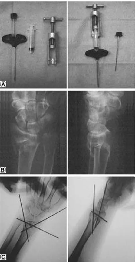
Fig. 1. A. Vertebroplasty Kit for injection of bone cement. B. Preoperative X-ray.
C. Under the fluoroscopic image intensifier, closed reduction and percutaneous pinning.
토의 및 결론
원위 요골 골절과 골다공증이 동반된 경우 단순 석고 고정, 내고정 및 외고정 방법, 경피적 핀 삽입술 등의 방 법을 통하더라도 견고한 고정을 얻기 어려워 치료에 결 국 장기간 부목 고정이나 부정유합을 초래하여 그 치료 결과가 좋지 못한 경우가 많다. 이를 극복하기 위해 Norian skeletal repair system(SRS)(Norian Corporation, Cupertino, California) 또는 골시멘 트( P M M A )를 주입하는 방법이 보고되고 있다3 , 4 ). Norian SRS는 cancellous bone cement로 성상은 Calcium phosphate(carbonated apatite)이며
을 나타내며, biocompatible하여 재형성이 가능한 반 면 전단력에 약하고, 흡수가 느리며, 골유도의 기능은 없 다1 ). Jupiter 등은 2 0례의 원위 요골 골절에 대해 Norian SRS를 경피적으로 주입한 후 6주간 단상지 석 고고정으로 치료하고 6개월 이상 추시 관찰이 가능한 5 례에 대해 5례 모두 양호 이상의 결과를 보였으며 골 밖 으로 Norian SRS가 유출된 3례에서는 6개월 째 흡수 된 소견을 보였다고 하였다3 ). Sanchez-Sotelo 등은 5 0세 이상 된 원위 요골 골절 환자 1 1 0명에 대해 Norian SRS를 관혈적으로 골 결손 부위에 넣고 2주간 단상지 석고 붕대 고정한 군과 도수 정복 후 6주간 석고 붕대고정을 한 군의 비교 실험에서 Norian SRS군이 더 우수한 결과를 보였다고 보고하였다4 ). 국내에서는 Norian SRS가 수입되지 않아 치료 방법으로 선택할 수 오진록・박희전・김두섭
Fig. 1. D. Insertion of puncture needle (arrow) into fracture site, percutaneously.
E. After bone cement injection through puncture needle.
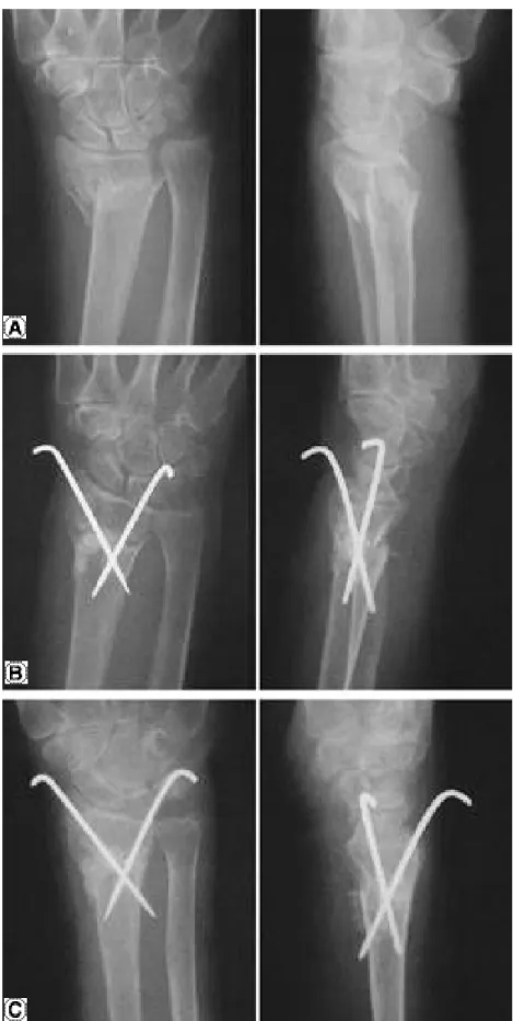
Fig. 2. A. Dorsal displaced comminution of distal radius. 72-year-old female.
B. Postoperative X-ray. C. Postoperative 6 weeks. Excellent result.
오진록・박희전・김두섭
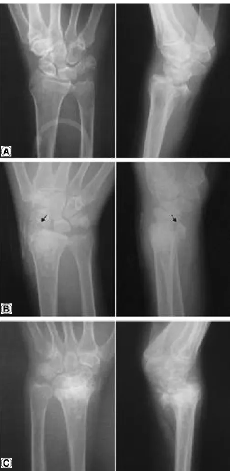
Fig. 3. A. Intraarticular fracture of distal radius. B. Leakage of bone cement int intraarticular space (arrow). C. Redisplacement of fracture. Poor result.
없다. 골시멘트는 Norian SRS와는 달리 비흡수성 물질 로서 골로 대치되지 않고 남아 있게되므로 골로 재형성 되는 경우에 비해 전체적인 골의 강도 면에서 떨어져 골 절되었던 부위가 다시 골절되는 경우에는 상대적으로 적 은 힘에 의해서도 골절이 일어날 수 있을 가능성이 있으 나 이는 생역학적 연구에 필요할 것으로 생각된다.
S c h m a l h o l z는 재전위된 C o l l e s’골절 환자에 대해 관 혈적 정복 후 골시멘트로 고정하여 치료한 군과 외고정 장치로 치료한 군과의 비교 연구에서 두 군간의 치료 결 과의 차이는 없었다고 보고하였으며, 골시멘트로 치료한 경우가 외고정으로 치료한 경우보다 재활 치료가 빠르 고, 환자에게 좀 더 편안하며, 합병증도 없었다고 하였 다. 또한 원위 요골 골절이 관절내 골절이거나, 심한 분 쇄 골절인 경우, 원위 척골 골절을 동반한 경우에는 골시 멘트를 쓰지 않는 것이 좋다고 하였다2 ).
저자는 골다공증과 원위 요골 골질이 동반된 환자에 대해 도수 정복 후 경피적 핀 삽입을 통한 골절부 고정 을 시행 후 골시멘트를 경피적으로 주입하였는데 그 이 유는 핀이 골간단부 골수강내에서 교차하는 부위에 골 시멘트를 주입함으로서 골 결손부를 메워주는 효과와 교차핀을 빠지지 않도록 안정화시키는 효과를 기대할 수 있다고 생각했기 때문이다. 주입된 골시멘트는 1 cc 이내로 하였고, 골시멘트가 피질골 밖으로 유출되지 않 도록 치약과 같은 정도의 유동성을 나타내는 시점에서 골시멘트를 주입하였다. 장기적인 추시는 하지 못했지 만 골 간단부위는 해면골이 많아 골시멘트와 피질골 사 이의 공간을 통해 골유합을 충분히 유도할 수 있으리라 판단된다. 저자가 시행한 시술방법에 몇 가지 주의를 기 울여야할 점이 있다. 첫째, 골시멘트의 관절내 누출 또 는 피질골 밖으로의 유출을 피해야 하며 특히 관절 내 골절이 있는 경우에는 이 시술을 하여서는 안된다. 둘
째, 삽입된 핀은 최소한 6주정도 유지시켜야하는데 그 이유는 골시멘트만으로는 수술 후 조기 능동적 완관절 운동을 지탱해낼 수 없고 저자의 경우 수술 직후 삽입되 었던 핀을 제거한 1례에서 재전위를 보였다. 셋째, 이 시술방법의 전재 조건으로 반드시 도수 정복을 통해 해 부학적 정복을 얻을 수 있어야 한다. 결론적으로 원위 요골의 관절 내 골절이 있는 경우를 제외하고, 도수정복 과 경피적 핀 삽입술로 해부학적 정복을 얻을 수 있다 면, 골다공증이 있는 요골 원위부 골절 환자에서 골 시 멘트와 K 강선을 이용하는 치료 방식도 고려해볼 만한 치료 방법으로 생각된다.
REFERENCES
01) Amy LL and Natban BP : Use of Bone-Graft Substitutes in Distal Radius Fractures, J Am Acad Orthop Surg 7:279- 290,1990.
02) A Schmalholz : External skeletal fixation versus cement fixation in the treatment of redislocated Colles’ fracture.
CORR 254:236-241,1990.
03) Jesse BJ, Steven W, Scott S, Colleen L, Charles P, Amy LL, Michelle VW, and Susanne TS : Repair of Five Radius Fractures with investigational Cancellous Bone Cement : A Preliminary Report. J Orthop Trauma 11:110- 116.1997.
04) J Sanchez-Sotelo, L Munuera, and R Madero : Treatment of Fractures of the Distal Radius with a Pemodellable Bone Cement. J Bone Joint Surg(Br) 82- B:856-863,2000.