J Korean Surg Soc 2013;84:346-352 http://dx.doi.org/10.4174/jkss.2013.84.6.346
ORIGINAL ARTICLE
JKSS JKSS JKSS
Journal of the Korean Surgical Society pISSN 2233-7903ㆍeISSN 2093-0488
Received March 4, 2013, Reviewed March 23, 2013, Accepted April 17, 2013 Correspondence to: Seung-Kee Min
Department of Surgery, Seoul National University Hospital, Seoul National University College of Medicine, 101 Daehak-ro, Jongno-gu, Seoul 110-744, Korea
Tel: +82-2-2072-2318, Fax: +82-2-766-3975, E-mail: [email protected]
Presented at the 2012 American Venous Forum; Orlando, FL. February 8–11, 2012.
cc Journal of the Korean Surgical Society is an Open Access Journal. All articles are distributed under the terms of the Creative Commons Attribution Non-Commercial License (http://creativecommons.org/licenses/by-nc/3.0/) which permits unrestricted non-commercial use, distribution, and reproduction in any medium, provided the original work is properly cited.
Reconstruction of portal vein and superior mesenteric vein after extensive resection for pancreatic cancer
Suh Min Kim, Seung-Kee Min, Daedo Park, Sang-Il Min, Jin-Young Jang, Sun-Whe Kim, Jongwon Ha, Sang Joon Kim
Department of Surgery, Seoul National University College of Medicine, Seoul, Korea
Purpose: Tumor invasion to the portal vein (PV) or superior mesenteric vein (SMV) can be encountered during the surgery for pancreatic cancer. Venous reconstruction is required, but the optimal surgical methods and conduits remain in controversies. Methods: From January 2007 to July 2012, 16 venous reconstructions were performed during surgery for pan- creatic cancer in 14 patients. We analyzed the methods, conduits, graft patency, and patient survival. Results: The involved veins were 14 SMVs and 2 PVs. The operative methods included resection and end-to-end anastomosis in 7 patients, wedge resection with venoplasty in 2 patients, bovine patch repair in 3 patients, and interposition graft with bovine patch in 1 patient. In one patient with a failed interposition graft with great saphenous vein (GSV), the SMV was reconstructed with a prosthetic interposition graft, which was revised with a spiral graft of GSV. Vascular morbidity occurred in 4 cases; occlusion of an interposition graft with GSV or polytetrafluoroethylene, segmental thrombosis and stenosis of the SMV after end-to-end anastomosis. Patency was maintained in patients with bovine patch angioplasty and spiral vein grafts. With mean follow-up of 9.8 months, the 6- and 12-month death-censored graft survival rates were both 81.3%. Conclusion: Many of the involved vein segments were repaired primarily. When tension-free anastomosis is impossible, the spiral grafts with GSV or bovine patch grafts are good options to overcome the size mismatch between autologous vein graft and portomesen- teric veins. Further follow-up of these patients is needed to demonstrate long-term patency.
Key Words: Portal vein, Superior mesenteric vein, Reconstruction, Pancreaticoduodenectomy
INTRODUCTION
Tumor invasion or adhesion to the portal vein (PV) or superior mesenteric vein (SMV) can be encountered dur- ing the surgery for pancreatic cancer. Venous resection and reconstruction are required for complete resection of
the tumor in these cases. Since extensive resection and ve- nous reconstruction was found not to increase the post- operative morbidity and mortality, en bloc resection with involved vein was performed in many cases [1-4].
Reconstruction of the PV or SMV is a challenge for the vascular surgeon because of the lack of size-matched au-
togenous conduit. In addition, concerns about graft in- fection have restricted the use of prosthetic grafts during the intra-abdominal surgery [2]. Reconstructions using various autogenous veins have been performed with dif- ferent methods for size-matching [5-10]. Each method, however, has limitations and the optimal conduit and sur- gical methods remains a controversy.
The purpose of this study was to analyze the result of re- construction of the PV and SMV during extensive re- section for pancreatic cancer in terms of the surgical meth- ods, conduits, graft patency and risk factors for graft occlusion.
METHODS
All patients who were diagnosed with pancreatic can- cer and underwent reconstruction of the PV or SMV after en bloc tumor resection at our institution between January 2007 and July 2012 were identified. Retrospective review of medical records and radiologic studies was performed after the approval by the Institutional Review Board (H-1205-110-411). Information included baseline demo- graphics and clinical characteristics, primary malignancy as determined by pathologic examination, surgical meth- ods, conduits and pathologic results of the involved veins.
Vascular complications and other causes of morbidities were recorded. All the patients were evaluated with com- puted tomographic (CT) scan within postoperative 3 weeks. The results of postoperative imaging study were also reviewed.
Graft patency was determined by CT scan in all cases but one case, in which it was determined by a second-look operation. Severe stenosis was defined as >50% narrow- ing of the luminal diameter. If distal blood flow was main- tained despite severe stenosis, the graft was defined as patent. If total occlusion or segmental thrombosis disturb- ing blood flow had occurred, the graft was defined as failed. Mortality was determined using recent medical re- cords and government database.
Venous resection and reconstruction were performed according to standard vascular techniques [4]. The tumor was dissected and the extent of venous invasion was
determined. The proximal and distal portions of the in- volved vein were clamped and en bloc tumor resection with the involved vein was performed. The surgical meth- ods for reconstruction and the conduits used were tailored according to the vein involved and the extent of resection.
Consultation for venous reconstruction was usually per- formed preoperatively according to CT images. Preoper- ative CT scan was also reviewed by a vascular surgeon to analysis the diameter of veins and the extents of involved segments. When primary repair seemed to be difficult, preoperative mapping of great saphenous vein (GSV) was done. Statistical analyses were performed using Fisher ex- act test, the chi-square test and the Mann-Whitney test.
The Kaplan-Meier method was used to calculate graft and patient survival rates. A P-value < 0.05 was considered statistically significant. All statistical analyses were per- formed using the IBM SPSS ver. 18.0 (IBM Co., Armonk, NY, USA).
RESULTS
Demographics
A total of 330 patients were diagnosed as pancreatic can- cer and underwent surgery, mainly pancreaticoduodenec- tomy, during the study period. Among them, 16 venous re- constructions were performed in 14 patients. Median pa- tient age was 66 years (range, 51 to 73 years). Seven male and 7 female patients were included. All the patients were diagnosed with stage II (n = 10) or III (n = 4) pancreatic cancers. The involved veins were the SMV in 14 cases and the PV in 2 cases. The demographics and surgery-related characteristics are summarized in Table 1.
Operative techniques
Of 14 patients, 10 underwent pancreaticoduodenec- tomy, 3 total pancreatectomy and 1 distal pancreatectomy.
Surgical methods for the PV or SMV reconstruction in- cluded segmental resection with end-to-end anastomosis in 7 patients and wedge resection with venoplasty in 2 patients. In 3 patients, lateral resection and bovine patch repair was performed (Fig. 1). In 1 patient, segmental re- section of the SMV and interposition graft with bovine
Table 1. Summary of patients, methods of the surgery and the results
No. Age (yr) Sex Name of
operation Involved vein Surgical method for
venous reconstruction Results of CT scan F/U (mo)
1 73 F PD SMV SR & EEA No stenosis 8
2 55 F TP SMV SR & EEA Mild stenosisa) 16
3 63 F PD SMV SR & EEA Segmental occlusiona) 6
4 68 F PD SMV SR & EEA Severe stenosis 10
5 62 F TP SMV SR & EEA No stenosis 22
6 51 M PD PV Wedge resectionb) No stenosis 18
7 68 F PD SMV Interposition graft with GSV Total occlusionc) 6
8 68 F PD SMV Interposition graft with PTFE Total occlusionc) 6
9 68 F PD SMV Spiral graft with GSV No stenosisa) 6
10 60 M PD PV Bovine patch angioplasty Mild stenosis 12
11 72 F PD SMV Bovine patch angioplasty Mild stenosisc) 8
12 52 M PD SMV SR & EEA No stenosis 10
13 64 F PD SMV Wedge resection Mild stenosis 11
14 66 M PD PV SR & EEA No stenosisc) 9
15 71 M DP SMV Bovine patch angioplasty Mild stenosis 4
16 73 M PD SMV Interposition graft with bovine patch No stenosisc) 4
CT, computed tomography; F/U, follow-up; PD, pancreaticoduodenectomy; SMV, superior mesenteric vein; SR & EEA, segmental resection and end-to-end anastomosis; TP, total pancreatectomy; PV, portal vein; GSV, great saphenous vein; PTFE, polytetrafluoroethylene; DP, distal pancreatectomy.
a)Intra-abdominal fluid collection. b)Wedge resection with venoplasty. c)Anticoalugation therapy or use of antiplatelet agent.
Fig. 1. The superior mesenteric vein (SMV) and portal vein (PV) reconstruction with bovine patch angioplasty.
Fig. 2. The superior mesenteric vein (SMV) reconstruction with interposition graft with bovine patch. PV, portal vein.
patch was performed (Fig. 2). In one patient, spiral graft with GSV was performed after the failure of interposition graft with GSV and polytetrafluoroethylene (PTFE) (Fig.
3). Final pathologic examinations of the resected venous segment showed venous invasion by the tumor in 11 patients. In 3 patients, no tumor cells were detected in the resected veins. Negative margins were obtained in all patients.
Patency of the revascularization
All patients were evaluated by CT scans within 3 weeks of the surgery and every 3 to 6 months thereafter. Four cas- es of vascular complications were observed in 3 patients, a segmental thrombosis of the SMV, severe stenosis of the SMV, and bowel edema after interposition graft with GSV
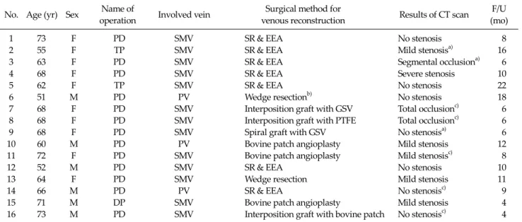
Fig. 3. Schematic illustrations of superior mesenteric vein (SMV) reconstruction with a spiral graft with a great saphenous vein. SMA, superior mesenteric artery; PV, por- tal vein.
and an occlusion of PTFE interposition graft. The first pa- tient a 63-year-old woman (Table 1, case no. 3) underwent segmental resection with end-to-end anastomosis of the SMV. On follow-up CT scan, a 4.5 cm segmental occlusion with severe stenosis was detected. Loculated fluid collec- tion was observed around the graft. The cause of occlusion was thought to be anastomotic tension and inflammation due to complicated fluid collection. But there was no clin- ical symptom associated with the stenosis of reconstruct- ed SMV.
The second patient, a 68-year-old woman (case no. 4) underwent segmental resection and end-to-end anasto- mosis of the SMV. Owing to immediate occlusion with thrombosis, further resection and reanastomosis were performed. Severe stenosis was detected on a CT scan 3 days after the operation. But there was no clinical symptom.
The third patient, a 68-year-old woman (case no. 7) un- derwent segmental resection of the 4-cm length SMV and reconstruction with a GSV interposition graft. Because of the size mismatch between the SMV and the GSV graft, bowel congestion developed. Therefore, an interposition graft with 8-mm PTFE (case no. 8) was made and delayed closure of abdominal wall was planned. Two days later, however, graft occlusion was observed during a second look operation. To overcome the size discrepancy and pos-
sible infectious complication, the PV was reconstructed with a spiral graft of GSV (case no. 9).
In all of these patients, thrombosis or stenosis was ob- served within 1 week after the operation. None of these pa- tients showed additional delayed graft failure during fol- low-up. With mean follow-up of 9.8 months (median, 8.5 months), the 6- and 12-month death-censored graft pa- tency rates were both 81.3%. Three patients died during the study period. The 6- and 12-month patient survival rates were 71.4% and 55.1%, respectively.
Risk factors
Of the 3 patients who experienced complicated fluid collection around the graft, one patient who underwent end-to-end anastomosis developed segmental occlusion with severe stenosis.
Postoperative anticoagulation therapy was done in 4 patients with low molecular weight heparin (LMWH) or antiplatelet agents. Patients underwent end-to-end anas- tomosis, patch angioplasty, and interposition graft with bovine patch were included and target veins remained patent. They were treated with LMWH for a week, fol- lowed by aspirin.
Two of 3 patients with vascular morbidity after re- construction underwent end-to-end anastomosis. The oth- er patients underwent interposition graft with PTFE fol-
lowed by GSV. All patients who underwent reconstruction with a bovine patch, spiral graft, or wedge resection with venoplasty remained patent.
DISCUSSION
Tumor invasion into the PV or SMV can be met occa- sionally during surgery for pancreatic cancer. Vascular surgeons are asked to reconstruct the vein appropriately, but it is quite difficult to decide the optimal surgical meth- od and conduit for each patient. When reconstruction with primary repair is impossible, a suitable size-matched con- duit is required. GSV is the most widely used autogenous vein graft, but it is too small in diameter to fit the PV or SMV segments. The use of prosthetic grafts is limited due to the concerns about infection, especially during con- taminated abdominal surgery.
Various types of autogenous veins have been used.
Fleming et al. [7] reported that the superficial femoral vein was an excellent size-matched conduit for reconstruction of the SMV or PV without serious complications asso- ciated with venous insufficiency in the leg. The patency of reconstruction of the PV or SMV using the femoral vein or GSV reported by Lee et al. [11] was 88% at mean follow-up of 5 months with only a few patients developing mild low- er leg edema. Suzuki et al. [10] demonstrated that re- construction of the inferior vena cava (IVC) or PV with the left renal vein was durable and safe method without ad- verse effects on early and long-term renal function. A case report described PV interposition with a cold-preserved homologous iliac vein graft obtained from a deceased do- nor [12]. To overcome size discrepancy, the gonadal vein had been customized by cutting longitudinally and sutur- ing into a sheet or tube-like graft [5]. Despite of many ef- forts, the best option for PV and SMV reconstruction dur- ing surgery for pancreatic cancer remains unclear.
Five types of surgical method were used in our patients, with resection of the involved venous segment and re- construction with primary repair being the most common.
But sometimes tension-free anastomosis was impossible.
Wedge resection with venoplasty was possible when only the lateral wall of the vein was invaded by the tumor.
When the proportion of the circumference of the sacrificed vein was extensive, there was a risk of stenosis. If the in- volvement was more than 20% of the circumference of the vein, we tried to use a bovine patch for lateral wall reconstruction. Three patients underwent bovine patch angioplasty and any occlusion or significant stenosis was not detected on follow-up CT scans. Bovine patches are widely used in the revascularization of the carotid or fem- oral artery, but have not been widely used in venous reconstruction. Only a few literatures commented the du- rability of the bovine patch in venous reconstruction. A study of IVC reconstruction in patients with renal cell car- cinoma found that 8 of 17 patients with bovine patch an- gioplasty remained patent at a mean follow-up of 18 months [13].
Bovine patches have many advantages for anastomosis, including easier handling, no need for additional incisions to harvest the GSV or superficial femoral vein, reasonable costs. Patches in various sizes are readily available for off-the-shelf use. They also have the advantage of strong durability, excellent biocompatibility, and low rate of in- fection [14,15]. One complication of bovine grafts, pseu- doaneurysm, thought to be caused by graft deterioration [16]. But this is expected to occur rarely in venous anasto- mosis due to reduced pressure and mechanical stress.
Little is known to date about the outcomes of venous re- pair with bovine grafts. Although the follow-up time of the present study was relatively short, bovine patch angio- plasty was found to be patent without symptoms.
Considering the advanced stages of the original tumors and limited life expectancy of the patients, prompt recov- ery after surgery seems to be more important to the patient than long-term patency of the target vein.
If the tumor involvement was more extensive (i.e., the involved segment was more than a half of the circum- ference), we tried to reconstruct the PV or SMV with inter- position graft. In arterial bypass of lower extremities, GSV and PTFE are the widely used conduits. However, GSV is too small to overcome the size-mismatch with the PV or SMV. Vascular surgeons are also reluctant to use PTFE graft during contaminated intra-abdominal surgery.
Although there were a few studies with acceptable results of PTFE graft in selected patients [1,2], the concerns for in-
fection and thrombosis in contaminated intra-abdominal surgery still exist. As an alternative, bovine patch or spiral graft can be used.
One of our patients underwent a spiral graft with a GSV for reconstruction of the SMV during pancreaticoduo- denectomy. Spiral saphenous venous grafts have been used as substitutes for large-diameter veins, such as the in- ternal jugular vein and vena cava [17,18]. Chiu et al. [6] rec- ommended that PV reconstruction with spiral graft could be considered prior to other methods. After calculating the required lengths, the harvested GSV can be wrapped around a chest tube and sutured to make a spiral compo- site [6]. This type of graft can be tailored to fit a vessel of any size and easy to handle. We utilized this method to sal- vage a failed interposition graft of the SMV. During the ini- tial operation, the SMV was reconstructed with a GSV, but severe bowel congestion developed due to size mismatch.
An interposition graft with PTFE was performed, and a second-look operation was planned. When the PTFE graft was found to be occluded, a spiral vein graft was per- formed, resulting in the restoration of portal flow and an immediate improvement of bowel edema. A spiral GSV graft may be an ideal conduit in such circumstances, over- coming the drawbacks of small-sized autogenous vein and prosthetic grafts. Despite intra-abdominal fluid col- lection and hematoma compressing the graft, it remained patent for 6 months in this patient.
Owing to the small sample size, our ability to determine the risk factors for vascular complications was limited.
However, our findings provide some clues about factors related to stenosis or thrombosis. Of the 2 patients who ex- perienced severe stenosis or thrombosis, one had intra-ab- dominal fluid collection around the reconstructed vein.
Stenosis was likely caused by adjacent localized fluid col- lection compressing the graft and inducing inflammation.
The other underwent reanastomosis immediately after the first procedure due to immediate thrombosis. Increased tension after further resection and reanastomosis was thought to be the cause of stenosis. Patients who under- went reconstruction with a spiral graft or bovine patch re- mained patent without stenosis. Primary repair could cause tension or stenosis in the reconstructed vein, espe- cially in patients with adjacent fluid collection. We think
angioplasty or interposition graft with bovine patch can be used easily and widely to prevent anastomotic tension and stenosis.
There are no standard guidelines for anticoagulation therapy in patients who undergo venous reconstruction.
In a study of the durability of 64 PV reconstructions by Smoot et al. [19], no significant difference in thrombosis rate was observed between who did and those did not re- ceive anticoagulation. Most patients remained patent without the use of warfarin or aspirin, and that anti- coagulation therapy did not seem to influence outcomes.
Because of the high flow and the absence of valves in the portomesenteric vein, the risk for thrombosis seemed to be low. However, endothelial injury occurred during surgery and cancer-related hypercoagulability may cause venous thrombosis. Comparative studies on outcomes in patients with and those without anticoagulation are needed to es- tablish a standardized protocol.
This study had several limitations. First, it involved a small number of patients, limiting statistical analysis. Second, the mean follow-up period was relatively short. However, since the expected survival of this group of patients is about 10 to 18 months, good results after 6 months are encouraging.
In conclusion, many of the involved vein segments dur- ing surgery for pancreatic cancer can be reconstructed with end-to-end anastomosis or wedge resection with venoplasty. When tension-free anastomosis or venoplasty without stenosis is doubtful, angioplasty with a bovine patch graft are good options to overcome the size mis- match or resultant stenosis. If the tumor involvement was more extensive interposition graft with bovine patch or spiral graft with a GSV can be tried. Revascularization methods and intra-abdominal complicated fluid collec- tion may be related to graft occlusion and severe stenosis.
Further follow-up of these patients and evaluation of ad- ditional patients are required to determine long-term graft patency and the risk factors for thrombosis or stenosis.
CONFLICTS OF INTEREST
No potential conflict of interest relevant to this article was reported.
REFERENCES
1. Stauffer JA, Dougherty MK, Kim GP, Nguyen JH. Interpo- sition graft with polytetrafluoroethylene for mesenteric and portal vein reconstruction after pancreaticoduodenec- tomy. Br J Surg 2009;96:247-52.
2. Chu CK, Farnell MB, Nguyen JH, Stauffer JA, Kooby DA, Sclabas GM, et al. Prosthetic graft reconstruction after por- tal vein resection in pancreaticoduodenectomy: a multi- center analysis. J Am Coll Surg 2010;211:316-24.
3. Hemming AW, Reed AI, Langham MR Jr, Fujita S, Howard RJ. Combined resection of the liver and inferior vena cava for hepatic malignancy. Ann Surg 2004;239:712-9.
4. DiPerna CA, Bowdish ME, Weaver FA, Bremner RM, Jabbour N, Skinner D, et al. Concomitant vascular proce- dures for malignancies with vascular invasion. Arch Surg 2002;137:901-6.
5. Yamamoto Y, Sakamoto Y, Nara S, Ban D, Esaki M, Shimada K, et al. Reconstruction of the portal and hepatic veins using venous grafts customized from the bilateral gonadal veins. Langenbecks Arch Surg 2009;394:1115-21.
6. Chiu KM, Chu SH, Chen JS, Li SJ, Chan CY, Chen KS.
Spiral saphenous vein graft for portal vein reconstruction in pancreatic cancer surgery. Vasc Endovascular Surg 2007;41:149-52.
7. Fleming JB, Barnett CC, Clagett GP. Superficial femoral vein as a conduit for portal vein reconstruction during pancreaticoduodenectomy. Arch Surg 2005;140:698-701.
8. Ohwada S, Hamada K, Kawate S, Sunose Y, Tomizawa N, Yamada T, et al. Left renal vein graft for vascular re- construction in abdominal malignancy. World J Surg 2007;
31:1215-20.
9. Sasaki R, Fujita T, Takeda Y, Hoshikawa K, Takahashi M, Funato O, et al. Portal vein reconstruction using a left renal vein graft for a patient with hilar cholangiocarcinoma.
Hepatogastroenterology 2007;54:1919-21.
10. Suzuki T, Yoshidome H, Kimura F, Shimizu H, Ohtsuka M, Kato A, et al. Renal function is well maintained after use of left renal vein graft for vascular reconstruction in hep- atobiliary-pancreatic surgery. J Am Coll Surg 2006;202:87- 92.
11. Lee DY, Mitchell EL, Jones MA, Landry GJ, Liem TK, Sheppard BC, et al. Techniques and results of portal vein/superior mesenteric vein reconstruction using femo- ral and saphenous vein during pancreaticoduodenectomy.
J Vasc Surg 2010;51:662-6.
12. Hwang S, Ha TY, Jung DH, Park JI, Lee SG. Portal vein in- terposition using homologous iliac vein graft during ex- tensive resection for hilar bile duct cancer. J Gastrointest Surg 2007;11:888-92.
13. Hyams ES, Pierorazio PM, Shah A, Lum YW, Black J, Allaf ME. Graft reconstruction of inferior vena cava for renal cell carcinoma stage pT3b or greater. Urology 2011;78:838-43.
14. Kim JH, Cho YP, Kwon TW, Kim H, Kim GE. Ten-year com- parative analysis of bovine pericardium and autogenous vein for patch angioplasty in patients undergoing carotid endarterectomy. Ann Vasc Surg 2012;26:353-8.
15. Ladowski JM, Ladowski JS. Retrospective analysis of bo- vine pericardium (Vascu-Guard) for patch closure in car- otid endarterectomies. Ann Vasc Surg 2011;25:646-50.
16. Lin PH, Bush RL, Lumsden AB. Successful stent-graft ex- clusion of a bovine patch-related carotid artery pseudo- aneurysm. J Vasc Surg 2003;38:396.
17. Leafstedt SW, Rubenstein RB, Pallanch JF, Wilder WH.
Spiral saphenous vein graft for replacement of internal jug- ular vein: a series of case reports. Angiology 1985;36:827- 31.
18. Gloviczki P, Pairolero PC, Cherry KJ, Hallett JW Jr. Recon- struction of the vena cava and of its primary tributaries: a preliminary report. J Vasc Surg 1990;11:373-81.
19. Smoot RL, Christein JD, Farnell MB. Durability of portal venous reconstruction following resection during pan- creaticoduodenectomy. J Gastrointest Surg 2006;10:1371-5.