Associating liver partition and portal vein ligation for staged hepatectomy (ALPPS) has recently been introduced as a new surgical technique to increase future liver remnant in patients with marginal liver volume contemplating major liver resection. We herein present two cases. Case 1: a 68-year-old male patient with colorectal liver metastasis was referred to our department. The future liver remnant (FLR) was 22%. We performed first-stage ALPPS and colorectal surgery concurrently and second stage operation was performed 8 days later. The patient discharged 28 days after the first-stage ALPPS procedure. Case 2: a 69-year-old male patient with a huge hepatic mass was referred for hepatic surgery. The FLR was 19%. After the first stage of the ALPPS procedure, acute renal failure and posthepatectomy liver failure occurred. The patient began to recover on the 5th postoperative day. At 10 days after the first stage, the patient completed the second-stage procedure. The patient discharged 23 days after the first-stage ALPPS procedure. So far many studies are currently underway to identify factors associated with the morbidity and mortality of the ALPPS procedure, it is necessary to continue follow-up studies and observe the results. (Ann Hepatobiliary Pancreat Surg 2017;21:151-156)
Key Words: Posthepatectomy liver failure; Future liver remnant; Staged hepatectomy
Received: July 21, 2017; Revised: August 2, 2017; Accepted: August 11, 2017 Corresponding author: Dong Hoon Shin
Department of Surgery, Kosin University College of Medicine, 34 Amnam-dong, Seo-gu, Busan 602-703, Korea Tel: +82-51-990-6462, Fax: +82-51-246-6093, E-mail: [email protected]
Copyright Ⓒ 2017 by The Korean Association of Hepato-Biliary-Pancreatic Surgery
This is an Open Access article distributed under the terms of the Creative Commons Attribution Non-Commercial License (http://creativecommons.org/
licenses/by-nc/4.0) which permits unrestricted non-commercial use, distribution, and reproduction in any medium, provided the original work is properly cited.
Annals of Hepato-Biliary-Pancreatic Surgery ∙ pISSN: 2508-5778ㆍeISSN: 2508-5859
INTRODUCTION
Surgical resection is the only treatment for patients with primary or metastatic hepatic malignancies. The treatment is especially recommended in the absence of extrahepatic malignancies. However, this approach is limited by the amount of functional future liver remnant (FLR) after hepatic resection. Surgeons want to preserve adequate FLR to avoid posthepatectomy liver failure. Removal of up to 75% of the total liver volume is feasible in a young patient with normal liver function. In the cases of under- lying liver disease, such as chemotherapy-related injury or cirrhosis, an FLR of 40% or greater is required.1,2
Associating liver partition and portal vein ligation for staged hepatectomy (ALPPS) has been introduced re- cently, as a new surgical technique to increase FLR in pa- tients with marginal liver volume contemplating major liv- er resection.3 The main indication for ALPPS is extensive
bilobar colorectal liver metastases with an FLR less than 25%.4
The researchers hereby report two cases of ALPPS. The first case concerns simultaneous ALPPS and colorectal surgery, for a patient with extensive bilobar colon cancer liver metastases and a small predicted FLR. The second case concerns a patient with a mass larger than 15 centi- meters, who underwent an ALPPS procedure.
CASE
Case 1
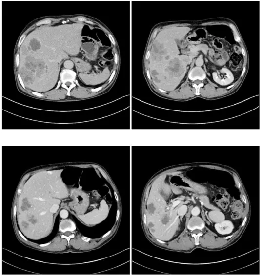
A healthy and asymptomatic 68-year-old male patient was referred to our institution with synchronous hepatic metastasis of left colon cancer. The initial imaging studies showed seven metastases in the right lobe of liver and two metastases in the left lobe. The largest, which was in seg- ment 6, measured 6.5 cm×5.5 cm×6 cm (Fig. 1). The
Fig. 1. Preoperative computed tomography shows the largest metastasis located at the seg- ment 6, measuring 6.5 cm×5.5 cm×6 cm.
Fig. 2. Preoperative computed tomography after 4 months, shows that the sizes of the hep- atic metastases decreased, with the largest one measuring 3.5 cm×3 cm.
physicians of the department of gastroenterology planned treatment with combination of bevacizumab and FOLFIRI (5-fluorouracil, leucovorin, and irinotecan) therapy. Over four months, the patient received seven cycles of bev- acizumab and FOLFIRI combination therapy. After four months, the sizes of the hepatic metastases were de- creased, with the largest one measuring 3.5 cm×3 cm (Fig.
2). To achieve hepatic clearance, a right hemihepatectomy with partial hepatectomy in left lobe was necessary.
The total liver volume (TLV) was 1,536 ml, the volume of the left lobe, and the FLR volume was 340 ml (22%).
Liver function evaluation revealed albumin 3.9 g/dl, crea- tinine 1.25 mg/dl, international normalized ratio (INR) 0.89, serum bilirubin 0.40 mg/dl, and indocyanine green retention rate at 15 minutes (ICG-R15) 11.5%. The patient had no ascites and a platelet count of 272×103/mm3. Therefore, the researchers decided to achieve sufficient hypertrophy through ALPPS procedure. Three weeks after
the last cycle of chemotherapy, the first stage of ALPPS with left hemicolectomy was performed.
The operation started with a left hemicolectomy for left colon cancer, followed by ALPPS. After identification of the portal structures, the right portal vein was transected.
The right hepatic artery and right hepatic vein were marked with vessel loops. The parenchyma was trans- ected, through a demarcated line, for the right hemiliver and the right border of the middle hepatic vein.
Enucleation was performed for lesions on the left lobe.
Blood loss in the first stage was 650 ml (Figs. 3 and 4).
Computed tomography (CT) volumetry on day 7 after the first stage revealed 35% FLR. Therefore, we per- formed second-stage ALPPS on day 8 following first-stage ALPPS (Fig. 5). There were no intraoperative complica- tions and the blood loss was less than 50 ml. The post- operative histology revealed seven metastatic ad- enocarcinomas, with the largest one measuring 5.5 cm×3.3
Fig. 6. Gross photograph of the specimen revealed seven meta- static adenocarcinomas, with the largest one measuring 5.5 cm×3.3 cm.
Fig. 5. Computed tomography volumetry on day 7, after the first-stage operation revealed future liver remnant volume was 35% of the total liver volume.
Fig. 4. Intraoperative photograph shows that the right hepatic artery and right hepatic vein were marked with vessel loops.
Fig. 3. Intraoperative photograph shows the extents of liver resection were marked.
gastric carcinoma and histologic type was hepatoid adenocarcinoma. The cancer stage was T2N0M0, stage I.
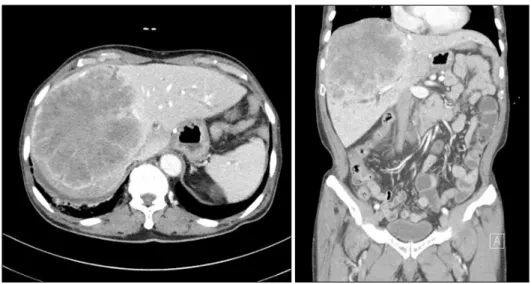
A contrast-enhanced CT revealed a large tumor, measur- ing 16 cm×15 cm in segments 5-8, middle hepatic vein invasion, and no signs of liver cirrhosis. Liver function evaluation revealed albumin at 3.6 g/dl, creatinine at 0.66 mg/dl, international normalized ratio (INR) of 1.18, and
Fig. 7. Computed tomography volumetry showed that the total liver volume was 2,044 ml and the future liver remnant volume was 385 ml (19% of the total liver volume).
Fig. 8. Abdominal CT showed significant hypertrophy of the left lateral section.
serum bilirubin at 1.08 mg/dl. The patient had no ascites, no encephalopathy, and a platelet count of 272×103/mm3. The alpha-fetoprotein (AFP) level was 10,264 ng/ml and the prothrombin induced by vitamin K absence-II (PIVKA-II) level was 2,869 mAU/ml. The total liver vol- ume was 2,044 ml, and the FLR volume was 385 ml (19%) (Fig. 7). Based on these findings, the researchers decided to achieve sufficient hypertrophy through an ALPPS procedure.
After a preoperative evaluation, the patient underwent surgery. During the first-stage operation, the right portal vein was dissected and divided with a vascular stapler.
Transection of the liver parenchyma was performed at 1 cm on the right side of the falciform ligament, using a Cavitron ultrasonic surgical aspirator. An intermittent Pringle maneuver was performed during the parenchyma transection. The middle hepatic vein was divided during the transection, preserving the right and left hepatic veins.
Blood loss during the first-stage ALPPS procedure was 1,500 ml. After the first stage of the ALPPS procedure, acute renal failure and posthepatectomy liver failure oc- curred and continuous renal replacement therapy was necessary. The patient began to recover on the 5th post- operative day.
At nine days postoperatively, an abdominal CT showed significant hypertrophy of the left lateral section (Fig. 8).
The second operation started with the same abdominal in- cision and warm saline irrigation. The plastic bag was re- moved from the right extended lobe of liver. The right hepatic artery was then ligated and divided. The right bile duct and right hepatic vein were divided using vascular
staplers. Two silicone drains were placed and the abdo- men was closed.
Histopathology confirmed a 17 cm×14 cm×11 cm-sized probably metastatic hepatoid adenocarcinoma, from the stomach and presence of vascular invasion (Fig. 9). After the second procedure, the patient developed moderate as- cites, mild elevation of serum bilirubin (3.51 mg/dl), and mild prolongation of prothrombin time (21.1 sec). The re- maining postoperative course was uneventful and the pa- tient was discharged 23 days, after the first procedure.
DISCUSSION
ALPPS has recently been developed to induce rapid liv- er hypertrophy to reduce posthepatectomy liver failure. In 2012, Schnitzbauer et al.3 reported that the technique of
Fig. 9. Histopathology con- firmed a 17 cm×14 cm×11 cm probably metastatic hepatoid ad- enocarcinoma from the stomach.
“right portal vein ligation combined with in situ splitting, induces rapid left lateral liver lobe hypertrophy enabling 2-staged extended right hepatic resection in small-for-size settings”. Also in 2012, de Santibañes and Clavien pro- posed the acronym ALPPS, for this novel surgical techni- que of associating liver partition and portal vein ligation for staged hepatectomy.5 ALPPS is still considered to be in an early developmental phase and its surgical in- dications and techniques have not yet been standardized.
The study’s second case showed posthepatectomy liver failure and acute renal failure, after the first stage of ALPPS. Although these recovered after postoperative day 5, they were considered complications of Clavien-Dindo grade III or more.6 Some clinicians oppose the ALPPS procedure, due to its high morbidity and mortality. The ini- tial experience of ALPPS was impeded by a high incidence of postoperative complications such as bile leakage, in- fections, and high mortality.7 Although the short-term re- sults of ALPPS are improving due to the development of improved surgical techniques and accumulated experience, morbidity rates after ALPPS are 15.3-100%. These rates are exhibited with ≥Clavien-Dindo grade III morbidity rates of 13.6-44%. Mortality rates are reported to be 0-29%.6
Since many studies are currently underway to identify the factors associated with the morbidity and mortality of the ALPPS procedure, it is necessary to follow-up with additional studies. The first report of the International ALPPS Registry presented a 90-day mortality of 9% and a severe complication rate (Clavien-Dindo grade IIIb) of 27% in 202 patients. However, postoperative outcomes were significantly better in patients younger than 60 years and in those with colorectal liver metastasis (CRLM).4
In the first case, the researchers successfully performed ALPPS and colorectal surgery concurrently in a patient over 60 years of age. However, Kerollos et al. reported a severe complication rate of 23%, and 90-day mortality of 15% in a study of both ALPPS and colorectal surgery.8 The use of ALPPS for CRLM is expected to increase.
Since ALPPS surgery for CRLM is performed in patients with no steatosis and no cirrhotic liver, liver hypertrophy and functional recovery after liver resection should be su- perior to those exhibiting cirrhotic liver. Research to reach consensus on a standardized surgical technique is neces- sary to improve results, after simultaneous ALPPS and colorectal surgery. Evidence-based criteria are essential to realize the potential advantages of ALPPS without in- creased morbidity or mortality.
REFERENCES
1. Loss M, Jung EM, Scherer MN, Farkas SA, Schlitt HJ. Surgical treatment of liver metastases. Chirurg 2010;81:533-541.
2. Clavien PA, Petrowsky H, DeOliveira ML, Graf R. Strategies for safer liver surgery and partial liver transplantation. N Engl J Med 2007;356:1545-1559.
3. Schnitzbauer AA, Lang SA, Goessmann H, Nadalin S, Baumgart J, Farkas SA, et al. Right portal vein ligation combined with in situ splitting induces rapid left lateral liver lobe hypertrophy en- abling 2-staged extended right hepatic resection in small-for-size settings. Ann Surg 2012;255:405-414.
4. Schadde E, Ardiles V, Robles-Campos R, Malago M, Machado M, Hernandez-Alejandro R, et al. Early survival and safety of ALPPS: first report of the International ALPPS Registry. Ann Surg 2014;260:829-836; discussion 836-838.
5. de Santibañes E, Alvarez FA, Ardiles V. How to avoid post- operative liver failure: a novel method. World J Surg 2012;36:
125-128.
6. Lau WY, Lai EC, Lau SH. Associating liver partition and portal vein ligation for staged hepatectomy: the current role and development. Hepatobiliary Pancreat Dis Int 2017;16:17-26.
7. Schadde E, Ardiles V, Slankamenac K, Tschuor C, Sergeant G,
Amacker N, et al. ALPPS offers a better chance of complete re- section in patients with primarily unresectable liver tumors com- pared with conventional-staged hepatectomies: results of a multi- center analysis. World J Surg 2014;38:1510-1519.
8. Wanis KN, Buac S, Linecker M, Ardiles V, Tun-Abraham ME, Robles-Campos R, et al. Patient survival after simultaneous ALPPS and colorectal resection. World J Surg 2017;41:1119-1125.