ISSN 2234-3806 • eISSN 2234-3814
http://dx.doi.org/10.3343/alm.2015.35.3.379 www.annlabmed.org 379
Ann Lab Med 2015;35:379-381
http://dx.doi.org/10.3343/alm.2015.35.3.379
Letter to the Editor
Clinical Microbiology
First Case of Pulmonary Mycobacterium
parascrofulaceum Infection in a Patient With Bronchiectasis in Korea
Kyoung-Bo Kim, M.D.1, Sung-Gyun Park, M.D.1, Jae-Seok Park, M.D.2, Wonmok Lee, M.D.1, Jung-Sook Ha, M.D.1, Nam-Hee Ryoo, M.D.1, Dong-Seok Jeon, M.D.1, and Jae-Ryong Kim, M.D.1
Departments of Laboratory Medicine1 and Internal Medicine2, Keimyung University School of Medicine, Daegu, Korea
Dear Editor
Mycobacterium parascrofulaceum is a member of slow-growing scotochromogenic nontuberculous mycobacteria (NTM) that can cause opportunistic infections in immunocompromised patients [1]. Only a few clinical infections with this strain have been re- ported to date [1-4]. We describe a case of M. parascrofulaceum pulmonary infection in a patient with bronchiectasis. To our knowledge, this is the first case of M. parascrofulaceum infection in Korea.
A 65-yr-old man was referred to our hospital because of recur- rent hemoptysis and prulent sputum for two days. He had a his- tory of stomach cancer that had been treated by using total gas- trectomy with Billroth II anastomosis and chemotherapy seven years ago; at that time, he was diagnosed as having bronchiecta- sis. Our laboratory tests revealed peripheral blood leukocyte counts of 6.48×10⁹/L with 81.2% neutrophils. Chest computed tomography revealed no interval changes in the preexisting bron- chiectasis of the lower left lobe of the lung. Nonetheless, new peribronchial infiltration and centrilobular nodules were found in the middle right lobe and lower left lobe. Acid-fast bacillus (AFB) staining, qualitative real-time PCR for Mycobacterium tuberculo- sis (COBAS TaqMan MTB Test; Roche, Basel, Switzerland), and
nested PCR for NTM (MTB & NTM PCR Kit; BioSewoom Inc., Seoul, Korea) in the expectorated-sputum specimens all tested negative. Sputum cultures in liquid media were performed in triplicate by using the Mycobacteria Growth Indicator Tube Sys- tem (MGIT, Becton, Dickinson and Company, Franklin Lakes, NJ, USA), and positive results were obtained from two cultures.
For species identification, a PCR-based reverse line blot hybrid- ization assay for the ITS gene (AdvanSure Mycobacteria GenoB- lot Assay; LG Life Sciences, Seoul, Korea) was performed on both culture isolates, revealing poor discrimination of species (Mycobacterium lentiflavum/Mycobacterium genavense).
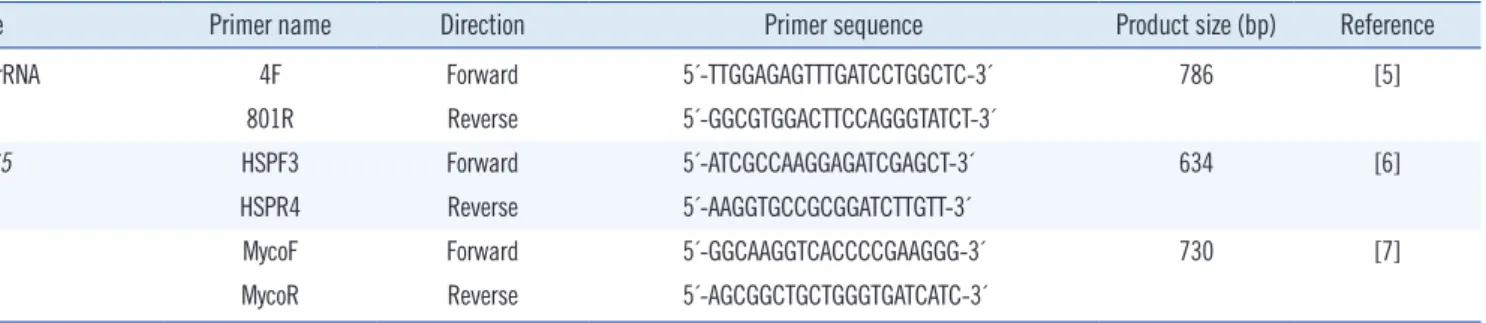
For definitive species identification, sequencing analysis of the 16S rRNA, hsp65, and rpoB genes was performed. Primers used for PCR are shown in Table 1 [5-7]. 16S rRNA sequences ob- tained were searched against publicly available databases by us- ing the basic local alignment search tool (BLAST) [8] and Ez- Taxon [9]. When the sequences were submitted to BLAST, both culture isolates revealed 99.74% similarity (767/769 bp) with M.
parascrofulaceum HSC68. The next closest match was M. para- scrofulaceum ATCC BAA-614 with similarity of 99.61% (776/779 bp). In EzTaxon, the isolates exhibited 99.36% similarity (773/778 bp) with M. parascrofulaceum ATCC BAA-614 followed by M. eu-
Received: August 13, 2014
Revision received: November 10, 2014 Accepted: February 7, 2015
Corresponding author: Wonmok Lee
Departments of Laboratory Medicine, Keimyung University School of Medicine, 56 Dalseong-ro, Jung-gu, Daegu 700-712, Korea Tel: +82-53-250-7733, Fax: +82-53-250-7275
E-mail: [email protected]
© The Korean Society for Laboratory Medicine.
This is an Open Access article distributed under the terms of the Creative Commons Attribution Non-Commercial License (http://creativecommons.org/licenses/by-nc/3.0) which permits unrestricted non-commercial use, distribution, and reproduction in any medium, provided the original work is properly cited.
Kim K-B, et al.
Pulmonary M. parascrofulaceum infection
380 www.annlabmed.org http://dx.doi.org/10.3343/alm.2015.35.3.379 ropaeum FI-95228 with similarity of 98.94% (750/758 bp). In the
BLAST search, sequences of hsp65 and rpoB genes revealed 99.83% (602/603 bp) and 99.32% (740/745 bp) similarity with M. parascrofulaceum, respectively. The other best matches were Mycobacterium sp. MOTT-01 (99.83%, 602/603 bp) and Myco- bacterium sp. MOTT-27 (99.06%, 739/746 bp), respectively. A phylogenetic tree, which was constructed using the neighbor- joining method by means of the MEGA software, version 5.05 (http://www.megasoftware.net), identified this isolate as M. para- scrofulaceum (Fig. 1). Although there was a possibility of coloni- zation of the respiratory tract, the patient was diagnosed as hav-
ing M. parascrofulaceum pulmonary infection according to his newly developed radiological images. At diagnosis, he had only a mild cough with no progression according to chest radiography.
Therefore, he did not receive long-term antibiotic therapy. Addi- tional sputum cultures one year after the initial sputum analysis yielded negative results. Currently, he exhibits no symptomatic aggravation and regularly visits our hospital (and does not take medication). Therefore, it is possible that this isolate, M. para- scrofulaceum, caused a transient infection rather than a progres- sive disease or colonization. We need long-term follow-up and re- peat sputum analysis to confirm either the progressive disease or Table 1. Primers and amplicon sizes for PCR analysis of the 16s rRNA, hsp65, and rpoB genes
Gene Primer name Direction Primer sequence Product size (bp) Reference
16s rRNA 4F Forward 5´-TTGGAGAGTTTGATCCTGGCTC-3´ 786 [5]
801R Reverse 5´-GGCGTGGACTTCCAGGGTATCT-3´
hsp65 HSPF3 Forward 5´-ATCGCCAAGGAGATCGAGCT-3´ 634 [6]
HSPR4 Reverse 5´-AAGGTGCCGCGGATCTTGTT-3´
rpoB MycoF Forward 5´-GGCAAGGTCACCCCGAAGGG-3´ 730 [7]
MycoR Reverse 5´-AGCGGCTGCTGGGTGATCATC-3´
Fig. 1. The neighbor-joining phylogenetic tree based on the 16S rRNA gene sequences of our Mycobacterium parascrofulaceum isolate and 19 similar microorganisms. The scale bar corresponds to 0.2% sequence divergence.
Kim K-B, et al.
Pulmonary M. parascrofulaceum infection
http://dx.doi.org/10.3343/alm.2015.35.3.379 www.annlabmed.org 381
transient infection.
There are >150 species comprising the genus Mycobacte- rium, with a significant increase in the number of newly identi- fied species recently. The latter phenomenon can be attributed in part to development of molecular techniques enabling accurate identification via detection of differences in the 16S rRNA gene [10]. M. parascrofulaceum is one of the species discovered re- cently after sequence-based identification and was previously known as the “MCRO 33” group of mycobacteria (GenBank ac- cession No. AF152559). Discrimination of M. parascrofulaceum from other NTM is difficult because it shows phenotypic charac- teristics similar to those of Mycobacterium scrofulaceum and is genotypically close to Mycobacterium simiae [1]. Direct 16S rRNA gene sequence analysis is a powerful tool for confirming this strain; additional sequence analysis of the hsp65, rpoB, and ITS genes may be useful for unequivocal identification [10].
As the genetic diversity of NTM continues to be elucidated and new species are discovered, more accurate and specific di- agnostic tests will be necessary for NTM identification. Multigene sequence analysis may be needed to discriminate NTM species accurately.
Authors’ Disclosures of Potential Conflicts of Interest
No potential conflicts of interest relevant to this article were re- ported.
REFERENCES
1. Turenne CY, Cook VJ, Burdz TV, Pauls RJ, Thibert L, Wolfe JN, et al. My- cobacterium parascrofulaceum sp. nov., novel slowly growing, scoto- chromogenic clinical isolates related to Mycobacterium simiae. Int J Syst Evol Microbiol 2004;54:1543-51.
2. Tortoli E, Chianura L, Fabbro L, Mariottini A, Martín-Casabona N, Maz- zarelli G, et al. Infections due to the newly described species Mycobac- terium parascrofulaceum. J Clin Microbiol 2005;43:4286-7.
3. Teruya H, Tateyama M, Hibiya K, Tamaki Y, Haranaga S, Nakamura H, et al. Pulmonary Mycobacterium parascrofulaceum infection as an im- mune reconstitution inflammatory syndrome in an AIDS patient. Intern Med 2010;49:1817-21.
4. Liu G, Wang GR, Yu X, Liang Q, Mu J, Shang YY, et al. Bacteriological characterization of a Mycobacterium parascrofulaceum strain isolated from a Chinese pneumonia patient. Int J Infect Dis 2014;25:82-7.
5. Simmon KE, Mirrett S, Reller LB, Petti CA. Genotypic diversity of anaer- obic isolates from bloodstream infections. J Clin Microbiol 2008;46:
1596-601.
6. Kim H, Kim SH, Shim TS, Kim MN, Bai GH, Park YG, et al. Differentia- tion of Mycobacterium species by analysis of the heat-shock protein 65 gene (hsp65). Int J Syst Evol Microbiol 2005;55:1649-56.
7. Adékambi T, Colson P, Drancourt M. rpoB-based identification of non- pigmented and late-pigmenting rapidly growing mycobacteria. J Clin Microbiol 2003;41:5699-708.
8. Altschul SF, Madden TL, Schäffer AA, Zhang J, Zhang Z, Miller W, et al.
Gapped BLAST and PSI-BLAST: a new generation of protein database search programs. Nucleic Acids Res 1997;25:3389-402
9. Chun J, Lee JH, Jung Y, Kim M, Kim S, Kim BK, et al. EzTaxon: a web- based tool for the identification of prokaryotes based on 16S ribosomal RNA gene sequences. Int J Syst Evol Microbiol 2007;57:2259-61.
10. Clinical and Laboratory Standards Institute. Interpretive criteria for iden- tification of bacteria and fungi by DNA target sequencing. Approved guideline, MM18-A. Wayne, PA: Clinical and Laboratory Standards In- stitute, 2008.