Effects of Combination Therapy of Alendronate and Hormonal Therapy on Bone Mineral Density in Postmenopausal Korean Women: Multicenter, Randomized Controlled Clinical Trial
This study evaluated the effects of combination treatment with alendronate (ALEN) and hormone therapy (HT) on bone mineral density (BMD) in postmenopausal Korean women.
This multicenter, randomized, controlled clinical trial enrolled 344 postmenopausal women with low BMD. The women received HT (0.625 mg/day of conjugated equine estrogen and 2.5 mg/day of medroxyprogesterone acetate) alone or in combination with ALEN (10 mg/
day) for 1 year. Changes in BMD and biochemical markers of bone turnover were evaluated. Data from 203 women (HT alone, 99; combination treatment, 104) who completed this study were analyzed. BMD at the lumbar spine and total hip increased significantly in both treatment groups after 1 year. There were no significant differences between HT alone vs. the combination of ALEN and HT in mean BMD increase at the lumbar spine (6.9% vs. 7.9%) and total hip (3.7% vs. 3.8%). Combined therapy suppressed serum osteocalcin and urinary deoxypyridinoline to a greater extent than HT alone. In conclusion, compared to HT alone, combination treatment with ALEN and HT for 1 year did not offer a benefit in BMD in postmenopausal Korean women with low BMD.
Keywords: Osteoporosis; Bone Mineral Density; Combination Therapy; Alendronate;
Hormone Therapy Byung-Koo Yoon,1* Dong-Yun Lee,1*
Man Chul Park,2 Soo Hyun Cho,3 Hyoung Moo Park,4 and Young Min Choi5,6
1Department of Obstetrics and Gynecology, Sungkyunkwan University School of Medicine, Seoul, Korea; 2Department of Obstetrics and Gynecology, Hallym University College of Medicine, Anyang, Korea; 3Department of Obstetrics and Gynecology, Hanyang University School of Medicine, Seoul, Korea; 4Department of Obstetrics and Gynecology, Chung-Ang University College of Medicine, Seoul, Korea; 5Department of Obstetrics and Gynecology, Seoul National University College of Medicine, Seoul, Korea; 6The Institute of Reproductive Medicine and Population, Medical Research Center, Seoul National University College of Medicine, Seoul, Korea
* Byung-Koo Yoon and Dong-Yun Lee contributed equally to this work.
Received: 21 September 2016 Accepted: 19 February 2017 Address for Correspondence:
Young Min Choi, MD
Department of Obstetrics and Gynecology, Seoul National University College of Medicine, 103 Daehak-ro, Jongno-gu, Seoul 03080, Korea
E-mail: [email protected]
Funding: This work was supported in part by a grant from the Seoul National University Hospital Research Fund (06-1999- 055), the Samsung Medical Center Research Funds (PH01991081 and PHO1133361), and the IN-SUNG Foundation for Medical Research (C-A5-811-1).
https://doi.org/10.3346/jkms.2017.32.6.992 • J Korean Med Sci 2017; 32: 992-998
INTRODUCTION
Estrogen insufficiency is the main cause of involutional osteoporosis in women. In- creased bone turnover and an imbalance between bone resorption and formation re- sults in accelerated loss of bone mass in early postmenopausal women. The ensuing secondary hyperparathyroidism and decrease in bone formation are also associated with estrogen deficiency (1).
Hormone therapy (HT) prevents bone loss, improves bone mineral density (BMD) (2,3), and reduces the incidence of vertebral and non-vertebral fractures in both osteo- penic and osteoporotic women (4-6). HT is an established treatment for osteoporosis in postmenopausal women. Bisphosphonate, which inhibits osteoclast activity and decreases bone turnover at sites of bone resorption, is another established treatment for osteoporosis (7,8). However, the mechanisms of actions for bone health are some- what different; whereas bisphosphonate predominantly acts on osteoclasts (9), estro- gen also has effects on osteoblasts and osteocytes (10). It has been suggested that es- trogen might have an anabolic effect on bone (11,12), and also has favorable effects on bone quality (13).
Suboptimal or no response to treatment is an important issue in clinical practice that may result from poor adherence to treatment, co-morbidities, insufficient calcium and vitamin D, malabsorption, erroneous dose or interval, or lack of drug efficacy (14). In- deed, 20%–30% of postmenopausal women may experience bone loss even with HT (15). Therefore, in some situations, such as severe osteoporosis or failure to achieve an optimal response to either HT or bisphosphonate alone, additional benefit from a com- bination of the 2 treatments might be expected because of their different mechanisms Obstetrics & Gynecology
2017-03-16 https://crossmark-cdn.crossref.org/widget/v2.0/logos/CROSSMARK_Color_square.svg
of action. Indeed, the combination of HT and bisphosphonate produced a greater increase in BMD over either treatment alone in several studies (16-19). However, data on the effect of combi- nation therapy are mixed (16-23). Moreover, the efficacy of the combined therapy for fracture prevention has not yet been proven.
Ethnic differences in treatment response might exist. As Asians usually have lower BMD than Caucasians, mainly due to their smaller bone size (19), osteoporosis treatment could produce stronger effects on BMD in Asian women than in Caucasians.
To date, few randomized studies combining bisphosphonate with HT have been reported in Asian countries (24). In a recent study, no difference in BMD gain was reported by the addition of alendronate (ALEN) for 1 year to ongoing HT before ALEN use in Korean women (25). The current study was conducted to evaluate the effects of combining ALEN with HT in postmeno- pausal Korean women who had low BMD.
MATERIALS AND METHODS Study participants
A total of 344 postmenopausal women (mean age, 59.1 years) participated in this multicenter clinical trial conducted at 5 uni- versity hospitals from September 1999 to June2003. Women were considered postmenopausal if duration of amenorrhea was ≥ 12 months or if the serum level of follicle stimulating hormone was
> 40 IU/L.
Only women with BMD at least 2 standard deviations (SD) lower at the lumbar spine or total hip compared with the mean bone mass of normal young Korean women by dual-energy X- ray absorptiometry (DXA) were considered for inclusion. Wom- en were excluded from the study if they had a history of diseas- es or if they were taking medications (including HT or ALEN) that might affect bone metabolism within 1 year before enroll- ment. Women were also excluded if they had contraindications for ALEN or HT.
Study design
Patients were randomly assigned to receive either HT alone (173 patients) or HT + ALEN (171 patients) for one year in a 1:1 ratio.
The allocation of treatment was based on randomization codes created by SAS program (SAS Institute, Cary, NC, USA) within the same study center. No other specific randomization stratifi- cation factor was applied. All women received 0.625 mg/day of conjugated equine estrogen (CEE; Pfizer Inc., Seoul, Korea) and 2.5 mg of medroxyprogesterone acetate (MPA; Pfizer Inc.). In the combination group, 10 mg/day of ALEN (Fosamax; MSD, Seoul, Korea) was given immediately after the patient woke up.
Participants were educated to take ALEN with plenty of plain water, and to maintain an upright position for at least 30 min- utes afterwards. Calcium supplementation (CaCO3, 500 mg bid) and regular exercise were also encouraged.
The primary end point for efficacy was the change in lumbar spine BMD. The secondary end point was the change in total hip BMD and biochemical markers of bone turnover.
BMD
BMD was measured at the second to fourth vertebrae of the lum- bar spine and at the hip by DXA at each hospital. Bone densitom- etry was performed at study enrollment and after 12 months of treatment using the same device.
Biochemical markers
Samples were collected at 0, 3, 6, and 12 months in the morning after an overnight fast. As a marker of bone formation, serum osteocalcin (OC) was measured using an enzyme-linked immu- nosorbent assay (ELISA) kit. Urinary deoxypyridinoline (DPD), a marker of bone resorption, was assessed using an ELISA kit and corrected for creatinine level.
Statistical analysis
Statistical analyses were performed using Predictive Analytics SoftWare (PASW) statistics 20 (SPSS Inc., Chicago, IL, USA). At least 224 patients were required to achieve a power of 80% and an alpha of 0.05 to detect a 1.5% difference in the mean change in BMD at the lumbar spine. Considering dropping out of the study, it was initially aimed to enroll a total of 350 participants (70 for each center) into the study.
Data are shown as the mean ± SD or number (percent). T- tests, χ2 test, or Fisher exact test were used to compare the base- line characteristics, the proportion of participants with no BMD increase, and adverse effects. Changes in BMD within and be- tween the groups were evaluated by paired or Student’s t-tests, respectively. In addition, t-tests and repeated-measures analy- sis of variance were used to evaluate the changes in bone turn- over markers within and between the groups. Correlations of changes in BMD between the 2 groups were evaluated using regression analysis after adjusting for age, reproductive history, body mass index (BMI), history of HT, and baseline BMD. A P value < 0.05 was considered statistically significant.
Ethics statement
The present study protocol was reviewed and approved by the Institutional Review Board of Samsung Medical Center (No. 1999- 12-04). Informed consent was submitted by all subjects when they were enrolled. This trial was not registered because the reg- istry system was not available when it was investigated.
RESULTS
Characteristics of study participants
Among 344 postmenopausal women who were enrolled, 203 (59%) completed this study including 99 in the HT alone group
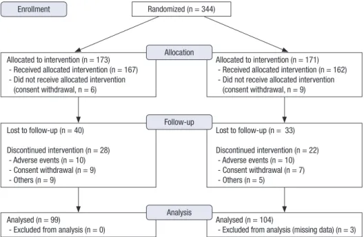
(57.2%) and 104 in the HT and ALEN group (60.8%) (Fig. 1). The dropout rate was comparable between groups. The baseline cha- racteristics of the participants are shown in Table 1. There was no statistical difference in any of the variables analyzed between the 2 groups, and the characteristics were not different between women who dropped out and those who completed treatment.
We analyzed data for women who completed the study protocol.
BMD changes
BMD at the lumbar spine and total hip increased significantly after 1 year of treatment in both the HT alone and combination group. The 2 groups did not show a statistically significant dif-
Table 1. Baseline characteristics of participants
Characteristics Complete (n = 203) Drop-out (n = 142)
HT alone (n = 99) HT + ALEN (n = 104) P HT alone (n = 74) HT + ALEN (n = 68) P
Age, yr 58.8 ± 5.9 59.4 ± 6.4 0.502 58.7 ± 7.2 60.5 ± 6.1 0.256
Age at menarche, yr 16.5 ± 2.0 16.8 ± 2.0 0.565 16.7 ± 1.5 16.4 ± 1.6 0.459
Age at menopause, yr 49.2 ± 4.3 48.6 ± 4.8 0.346 49.8 ± 3.9 49.0 ± 6.0 0.376
Years since menopause, yr 9.3 ± 6.7 10.4 ± 6.9 0.270 8.8 ± 7.9 11.5 ± 9.0 0.199
Parity, No. 3.2 ± 1.6 3.2 ± 1.4 0.941 3.3 ± 1.3 3.4 ± 1.3 0.688
BMI, kg/m2 23.8 ± 2.9 23.2 ± 3.1 0.235 23.7 ± 3.2 24.8 ± 2.6 0.156
Type of menopause 0.330 0.588
Surgical 15 (15.2) 11 (10.6) 11 (14.9) 8 (11.8)
Natural 84 (84.8) 93 (89.4) 63 (85.1) 60 (88.2)
History of HT 0.402 0.964
Never 80 (80.8) 79 (76.0) 59 (79.7) 54 (79.4)
Ever 19 (19.2) 25 (24.0) 15 (20.3) 14 (20.6)
BMD, g/cm2
Lumbar spine 2–4 0.790 ± 0.085 0.776 ± 0.095 0.284 0.783 ± 0.136 0.792 ± 0.110 0.671
Total hip 0.755 ± 0.109 0.728 ± 0.131 0.127 0.734 ± 0.137 0.745 ± 0.106 0.702
T-score of BMD
Lumbar spine 2–4 −2.6 ± 0.7 −2.7 ± 0.8 0.367 −2.7 ± 1.1 −2.7 ± 1.0 0.825
Total hip −1.4 ± 0.7 −1.6 ± 0.8 0.089 −1.4 ± 1.0 −1.5 ± 0.8 0.651
Bone turnover marker
OC, ng/mL 17.1 ± 12.1 15.2 ± 10.4 0.251 14.2 ± 10.6 15.0 ± 9.2 0.659
DPD, nM/mMCr 7.9 ± 3.2 7.9 ± 3.3 0.998 8.7 ± 5.7 8.4 ± 4.3 0.723
Data are presented as mean ± SD or number of participants (%).
HT = hormone therapy, ALEN = alendronate, BMI = body mass index, BMD = bone mineral density, DPD = deoxypyridinoline, OC = osteocalcin, SD = standard deviations.
Fig. 1. Flow diagram of the study.
Enrollment
Allocated to intervention (n = 173) - Received allocated intervention (n = 167) - Did not receive allocated intervention
(consent withdrawal, n = 6)
Allocated to intervention (n = 171) - Received allocated intervention (n = 162) - Did not receive allocated intervention
(consent withdrawal, n = 9) Allocation
Lost to follow-up (n = 40) Discontinued intervention (n = 28) - Adverse events (n = 10) - Consent withdrawal (n = 9) - Others (n = 9)
Lost to follow-up (n = 33) Discontinued intervention (n = 22) - Adverse events (n = 10) - Consent withdrawal (n = 7) - Others (n = 5)
Follow-up
Analysed (n = 99)
- Excluded from analysis (n = 0)
Analysed (n = 104)
- Excluded from analysis (missing data) (n = 3) Analysis
Randomized (n = 344)
Table 3. Adverse effects of treatment on participants
Adverse effects HT alone
(n = 99) HT + ALEN (n = 104) Participants with any adverse effects 30 (30.3) 36 (34.6) Problems experienced (multiple choices)
Mastalgia 11 (12.2) 13 (12.5)
Vaginal spotting/bleeding 10 (11.1) 8 (7.7)
GI trouble 5 (5.5) 11 (10.6)
Musculoskeletal pain 3 (3.3) 3 (2.9)
Fracture 0 (0.0) 2 (1.9)
Others 20 (20.2) 25 (24.0)
Data are presented as number of participants (%); There was no statistical difference between groups.
HT = hormone therapy, ALEN = alendronate, GI = gastrointestinal.
Table 2. Proportion of participants with no BMD increase in the 2 treatment groups
Sites HT alone (n = 99) HT + ALEN (n = 104)
Lumbar spine 12 (12.1) 11 (10.6)
Total hip 17 (17.2) 22 (21.1)
Either 27 (27.3) 29 (27.9)
Data are presented as number of participants (%); There was no statistical difference between groups.
BMD = bone mineral density, HT = hormone therapy, ALEN = alendronate.
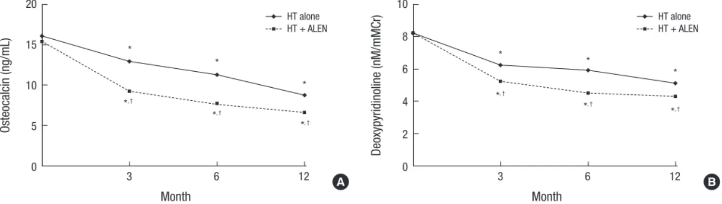
Fig. 3. Mean changes in biochemical markers of bone turnover. (A) OC, (B) DPD. Combination therapy suppressed serum levels of osteocalcin and urinary DPD by a significantly greater extent than HT alone at each time point.
OC = osteocalcin, DPD = deoxypyridinoline, HT = hormone therapy, ALEN = alendronate.
*P < 0.05 vs. baseline; †P < 0.05 vs. HT alone.
Osteocalcin (ng/mL)
Month
3 6 12 20
15
10
5
0
*
*
*
*, †
*, †
*, † HT alone HT + ALEN
Deoxypyridinoline (nM/mMCr)
Month
3 6 12 10
8 6 4 2 0
* *
*
*, †
*, † *, †
HT alone HT + ALEN
A B
Fig. 2. Mean percent changes in BMD. Differences between the 2 groups were not significant.
BMD = bone mineral density, HT = hormone therapy, ALEN = alendronate.
*P < 0.05 vs. baseline.
BMD change (%)
Lumbar spine Total hip
10 8 6 4 2 0
*
*
* *
HT alone (n = 99) HT + ALEN (n = 104)
ference in the mean change in BMD at the lumbar spine (6.9%
vs. 7.9%) and the total hip (3.7% vs. 3.8%) (Fig. 2). Age, baseline BMD, years since menopause, and history of HT did not affect the change in BMD after adjusting for variables, regardless of whether the women received ALEN (data not shown).
The proportion of participants with no BMD increase was sim- ilar in both groups, regardless of site (Table 2). When BMD re- sponse was stratified on baseline age or T-score using a cutoff of 60 or −2.5, respectively, the mean BMD change and the pro- portion of subjects with a reduction in BMD did not differ be-
tween the 2 groups at all sites tested (data not shown).
Changes in biochemical markers of bone turnover The baseline levels of markers of bone turnover were compara- ble between the 2 groups (Table 1). Fig. 3 presents mean values of serum OC and urinary DPD at each time point. Levels of bone formation and resorption markers decreased significantly com- pared with the baseline values with both treatments. The pat- tern of change over time was similar between the 2 treatments for both markers, but the combination therapy suppressed se- rum OC and urinary DPD to a significantly greater extent than HT alone at each time point. When bone turnover markers were stratified again on baseline age or T-score using the same cut- offs as described above, women aged ≥ 60 years or those with T-score > −2.5 showed similar suppression of serum OC and urinary DPD after treatment in both groups (data not shown).
Adverse effects
Table 3 presents adverse effects in study participants according to the treatment regimen. The total number of subjects who ex- perienced any adverse effects and the distribution of adverse
effects in both groups were similar.
DISCUSSION
This study demonstrated that concomitant treatment with HT and ALEN for 1 year did not provide a significant extra benefit in BMD over HT alone in postmenopausal Korean women with low BMD. However, the levels of bone turnover markers were reduced to a significantly greater extent in the combination ther- apy compared to HT alone.
Our finding is consistent with previous negative studies re- porting a comparable BMD response with 1-year combination therapy and hormonal therapy alone (16-19). However, other studies demonstrated a positive effect of combined therapy on BMD (22-24). Since the HT regimen (CEE + MPA) used in the current study was similar to those used in the previous studies (17,23), the BMD response to HT is one possible explanation for differences across the studies. In the present study, the increase in BMD after HT alone for 1 year at the lumbar spine (7%) or hip (3.8%) was greater than that in the positive studies (2.5%–4% at the lumbar spine and 2%–3% at the hip) (22-24), but similar to that of negative studies (16-19). Interestingly, BMD changes at 1 year of combination therapy in this study are similar to those achieved in the positive studies described above. When the BMD increase induced by HT alone is high, further BMD gain by the addition of ALEN to HT might be expected to be minimal.
The reason for different responses to HT, even among Asian women, is not clear and further studies are warranted. In fact, the magnitude of BMD increase by HT alone in the present study was comparable to that in previous reports in osteoporotic (26) and healthy (27) postmenopausal Korean women.
In addition, progestogen could have favorable independent effects on bone metabolism (3,28,29). The addition of MPA to CEE significantly increases spine BMD compared to CEE alone (3). In the present study, MPA was given continuously with CEE, contributing to the better BMD response to HT.
Age, initial BMD, and previous hormone use are significant variables affecting BMD response to HT (2). These variables, how- ever, had no influence in our study. This might be explained in part by the inclusion of study subjects with low BMD. The pro- portion of participants with no BMD increase in HT group in this study was within the range previously reported (15), and the proportions were not different between the 2 groups. Although a suboptimal response might be a possible indication of com- bination therapy, our results suggest that improvement of an inadequate response to HT by 1-year addition of ALEN should not be expected.
As BMD continues to increase at least for several years with either estrogen or bisphosphonate therapies, the study duration of the current study might be too short. Although the duration of 1 year might be useful for evaluating rapid responses to os-
teoporosis treatment, it might take longer to get final responses.
Indeed, positive responses were observed only after 2 to 3 years of ALEN in combination with HT (16,17,30). Of note, the type of bisphosphonate may also be an important factor; for example, the increase in BMD at the femoral neck was reported to be great- er with 1-year combination therapy using risedronate than with HT only (18).
Bone turnover markers are associated with changes in bone mass and fracture risk in postmenopausal women with osteo- porosis (31-33). In the current study, HT reduced levels of both DPD and OC by as early as 3 months in women with low BMD, which confirms previous reports (17,26). In addition, the com- bination therapy suppressed the bone turnover markers to a great- er degree than HT alone from 3 months of treatment onward, in agreement with most previous studies (17,24).
There is a growing concern over adverse events such as osteo- necrosis of jaw or atypical subtrochanteric fracture due to over- suppression of bone turnover with combination therapy. How- ever, previous studies showed that bone markers remained with- in the normal premenopausal range with combination therapy (34) and the addition of HT to etidronate prevented the bone mineralization defects associated with etidronate (35). More- over, no impairment of bone quality by combining bisphospho- nates with HT was found from bone morphometry data (34).
Importantly, improvements in the hip structure analysis indices were significantly greater with combination therapy than with either HT or ALEN monotherapy (36), which suggests a mecha- nism for potential fracture reduction. From these aspects, long- term data including fracture risk and adverse events are neces- sary to define the exact role of combination therapy.
This study has several limitations. First, our study was not de- signed to evaluate fracture risk and bone quality. Although com- bination therapy is expected to provide an additional fracture reduction from a logistic model (37), to date no study on com- bination therapy has been powered to detect a reduction in the risk of fracture. Second, the dropout rate in the present study was relatively high. Third, an ALEN-only arm was not included in the study, so it is not possible to separate the effect of com- bined therapy from that of ALEN alone. In addition, we did not measure and control for serum vitamin D levels.
In conclusion, 1-year combination therapy of ALEN and HT did not increase bone mass over HT alone in postmenopausal Korean women with low BMD. Further studies are needed to evaluate long-term changes in BMD and fracture risks when bis- phosphonate is added to HT.
DISCLOSURE
The authors have no potential conflicts of interest to disclose.
AUTHOR CONTRIBUTION
Conceptualization: Yoon BK, Choi YM. Data curation: Yoon BK, Lee DY. Formal analysis: Lee DY. Funding acquisition: Yoon BK, Choi YM. Investigation: Yoon BK, Lee DY, Park MC, Cho SH, Park HM, Choi YM. Writing - original draft: Yoon BK, Lee DY. Writing - review & editing: Yoon BK, Lee DY, Choi YM.
ORCID
Byung-Koo Yoon http://orcid.org/0000-0002-1326-6102 Dong-Yun Lee http://orcid.org/0000-0002-7540-0522 Man Chul Park http://orcid.org/0000-0001-9004-4792 Soo Hyun Cho http://orcid.org/0000-0001-9249-1019 Hyoung Moo Park http://orcid.org/0000-0003-0767-7711 Young Min Choi http://orcid.org/0000-0003-1245-0378
REFERENCES
1. Khosla S, Melton LJ 3rd, Riggs BL. The unitary model for estrogen defici
ency and the pathogenesis of osteoporosis: is a revision needed? J Bone Miner Res 2011; 26: 44151.
2. Effects of hormone therapy on bone mineral density: results from the post
menopausal estrogen/progestin interventions (PEPI) trial. The Writing Group for the PEPI. JAMA 1996; 276: 138996.
3. Lindsay R, Gallagher JC, Kleerekoper M, Pickar JH. Effect of lower doses of conjugated equine estrogens with and without medroxyprogesterone acetate on bone in early postmenopausal women. JAMA 2002; 287: 2668
76.
4. Cummings SR, Ettinger B, Delmas PD, Kenemans P, Stathopoulos V, Ver
weij P, MolArts M, Kloosterboer L, Mosca L, Christiansen C, et al. The ef
fects of tibolone in older postmenopausal women. N Engl J Med 2008;
359: 697708.
5. Anderson GL, Limacher M, Assaf AR, Bassford T, Beresford SA, Black H, Bonds D, Brunner R, Brzyski R, Caan B, et al. Effects of conjugated equine estrogen in postmenopausal women with hysterectomy: the Women’s Health Initiative randomized controlled trial. JAMA 2004; 291: 170112.
6. Cauley JA, Robbins J, Chen Z, Cummings SR, Jackson RD, LaCroix AZ, Le
Boff M, Lewis CE, McGowan J, Neuner J, et al. Effects of estrogen plus pro
gestin on risk of fracture and bone mineral density: the Women’s Health Initiative randomized trial. JAMA 2003; 290: 172938.
7. Black DM, Cummings SR, Karpf DB, Cauley JA, Thompson DE, Nevitt MC, Bauer DC, Genant HK, Haskell WL, Marcus R, et al. Randomised trial of effect of alendronate on risk of fracture in women with existing vertebral fractures. Fracture Intervention Trial Research Group. Lancet 1996; 348:
153541.
8. Karpf DB, Shapiro DR, Seeman E, Ensrud KE, Johnston CC Jr, Adami S, Harris ST, Santora AC 2nd, Hirsch LJ, Oppenheimer L, et al. Prevention of nonvertebral fractures by alendronate. A metaanalysis. Alendronate Os
teoporosis Treatment Study Groups. JAMA 1997; 277: 115964.
9. Reszka AA, Rodan GA. Mechanism of action of bisphosphonates. Curr Osteoporos Rep 2003; 1: 4552.
10. Khosla S. Update on estrogens and the skeleton. J Clin Endocrinol Metab
2010; 95: 356977.
11. Wang Q, Alén M, Nicholson PH, Halleen JM, Alatalo SL, Ohlsson C, Suom
inen H, Cheng S. Differential effects of sex hormones on peri and endo
cortical bone surfaces in pubertal girls. J Clin Endocrinol Metab 2006; 91:
27782.
12. Khastgir G, Studd J, Holland N, AlaghbandZadeh J, Fox S, Chow J. Ana
bolic effect of estrogen replacement on bone in postmenopausal women with osteoporosis: histomorphometric evidence in a longitudinal study. J Clin Endocrinol Metab 2001; 86: 28995.
13. Byrjalsen I, Leeming DJ, Qvist P, Christiansen C, Karsdal MA. Bone turn
over and bone collagen maturation in osteoporosis: effects of antiresorp
tive therapies. Osteoporos Int 2008; 19: 33948.
14. Lewiecki EM. Nonresponders to osteoporosis therapy. J Clin Densitom 2003; 6: 30714.
15. Komulainen M, Kröger H, Tuppurainen MT, Heikkinen AM, Honkanen R, Saarikoski S. Identification of early postmenopausal women with no bone response to HRT: results of a fiveyear clinical trial. Osteoporos Int 2000;
11: 2118.
16. Greenspan SL, Resnick NM, Parker RA. Combination therapy with hor
mone replacement and alendronate for prevention of bone loss in elderly women: a randomized controlled trial. JAMA 2003; 289: 252533.
17. Bone HG, Greenspan SL, McKeever C, Bell N, Davidson M, Downs RW, Emkey R, Meunier PJ, Miller SS, Mulloy AL, et al. Alendronate and estro
gen effects in postmenopausal women with low bone mineral density.
Alendronate/Estrogen Study Group. J Clin Endocrinol Metab 2000; 85:
7206.
18. Harris ST, Eriksen EF, Davidson M, Ettinger MP, Moffett Jr AH Jr, Baylink DJ, Crusan CE, Chines AA. Effect of combined risedronate and hormone replacement therapies on bone mineral density in postmenopausal wom
en. J Clin Endocrinol Metab 2001; 86: 18907.
19. Eviö S, Tiitinen A, Laitinen K, Ylikorkala O, Välimäki MJ. Effects of alen
dronate and hormone replacement therapy, alone and in combination, on bone mass and markers of bone turnover in elderly women with os
teoporosis. J Clin Endocrinol Metab 2004; 89: 62631.
20. Wimalawansa SJ. A fouryear randomized controlled trial of hormone re
placement and bisphosphonate, alone or in combination, in women with postmenopausal osteoporosis. Am J Med 1998; 104: 21926.
21. Lindsay R, Cosman F, Lobo RA, Walsh BW, Harris ST, Reagan JE, Liss CL, Melton ME, Byrnes CA. Addition of alendronate to ongoing hormone re
placement therapy in the treatment of osteoporosis: a randomized, con
trolled clinical trial. J Clin Endocrinol Metab 1999; 84: 307681.
22. Tiraş MB, Noyan V, Yildiz A, Yildirim M, Daya S. Effects of alendronate and hormone replacement therapy, alone or in combination, on bone mass in postmenopausal women with osteoporosis: a prospective, randomized study. Hum Reprod 2000; 15: 208792.
23. Palomba S, Orio F Jr, Colao A, di Carlo C, Sena T, Lombardi G, Zullo F, Mas
trantonio P. Effect of estrogen replacement plus lowdose alendronate treatment on bone density in surgically postmenopausal women with osteoporosis. J Clin Endocrinol Metab 2002; 87: 15028.
24. Tseng LN, Sheu WH, Ho ES, Lan HH, Hu CC, Kao CH. Effects of alendro
nate combined with hormone replacement therapy on osteoporotic post
menopausal Chinese women. Metabolism 2006; 55: 7417.
25. Min YK, Lee DY, Choi SJ, Kim JH, Choi D, Yoon BK. Effects of adding alen
dronate to ongoing hormone therapy on bone mineral density in post
menopausal Korean women: a randomized, doubleblind, placebocon
trolled clinical trial. Menopause 2013; 20: 7616.
26. Kim SW, Park DJ, Park KS, Kim SY, Cho BY, Lee HK, Shin CS. Early chang
es in biochemical markers of bone turnover predict bone mineral density response to antiresorptive therapy in Korean postmenopausal women with osteoporosis. Endocr J 2005; 52: 66774.
27. Kim JY, Shin KJ, Kim JH, Min YK, Choi D, Lee JH, Yoon BK. The effect of hormone replacement therapy on bone mineral density in Korean post
menopausal women: 2 year prospective cohort study. J Korean Soc Meno- pause 1999; 5: 1508.
28. Liu JH, Muse KN. The effects of progestins on bone density and bone me
tabolism in postmenopausal women: a randomized controlled trial. Am J Obstet Gynecol 2005; 192: 131623.
29. SeifertKlauss V, Prior JC. Progesterone and bone: actions promoting bone health in women. J Osteoporos 2010; 2010: 845180.
30. Greenspan SL, Emkey RD, Bone HG, Weiss SR, Bell NH, Downs RW, McK
eever C, Miller SS, Davidson M, Bolognese MA, et al. Significant differen
tial effects of alendronate, estrogen, or combination therapy on the rate of bone loss after discontinuation of treatment of postmenopausal osteo
porosis. A randomized, doubleblind, placebocontrolled trial. Ann In- tern Med 2002; 137: 87583.
31. DresnerPollak R, Parker RA, Poku M, Thompson J, Seibel MJ, Greenspan SL. Biochemical markers of bone turnover reflect femoral bone loss in el
derly women. Calcif Tissue Int 1996; 59: 32833.
32. Garnero P, SornayRendu E, Duboeuf F, Delmas PD. Markers of bone turn
over predict postmenopausal forearm bone loss over 4 years: the OFELY study. J Bone Miner Res 1999; 14: 161421.
33. Garnero P, Hausherr E, Chapuy MC, Marcelli C, Grandjean H, Muller C, Cormier C, Bréart G, Meunier PJ, Delmas PD. Markers of bone resorption predict hip fracture in elderly women: the EPIDOS Prospective Study. J Bone Miner Res 1996; 11: 15318.
34. Fadanelli ME, Bone HG. Combining bisphosphonates with hormone ther
apy for postmenopausal osteoporosis. Treat Endocrinol 2004; 3: 3619.
35. Wimalawansa SJ. Combined therapy with estrogen and etidronate has an additive effect on bone mineral density in the hip and vertebrae: fouryear randomized study. Am J Med 1995; 99: 3642.
36. Greenspan SL, Beck TJ, Resnick NM, Bhattacharya R, Parker RA. Effect of hormone replacement, alendronate, or combination therapy on hip struc
tural geometry: a 3year, doubleblind, placebocontrolled clinical trial. J Bone Miner Res 2005; 20: 152532.
37. Cummings SR, Karpf DB, Harris F, Genant HK, Ensrud K, LaCroix AZ, Black DM. Improvement in spine bone density and reduction in risk of verte
bral fractures during treatment with antiresorptive drugs. Am J Med 2002;
112: 2819.