J Korean Soc Radiol 2018;78(2):95-102 https://doi.org/10.3348/jksr.2018.78.2.95
INTRODUCTION
Breast cancer is one of the most common female malignan- cies. In the past decade, breast-specific gamma imaging (BSGI) with 99mTc-methoxyisobutylisonitrile (MIBI) has evolved for use in cancer detection and has complemented traditional im- aging methods. However, the exact uptake mechanism of
99mTc-MIBI is still under investigation. It is reported that 99mTc-MIBI is distributed inside tissues in proportion to the blood flow and is especially concentrated in malignant cells.
Approximately 90% of the tracer is stored within mitochondria (1). BSGI provides independent biological information specific to breast cancer (2-6).
MRI is an effective diagnostic tool for differentiation between
Correlation of Semi-Quantitative Breast-Specific Gamma Imaging Findings with Dynamic Contrast-Enhanced MRI Parameters
Assessed by a Computer-Aided Evaluation Program for Breast Cancer
유방암 환자에서 유방 특이 감마영상 검사의 반정량적 분석 및 유방 자기공명영상 검사의 컴퓨터보조진단 분석 결과의 비교 연구
Saemee Ahn, MD
1, Hye Ryoung Koo, MD
1*, Jeong Seon Park, MD
1, Juhee Moon, MD
2, Yun Young Choi, MD
3, Min Sung Chung, MD
4Departments of 1Radiology, 3Nuclear Medicine, 4General Surgery, Hanyang University College of Medicine, Seoul, Korea
2Department of Radiology, Hanyang University College of Medicine, Hanyang University Guri Hospital, Guri, Korea
Purpose: To investigate whether a correlation exists between the semi-quantitative breast-specific gamma imaging (BSGI) findings and dynamic contrast-enhanced (DCE) MRI parameters assessed by a computer-aided evaluation program.
Materials and Methods: Semi-quantitative index of the lesion to non-lesion (L/N) ratio in BSGI and DCE-MRI parameters was assessed by a computer-aided evalua- tion program, where 47 cases of invasive breast cancer were obtained. Correlations between the L/N ratio and DCE-MRI parameters were assessed by a computer-aided evaluation program. Tumor diameter (cm), angio-volume (cc), degree of initial peak enhancement (%), persistent enhancement proportion (%), and washout enhance- ment proportion (%) were analysed. The relationships between the L/N ratio and DCE-MRI parameters were evaluated by a univariate and multivariate regression analysis.
Results: The mean L/N ratio of the 47 tumors was 3.63 ± 2.19 (range: 1–13.1). The L/N ratio was higher in tumors with larger diameters (p < 0.001), increased angio- volume (p < 0.001), higher degree of initial peak enhancement (p = 0.005) and in- creased washout enhancement proportion (p = 0.004). In the multivariate regres- sion analysis, angio-volume (cc) and washout enhancement proportion (%) were associated with L/N ratio (p = 0.007 and p = 0.024, respectively).
Conclusion: There was a correlation between the semi-quantitative L/N ratio in BSGI and DCE-MRI parameters assessed by a computer-aided evaluation program for breast cancer.
Index terms Breast Neoplasm
Magnetic Resonance Imaging
Image Interpretation, Computer-Assisted Radionuclide Imaging
Received June 12, 2017 Revised July 22, 2017 Accepted September 29, 2017
*Corresponding author: Hye Ryoung Koo, MD Department of Radiology, Hanyang University College of Medicine, 222-1 Wangsimni-ro, Seongdong-gu, Seoul 04763, Korea.
Tel. 82-2-2290-9156 Fax. 82-2-2293-2111 E-mail: [email protected]
This is an Open Access article distributed under the terms of the Creative Commons Attribution Non-Commercial License (http://creativecommons.org/licenses/by-nc/4.0) which permits unrestricted non-commercial use, distri- bution, and reproduction in any medium, provided the original work is properly cited.
benign and malignant breast masses, preoperative evaluation in breast cancer patients, breast cancer screening and early predic- tion of the response to neoadjuvant chemotherapy (7, 8). Dy- namic contrast-enhanced (DCE) MRI can depict the distribu- tion of a contrast agent within a tumor over time and can non- invasively assess the tissue vasculature (9). Computer-aided detection (CAD) programs for DCE-MRI automate kinetic as- sessment and provide an easier way to interpret the patterns of contrast enhancement across a series of images (10, 11).
Several studies have demonstrated that BSGI and DEC-MRI have similar sensitivity and specificity in breast cancer detec- tion (12, 13). However, little is known about the correlation of 99mTc-MIBI uptake in BSGI with DCE-MRI parameters of breast cancer. Therefore, we aimed to investigate whether a cor- relation exists between the semi-quantitative BSGI findings and DCE-MRI parameters assessed by a computer-aided evaluation program for breast cancer.
The visual interpretation criteria for BSGI were suggested ear- lier (14). Images were categorized asnormal (score of 1), with no focal or diffuse uptake; benign (score of 2), with minimal patchy uptake; probably benign (score of 3), with minimal patchy uptake with some areas of more focal uptake; probably abnor- mal (score of 4), with mild focal radiotracer uptake; and abnor- mal (score of 5), with marked focal radiotracer uptake. These criteria are qualitative and rather subjective. The lesion to non- lesion (L/N) ratio for semi-quantitative analysisreflects the 99mTc-MIBI uptake level and is an important parameter of BSGI as a functional imaging (3).
MATERIALS AND METHODS
Patients
The Institutional Review Board of our hospital approved this retrospective study (2014-12-012); the requirement for informed consent was waived. Between January 2014 and December 2014, 113 patients who had suspicious mammography or ultrasound findings were imaged with BSGI. Among them, 47 patients with lesions that had been biopsied and were confirmed to be inva- sive breast cancer underwent breast DCE-MRI for initial stag- ing. One patient was later excluded due to non-physiological CAD parameter values. One patient had bilateral breast cancer.
Finally, this retrospective study involved 46 patients (mean age:
51 years, range: 32–68 years) with 47 index breast cancers. None of the patients received chemotherapy or radiation therapy prior to the study.
BSGI
BSGI examinations were performed after the patients were administered 20 mCi 99mTc-MIBI through an antecubital vein.
The patients were seated for the procedure, and craniocaudal (CC) and mediolateral oblique (MLO) view images were acquired using a high-resolution breast-specific gamma camera (Dilon 6800 gamma camera; Dilon Technologies, Newport News, VA, USA). Images were acquired for 10 min for each view.
Semi-quantitative analysis of BSGI was performed by two board-certified radiologists in consensus, one with 10 years of experience and one with 6 years of experience in breast imaging.
The readers were not blinded to the lesion site. The L/N ratio was considered as the semi-quantitative indices of BSGI.
For lesions, a region of interest (ROI) was manually drawn cov- ering the target lesion area. The maximum 9 pixel counts of the ROI were measured on different CC and MLO images (6). The higher value was chosen for lesion maximal uptake between the CC and MLO views. For non-lesions, another three circular ROIs, approximately 1 cm in diameter, were drawn in the con- tralateral trisectioned breast parallel to the base of the breast (anterior/ middle/ posterior), and the mean values of the three ROIs were averaged into a single value (Fig. 1). The L/N ratio was then calculated by dividing the maximum pixel counts of the lesion by the average non-lesion uptake. There were two cases in which the tumor (lesion) and background breast tissue (non-lesion) were not distinguishable. The L/N ratio for these cases was regarded as 1.
MRI Evaluation
All MR examinations were performed by using two 3.0-T MR imagers with a dedicated 8-channel breast coil (n = 20, Achieva, Philips, Best, Netherlands) or with a dedicated 16-channel breast coil (n = 26, Ingenia, Philips). The DCE-MR images were ob- tained by using the following image parameters: T1-weighted fast spoiled gradient-echo sequence (5.4/2.5; matrix, 440 × 426;
flip angle, 12°; field of view, 320 × 320 mm; section thickness, 1 mm; no gap) with one pre-contrast and four post-contrast dy- namic series obtained at 100, 200, 300, and 400 seconds after
injection of a contrast agent. A 0.2 mmol/kg dose of gadobutrol (Gadovist; Bayer-Healthcare, Berlin, Germany) was injected into an antecubital vein using an automated injector at a rate of 2 mL/sec, followed by a 20-mL saline flush after the contrast agent was injected.
The CAD analysis was performed by one radiologist who specialized in breast imaging and had 6 years of experience. For measurement of dynamic MR image parameters, pre-contrast and four post-contrast T1-weighted image series were trans- ferred to a computer-aided evaluation system (CADstream, ver- sion 4.1.3; Confirma, Kirkland, WA, USA). The system automati-
cally segmented the tumors into three dimensions and calculated the tumor diameter (maximal size of an enhancing lesion); an- gio-volume (total enhancing lesion volume); peak enhance- ment value (highest pixel signal intensity at the first post-con- trast series); and proportions of persistent, plateau, and washout enhancement components within a tumor.
Statistical Analysis
Correlations between the L/N ratio and DCE-MRI parameters assessed by a computer-aided evaluation program, including tu- mor diameter (cm), angio-volume (cc), degree of initial peak enhancement (%), persistent enhancement proportion (%), and washout enhancement proportion (%), were then analysed.
We used linear regression analysis to evaluate the association between the L/N ratio and DCE-MRI parameters. Variables with p < 0.05 in the univariate analysis were applied to a multivariate analysis to determine which variables were independently asso- ciated with L/N ratio.
The data were analysed using statistical software (SAS, version 9.2; SAS Institute, Cary, NC, USA). Statistical significance was defined as p < 0.05.
RESULTS
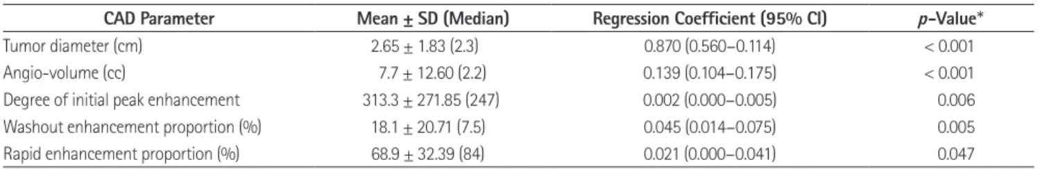
The mean L/N ratio of the 47 tumors was 3.63 ± 2.19 (range:
1–13.1). The DCE-MRI parameters assessed by a computer- aided evaluation program of the 47 tumors were as follows: the mean diameter of the tumor, 2.65 ± 1.83 cm (range 0.5–8.1, me- dian 2.3); angio-volume, 7.7 ± 12.60 cc (range 0.04–57.9, medi- an 2.2); degree of initial peak enhancement, 313.3 ± 271.85%
(range 99–1763, median 247); wash out enhancement propor- tion, 18.1 ± 20.71% (range 0–69, median 7.5), and rapid enhance- ment proportion, 68.9 ± 32.39% (range 0–100, median 84).
In the univariate analysis, the L/N ratio was significantly higher in tumors with larger diameter (p < 0.001), increased angio- volume (p < 0.001), higher degree of initial peak enhancement (p = 0.005), increased washout enhancement proportion (p = 0.004) and higher rapid enhancement proportion (p = 0.047) (Table 1).
In the multivariate regression analysis, angio-volume (cc) and washout enhancement proportion (%) were significantly asso- ciated with L/N ratio (Table 2). The estimated L/N ratio increas- Fig. 1. Lesion to non-lesion ratio of breast cancer in the left upper
outer breast. For lesions, the maximum ROI covering the lesion is drawn at the craniocaudal view and the mediolateral oblique view. For non-lesions, another three circular ROIs, approximately 1 cm in diam- eter, are drawn in the contralateral trisectioned breast, parallel to the base of the breast (anterior/middle/posterior).
ROI = region of interest
es 0.08 for each increase in angio volume (cc) and 0.03 for each increase in washout enhancement proportion (%), respectively.
There was a positive correlation between the semi-quantitative L/N ratio in BSGI and angio-volume (coefficient of determina- tion, R2 = 0.58) as well as washout enhancement proportion (co- efficient of determination, R2 = 0.16) (Figs. 2-5).
DISCUSSION
In this study, we investigated whether a correlation exists be-
tween the semi-quantitative BSGI findings and DCE-MRI pa- rameters assessed by a computer-aided evaluation program for 47 breast cancers. We observed that the L/N ratio was higher in tumors with larger diameter (p < 0.001) and increased angio- volume (p < 0.001). These results are similar to those of previ- ously published studies: 99mTc-MIBI uptake has been found to be positively related to tumor size. In a previous study of 102 breast cancers, the value of the semi-quantitative index expressed as the L/N ratio was independently related to tumor size (p = 0.002) and infiltration degree (p = 0.016). The study showed that the sensitivity of BSGI in diagnosing breast cancer was re- lated to tumor size: 100% for tumor size more than 2.0 cm and 89.1% for tumors less than or equal to 2.0 cm (15). Tadwalkar et al. also reported that BSGI detected all invasive breast can- cers that measured ≥ 7 mm regardless of grade (16). We had two pathology-confirmed breast cancer cases, considering no uptake in the MIBI scan; in other words, the L/N ratio was 1.
Table 1. Result of Univariate Regression Analysis between CAD Parameter and Lesion to Non-Lesion Ratio on Breast-Specific Gamma Imaging
CAD Parameter Mean ± SD (Median) Regression Coefficient (95% CI) p-Value*
Tumor diameter (cm) 2.65 ± 1.83 (2.3) 0.870 (0.560–0.114) < 0.001
Angio-volume (cc) 7.7 ± 12.60 (2.2) 0.139 (0.104–0.175) < 0.001
Degree of initial peak enhancement 313.3 ± 271.85 (247) 0.002 (0.000–0.005) 0.006
Washout enhancement proportion (%) 18.1 ± 20.71 (7.5) 0.045 (0.014–0.075) 0.005
Rapid enhancement proportion (%) 68.9 ± 32.39 (84) 0.021 (0.000–0.041) 0.047
*Linear regression analysis.
CAD = computer-aided detection, CI = confidence interval, SD = standard deviation Table 2. Results of Multivariate Regression Analysis between Com- puter-Aided Detection Parameter and Lesion to Non-Lesion Ratio on Breast-Specific Gamma Imaging
Regression Coefficient
(95% CI) p-Value*
Angio volume (cc) 0.084 (0.025–0.143) 0.007 Washout enhancement
proportion (%)
0.027 (0.004–0.049) 0.024
*Linear regression analysis.
CI = confidence interval
Fig. 2. Scatter plot of L/N ratio in breast-specific gamma imaging and angio-volume (coefficient of determination, R2 = 0.58).
L/N = lesion to non-lesion 20
15
10
5
0
L/N ratio
10
0 20 30 40 50 60
Angio volume (cc)
Fig. 3. Scatter plot of L/N ratio in breast-specific gamma imaging and washout enhancement proportion (coefficient of determination, R2 = 0.16).
L/N = lesion to non-lesion 20
15
10
5
0
L/N ratio
0 20 40 60
Washout (%)
A B
Fig. 4. Invasive ductal carcinoma in a 47 year-old woman.
A. Rt. cranio-caudal view of breast-specific gamma imaging. Lesion to non-lesion ratio is 3.97.
B. Angiomap and volume rendering image of the tumor obtained by computer-aided evaluation program in DCE-MRI (diameters: 2.9 × 2.0 × 2.2 cm, angio volume: 5.0 cc and washout enhancement proportion: 39%).
Fig. 5. Invasive ductal carcinoma in a 54 year-old woman.
A. Rt. cranio-caudal view of breast-specific gamma imaging. Lesion to non-lesion ratio is 8.99.
B. Angiomap and volume rendering image of the tumor obtained by computer-aided evaluation program in DCE-MRI (diameters: 6.9 × 5.1 × 8.1 cm, angio volume: 57.9 cc, washout enhancement proportion: 76%).
A B
These two breast cancer tumors were 0.6 cm and 0.8 cm in di- ameter. The sensitivity for subcentimeter cancer was 71.4%
(5/7) in our study. Further studies are needed for a more com- prehensive analysis of BSGI results with respect to tumor char- acteristics.
There are several studies regarding the existence of a correla- tion between the kinetic parameters on DCE-MRI and histo- pathologic prognostic factors of breast cancer. Baltzer et al. (17) reported that higher lesion volume and higher and earlier ini- tial enhancement were independent covariates that are predic- tive of higher tumor aggressiveness. In another study, numeric ratios obtained on gadolinium-enhanced dynamic MRI of the breast may be useful in predicting histologic-type breast carci- nomas, and there are three histologic prognostic factors used for invasive ductal carcinoma: modified Scarff-Bloom-Richard- son grade, microvessel density, and fibrotic focus (18). Bekis¸ et al. (19) reported that there was no statistically significant corre- lation between the degree of angiogenesis and early or delayed 99mTc-MIBI uptake and the washout index of 99mTc-MIBI in 24 invasive MIBI-positive breast lesions although there was a high correlation between angiogenesis and the washout index of 99mTc-MIBI in seven noninvasive breast lesions. We evalu- ated multiple DCE-MRI parameters that were assessed by CAD, such as tumor diameter, angio-volume, peak enhance- ment value, and proportions of persistent, plateau and washout enhancement components. Among these CAD parameters, in- creased uptake of 99mTc-MIBI in BSGI correlated with angio- volume (cc) and washout enhancement proportion (%) in the multivariate regression analysis. To the best of our knowledge, the present study is the first to correlate 99mTc-MIBI uptake in BSGI and DCE-MRI parameters for breast cancer. Further studies such as the relationship between 99mTc-MIBI uptake or ADC values and immunohistochemical prognostic factors are needed to investigate whether BSGI can be used as a non-inva- sive tool to study the aggressiveness of breast cancer.
The main limitation of the present study is its retrospective design, which unavoidably introduces selection bias and limits the general application of the results. The small sample size is also a limitation, as it is not sufficient for accurate statistical analysis.
In addition, this study lacked histopathologic correlation for breast cancer such as subtype, hormone receptor status or his- tologic grade which could have affected the result of 99mTc-
MIBI uptake in BSGI or DCE-MRI parameters. Further research for correlation between 99mTc-MIBI uptake and histopatholo- gy of breast cancer could be valuable. Limitations of this study include the fact that MRI was acquired on two different scan- ner-coil systems. The differences in spatial resolution and bound- ary detection for delineating ROIs of lesion could have affected the results of CAD parameters.
In conclusion, there was a correlation between the semi- quantitative L/N ratio in BSGI with DCE-MRI parameters as- sessed by a computer-aided evaluation program for breast can- cer. We observed that increased uptake of 99mTc-MIBI in BSGI was associated with increased angio-volume (cc) and washout enhancement proportion (%) in DCE-MRI.
Acknowledgments
This work was supported by the research fund of Hanyang University (HY-2014).
REFERENCES
1. Brem RF, Rechtman LR. Nuclear medicine imaging of the breast: a novel, physiologic approach to breast cancer de- tection and diagnosis. Radiol Clin North Am 2010;48:1055- 1074
2. Yoon HJ, Kim Y, Chang KT, Kim BS. Prognostic value of semi- quantitative tumor uptake on Tc-99m sestamibi breast- specific gamma imaging in invasive ductal breast cancer.
Ann Nucl Med 2015;29:553-560
3. Yu X, Hu G, Zhang Z, Qiu F, Shao X, Wang X, et al. Retrospec- tive and comparative analysis of (99m)Tc-Sestamibi breast specific gamma imaging versus mammography, ultrasound, and magnetic resonance imaging for the detection of breast cancer in Chinese women. BMC Cancer 2016;16:450 4. Specht JM, Mankoff DA. Advances in molecular imaging
for breast cancer detection and characterization. Breast Cancer Res 2012;14:206
5. Sun Y, Wei W, Yang HW, Liu JL. Clinical usefulness of breast- specific gamma imaging as an adjunct modality to mam- mography for diagnosis of breast cancer: a systemic review and meta-analysis. Eur J Nucl Med Mol Imaging 2013;40:
450-463
6. Tan H, Jiang L, Gu Y, Xiu Y, Han L, Wu P, et al. Visual and semi-
quantitative analyses of dual-phase breast-specific gam- ma imaging with Tc-99m-sestamibi in detecting primary breast cancer. Ann Nucl Med 2014;28:17-24
7. Kuhl CK, Mielcareck P, Klaschik S, Leutner C, Wardelmann E, Gieseke J, et al. Dynamic breast MR imaging: are signal intensity time course data useful for differential diagnosis of enhancing lesions? Radiology 1999;211:101-110 8. Orel SG, Schnall MD. MR imaging of the breast for the de-
tection, diagnosis, and staging of breast cancer. Radiology 2001;220:13-30
9. Jena A, Taneja S, Singh A, Negi P, Mehta SB, Sarin R. Role of pharmacokinetic parameters derived with high temporal resolution DCE MRI using simultaneous PET/MRI system in breast cancer: a feasibility study. Eur J Radiol 2017;86:
261-266
10. Aghaei F, Tan M, Hollingsworth AB, Qian W, Liu H, Zheng B.
Computer-aided breast MR image feature analysis for pre- diction of tumor response to chemotherapy. Med Phys 2015;
42:6520-6528
11. Dorrius MD, Jansen-van der Weide MC, van Ooijen PM, Pi- jnappel RM, Oudkerk M. Computer-aided detection in breast MRI: a systematic review and meta-analysis. Eur Radiol 2011;21:1600-1608
12. Kim BS. Usefulness of breast-specific gamma imaging as an adjunct modality in breast cancer patients with dense breast: a comparative study with MRI. Ann Nucl Med 2012;
26:131-137
13. Johnson N, Sorenson L, Bennetts L, Winter K, Bryn S, John- son W, et al. Breast-specific gamma imaging is a cost effec-
tive and efficacious imaging modality when compared with MRI. Am J Surg 2014;207:698-701; discussion 701 14. Brem RF, Floerke AC, Rapelyea JA, Teal C, Kelly T, Mathur V.
Breast-specific gamma imaging as an adjunct imaging mo- dality for the diagnosis of breast cancer. Radiology 2008;
247:651-657
15. Tan H, Zhang H, Yang W, Fu Y, Gu Y, Du M, et al. Breast-spe- cific gamma imaging with Tc-99m-sestamibi in the diagnosis of breast cancer and its semiquantitative index correlation with tumor biologic markers, subtypes, and clinicopatho- logic characteristics. Nucl Med Commun 2016;37:792-799 16. Tadwalkar RV, Rapelyea JA, Torrente J, Rechtman LR, Teal CB, McSwain AP, et al. Breast-specific gamma imaging as an adjunct modality for the diagnosis of invasive breast cancer with correlation to tumour size and grade. Br J Radiol 2012;
85:e212-e216
17. Baltzer PA, Vag T, Dietzel M, Beger S, Freiberg C, Gajda M, et al. Computer-aided interpretation of dynamic magnetic resonance imaging reflects histopathology of invasive breast cancer. Eur Radiol 2010;20:1563-1571
18. Narisada H, Aoki T, Sasaguri T, Hashimoto H, Konishi T, Mori- ta M, et al. Correlation between numeric gadolinium-en- hanced dynamic MRI ratios and prognostic factors and histologic type of breast carcinoma. AJR Am J Roentgenol 2006;187:297-306
19. Bekis¸ R, Degirmenci B, Aydin A, Ozdogan O, Canda T, Durak H. Correlation between 99mTc-MIBI uptake and angiogen- esis in MIBI-positive breast lesions. Nucl Med Biol 2005;32:
465-472
유방암 환자에서 유방 특이 감마영상 검사의 반정량적 분석 및 유방 자기공명영상 검사의 컴퓨터보조진단 분석 결과의 비교 연구
안새미
1· 구혜령
1* · 박정선
1· 문주희
2· 최윤영
3· 정민성
4목적: 유방암 환자에 있어서 반정량적 유방 특이 감마영상 검사와 컴퓨터 보조진단 분석을 이용한 유방 자기공명영상 검 사의 연관성을 보고자 하였다.
대상과 방법: 47명의 침습적 유방암 환자를 대상으로 유방 특이 감마영상 검사에서 병변과 비병변의 비(lesion to non- lesion raio; 이하 L/N ratio)와 역동적 조영증강 자기공명 매개변수의 연관성을 분석하였다. 역동성 조영증강 자기공명영 상의 매개변수는 컴퓨터보조진단 분석을 통하여 병변의 크기(cm), 혈관조영 부피(angio-volume, cc), 초기 조영증강 정 도(%), 지속적 조영증강 비율(%), 조영증강 감쇄 비율(%)의 항목을 분석하였다. 병변과 비병변의 비(L/N ratio)와 역동 적 조영증강 자기공명 매개변수를 대상으로 단변량/다변량 회귀분석을 시행하였다.
결과: 47개의 종양의 유방 특이 감마영상 검사에서 병변/비병변의 비(L/N ratio)의 평균값은 3.63 ± 2.19 (range:
1-13.1)였다. 병변/비병변의 비(L/N ratio)는 종양의 크기가 클수록(p < 0.001), 혈관조영 부피가 클수록(p < 0.001),
초기 조영증강 비율이 높을수록(p = 0.005), 조영증강 감쇄 비율이 높을수록(p = 0.004) 높게 나타났다. 다중회귀분석
결과, 혈관조영 부피(p = 0.007)와 조영증강 감쇄 비율(p = 0.024)이 유방 특이 감마영상 검사에서 병변/비병변의 비와 연관성을 보였다.
결론: 유방암 환자에서 컴퓨터보조진단 분석을 이용한 역동성 자기공명영상의 혈관조영 부피와 조영증강 감쇄 비율은 유 방 특이 감마영상 검사에서의 병변/비병변의 비와 연관성을 가진다.
한양대학교 의과대학 1영상의학교실, 3핵의학교실, 4외과학교실, 2한양대학교 의과대학 구리병원 영상의학과