www.krspine.org
Neurological Complications of Posterior Spinal Surgery:
Incidence and Clinical Features
Dong Ki Ahn, M.D. Ph.D., Jung Soo Lee, M.D., Won Shik Shin, M.D., Seong Min Yi, M.D., Ki Hyuk Koo, M.D.
J Korean Soc Spine Surg 2018 Mar;25(1):1-8.
Originally published online Marchr 31, 2018;
https://doi.org/10.4184/jkss.2018.25.1.1
Korean Society of Spine Surgery
Asan Medical Center 88, Olympic-ro 43 Gil, Songpa-gu, Seoul, 05505, Korea Tel: +82-2-483-3413 Fax: +82-2-483-3414
©Copyright 2017 Korean Society of Spine Surgery pISSN 2093-4378 eISSN 2093-4386
The online version of this article, along with updated information and services, is located on the World Wide Web at:
http://www.krspine.org/DOIx.php?id=10.4184/jkss.2018.25.1.1
This is an Open Access article distributed under the terms of the Creative Commons Attribution Non-Commercial License (http://
creativecommons.org/licenses/by-nc/4.0) which permits unrestricted non-commercial use, distribution, and reproduction in any medium, provided the original work is properly cited.
Journal of Korean Society of
Spine Surgery
Neurological Complications of Posterior Spinal Surgery: Incidence and Clinical Features
Dong Ki Ahn, M.D. Ph.D., Jung Soo Lee, M.D., Won Shik Shin, M.D., Seong Min Yi, M.D., Ki Hyuk Koo, M.D.
Seoul Sacred Heart General Hospital, Orthopedic Department Study design: Retrospective study.
Objectives: To identify clinical features and risk factors helpful for the prevention and early diagnosis of neurological complications.
Overview of Literature: Previous studies have investigated postoperative complications only for specific disease entities and did not present distinctive clinical features.
Materials and Methods: This was an observational study of patients who underwent posterior thoracolumbar spinal surgery in the orthopedic department of a single hospital over the course of 19 years (1995-2013). The incidence, cause, onset time, and risk factors of complications were investigated. Neurological deterioration was graded on a 5-point numeric scale: G1, increased leg pain or sensory loss, G2, unilateral motor weakness; G3, bilateral motor weakness; G4, cauda equina syndrome; and G5, complete paraplegia.
Results: Sixty-five cases out of 6574 (0.989%) developed neurological complications due to the following causes: epidural hematoma, 0.380%; instrumentation with inadequate decompression, 0.213%; mechanical injury, 0.167%; inadequate discectomy, 0.061%; and unknown cause, 0.167% (p=0.000). The grade of neurological deterioration was G1 in 0.167% of patients, G2 in 0.517%, G3 in 0.228%, G4 in 0.046%, and G5 in 0.030%. Neurological deterioration was most severe in patients who experienced epidural hematoma, followed by those in whom complications occurred due to instrumentation with inadequate decompression, unknown causes, mechanical injury, and inadequate discectomy, in order (p=0.009). Revision surgery was a significant risk factor (p=0.000; odds ratio, 2.741). The time that elapsed until symptom development was as follows, in order: unknown cause, 0.6 hours; epidural hematoma, 5.4 hours; mechanical injury, 6.6 hours; inadequate discectomy, 18.0 hours; and instrumentation with insufficient decompression, 36.0 hours (p=0.001).
Conclusions: The incidence of neurological complications in our cohort was 1%. Revision surgery increased the risk by 3 times.
Severe cases (cauda equina syndrome or complete paraplegia) rarely developed, occurring in 0.08% of patients. The major causes of neurological decline were epidural hematoma and instrumentation with inadequate decompression. Close observation in the early period was important for the diagnosis because most patients developed symptoms within 12 hours. Delayed diagnosis was most common in complications caused by instrumentation with inadequate decompression.
Key words: Spinal surgery, Nneurological complication, Incidence, Risk factors
Received: August 24, 2017 Revised: October 10, 2017 Accepted: December 19, 2017 Published Online: March 31, 2018 Corresponding author: Won Shik Shin, M.D.
ORCID ID: Won Shik Shin: https://orcid.org/0000-0002-4280-5793 Dong Ki Ahn: https://orcid.org/0000-0003-4075-3632 Jung Soo Lee: https://orcid.org/0000-0003-0401-084X Seong Min Yi: https://orcid.org/0000-0003-1900-5971 Ki Hyuk Koo: https://orcid.org/0000-0002-1926-3228 Department of Orthopedic Surgery, Seoul Sacred Heart Hospital, 259 Wangsan-ro, Dongdaemun-gu, Seoul, 02488 Korea
TEL: +82-2-966-1616, FAX: +82-2-968-2394 E-mail: dr.wonshik@hanmail.net
Introduction
According to the previous reports, 10~20% of patients undergoing spinal surgeries experience some adverse effects.1-2) Some of them are considered complications. While other kinds of complication are closely related to the patient’s comorbidities and often unavoidable, neurological complication (NC) is considered being influenced by surgical factors. It maybe preventable if surgeons can anticipate the risk or at least curable if the causes discovered within critical time in many cases.
NC is a disastrous condition for both patients and surgeons and previous studies focused on a specific disease entity have reported its incidence as 0~2%.3-7) Homogenous diseases or
Dong Ki Ahn et al Volume 25 • Number 1 • March 31 2018
www.krspine.org 2
operations would have a certain pattern of NC. However, average spinal surgeons are exposed to various conditions.
As far as we have researched, there have been a few previous studies mentioning the causes and distinctive clinical features of each one. In the present study, not only risk factors but also clinical manifestations were investigated. We hoped the results could give clues for prevention and early identification of NC for better management and consequence.
Materials and Methods
This was a retrospective observation study. Those who underwent posterior thoracolumbar surgeries for 19 years, between Jan. 1995 to Dec 2013 were reviewed through their medical records. Those who did not have eligible records for the investigated variables were excluded. Minor procedures under local anesthesia were excluded. The first author had worked at the hospital throughout the study period and collected the data prospectively. Repeated operations of the same patients were counted as different cases. Medical comorbidities, medications, drinking and smoking were not considered because those were not included in collected data items of the first author. Image tests were reviewed only in NC cases and all of them were done by the first author. The definition of NC was newly developed leg pain or sensory loss or motor weakness within 5 days after a spinal surgery. The time of symptom development was counted as the time when NC was discovered and recorded by doctors because the time when patient began to complain was not exact and sometimes vague. Particularly when symptom onset was insidious and retarded, the patients themselves did not notice exactly. The causes were investigated based on the records and postoperative image tests. The grade (G) of neurological deterioration was assessed by 5 numeric scales. G1 was increased leg pain or sensory loss, G2 was unilateral motor weakness, G3 was bilateral motor weakness, G4 was cauda equina syndrome and G5 was complete paraplegia. Operation site was classified as thoracic and lumbosacral areas. If the operation site crossed either side, it was classified as the site more segments were involved. Disease entities were classified as degenerative disease, herniated nucleus pulposus (HNP), trauma, deformity, infection, neoplasm and others. Total and each incidence according to the causes were analyzed. The denominator of
individual incidence was total number of operations during the index period. Total and each grade of neurological deterioration according to the causes were analyzed. Risk factors were investigated among the demographic and surgical factors. The examined variables were age, sex, preoperative diagnosis, whether revision or not, method and site of operation, whether instrumentation or not, and whether using suction drain or not. Those who did not have clear records about above variables were excluded. The documented odds ratios were calculated in multi-variable analysis. A revision surgery was defined as a reoperation of the same or adjacent segments. In statistical analysis, Chi square test was applied for the difference of etiological incidence, Kruskall Wallis test was applied for the difference of neurological deterioration, time of onset and degree of recovery. Fisher exact test was applied for the difference of incidence according to sex, site, instrumentation and revision. Multiple logistic regression test was applied for the risk factors. Significant level of P value was set as <0.05. SPSS for Windows software package (ver. 16.0;
SPSS Inc., Chicago, IL, USA) was used for the analysis.
Results
1. Incidences and causes
There were 7,224 cases of spine surgeries during the index period. Because of ineligibility of medical records, 650 cases were excluded and 6,574 cases (91%) were included in the present study. Average age was 56±13.7 years (11~87y).
There were 2,491 cases (38%) of male and 4,083 cases (62%) of female. There were 204 cases (3.1%) of thoracic region and 6,370 cases (96.9%) of lumbosacral region. Posterior instrumentation cases were 5,008 (76.2%). Revision cases were 761 (11.6%). Preoperative disease entity was degenerative disease in 4,187 cases (63.7%), HNP in 1,817 cases (27.6%), trauma in 234 cases (3.6%), deformity in 177 cases (2.7%), infection 57 (0.9%), neoplasm in 35 cases (0.5%) and others 65 cases (1.0%) (Table 1). There were 10 main operators, however, only the first author had done spine surgeries at the hospital throughout the study period and 4,858 cases (73.9%) were done by the first author. NC developed in 65 cases (0.989%). The causes were epidural hematoma (EH) in 25 cases (0.380%), instrumentation with inadequate decompression (IID) in 14 cases (0.213%), mechanical injury
(MI) in 11 cases (0.167%), inadequate discectomy (ID) in 4 cases (0.016%) and unknown in 11 cases (0.167%) (Fig.
1). There was a significant difference among the incidences according to the causes (p=0.001). In IID, 11 cases developed due to instrumentation with insufficient decompression and 5 of them were pedicle subtraction osteotomy for lumbar
kyphosis cases. Three cases were iatrogenic foraminal stenosis due to under-sized cages in posterior lumbar interbody fusion (PLIF). In MI, 4 cases were due to pedicle screw, 2 cases were by punch and a case by knife, a case by osteotome, a case by retractor, a case by gelfoam and a case while excising ossified meningioma.
2. Risk factors
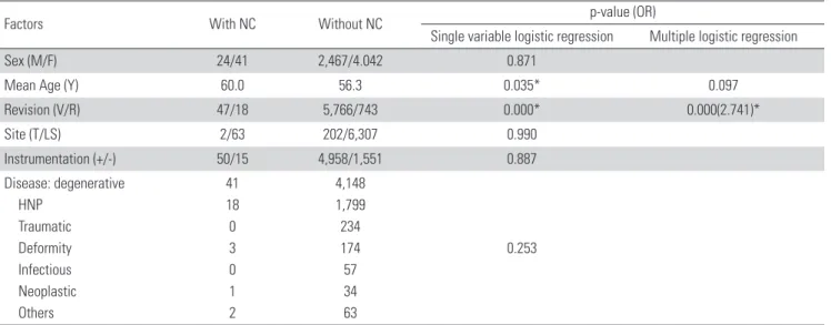
In risk factor analysis, old age and revision surgery significantly increased the risk in single variable analysis.
However, only revision surgery was approved significant in multiple logistic regression test (p=0.000, OR=2.741) (Table 1).
Table 1. Demography and risk factors.
Factors With NC Without NC p-value (OR)
Single variable logistic regression Multiple logistic regression
Sex (M/F) 24/41 2,467/4.042 0.871
Mean Age (Y) 60.0 56.3 0.035* 0.097
Revision (V/R) 47/18 5,766/743 0.000* 0.000(2.741)*
Site (T/LS) 2/63 202/6,307 0.990
Instrumentation (+/-) 50/15 4,958/1,551 0.887
Disease: degenerative HNP
Traumatic Deformity Infectious Neoplastic Others
41 18 0 3 0 1 2
4,148 1,799 234 174 57 34 63
0.253
*: Statistically significant
NC: Neurological complication, OR: Odds ratio, V/R: Virgin/Revision, T/LS: Thoracic/Lumbosacral.
Inadequate discectomy (0.06%)
Unknown (0.17%)
Epidural hematoma (0.38%)
Instrumentation with inadequate decompression (0.21%)
Mechanical injury (0.17%)
p=0.001
Fig. 1. Incidence and causes of neurological complications. The most common cause was epidural hematoma, followed by inadequate decom- pression and fusion, mechanical injury, unknown cause, and Instrumen- tation with inadequate decompression, in order. The difference in the incidence among them was significant.
p=0.009
Severity grade
Inadequate
discectomy Mechanical
injury Unknown Instrumentation with inadequate decompression
Epidural hematoma 5
4.5 4 3.5 3 2.5 2 1.5 1 0.5 0
Fig. 2. Neurological deterioration was most severe in cases of epidural hematoma and least severe in cases of inadequate discectomy.
Dong Ki Ahn et al Volume 25 • Number 1 • March 31 2018
www.krspine.org 4
3. Clinical manifestations
The degree of neurological deterioration was G1 in 11 cases (0.167%), G2 in 34 cases (0.517%), G3 in 15 cases (0.228%), G4 in 3 cases (0.046%) and G5 in 2 cases (0.030%). The degree according to the causes was the most severe in EH and the least in ID (p=0.009) (Fig. 2). The time of onset was 0.6 h in unknown, 5.4 h in EH, 6.6 h in MI, 18.0 h in ID and 36.0 h in IID. There was significant difference among them (p=0.001).
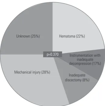
However, in subgroup analysis that developed symptoms immediately, just after the recovery from general anesthesia, all causes presented without significant difference (Fig. 3). The methods of treatment and final neurological sequalae were not analyzed in the present study.
Discussion
The greatest reason that makes a spinal surgery fearful would be NC. The incidences of NC have been reported as a wide range as 0-2.8%.3,5,8,9) Because most of the previous studies investigated a specific disease entity, the causes and clinical features were not diverse. Therefore it was not easy to estimate the general incidence of NC in overall spinal surgeries and they didn’t give any information to presume the causes according to the clinical manifestations. Cramer et al. studied a general
NC in single institute. However, their results showed very low incidence because they counted only severe NC at least more than paraparesis.10) In the present study, all neurological deficits including sensory change were regarded as NC. There were 10 main operators and the first author performed 74% of cases. Though there should have been learning curve effect, the annual incidences (which were not documented in the results) did not reduce as time went by. We thought it was due to the increased complexity of the operations and partially because it could not be prevented technically in some cases.
The most frequent cause was EH as 0.38% in the present study. All cases were confirmed by magnetic resonance image and immediate improvement by surgical removal of hematoma. Attention has been focused on antithrombotic therapy, however, there has been no agreed evidence yet.
The gross incidence has been reported with wide range as 0~0.7% in patients with antithrombotic medication11-13) and as 0.1~3.2% without antithrombotic medication.14-19) The gross incidence was not less in without antithrombotic medication group. All 25 EHs in the present study developed symptom within 24 hours and 8 cases did immediately as soon as they recovered from general anesthesia. Mostly unilateral or bilateral legs pain was the first sign and followed by motor weakness.
The degree of neurological deficit increased as time went by.
There was transient improvement in lateral decubitus position opposite to the affected site. It was supposed to be due to the reduced mass effect by gravity. But the improvement was only transient and did not improve eventually by conservative management. Though we used suction drains to prevent EH, it didn’t work. According to the previous reports, the effect of a suction drain is very untrustworthy,14, 20-25) however, there was a prospective study in which 89% of cases developed EH without suction drains while 36% of cases did with suction drains.26) The 3 EHs who received hematoma evacuation after more than 7 days were early cases. The prompt diagnosis and management were delayed because we were not aware of the clinical characteristics of EH. The patient who became paraplegia was 76 years old and had unknown hemophilia A.
He developed spontaneous EH after epidural steroid injection and transferred to our institute. At that time, he had only back and left leg pain and we did hematoma removal. Legs pain was improved immediately, however, it began again after 6 hours and complete paraplegia developed after 18 hours.
Inadequate discectomy (8%) p=0.370
Unknown (25%)
Instrumentation with inadequate decompression (17%) Mechanical injury (28%)
Hematoma (22%)
Fig. 3. Among the cases in which neurological complications immediately developed, all causes were found, with no significant differences.
Secondary hematoma removal was performed immediately and neurological deficit was recovered completely. We discovered belatedly that he had hemophilia A and his factor VIII was 28% of normal value.
IID was the second most common cause and there were 14 cases. Five were pedicle subtraction osteotomy cases in lumbar degenerative kyphosis and six were PLIF cases in hypolordosis.
While segmental lumbar lordosis was increased intentionally or unintentionally, magnitude of decompression was not sufficient. The patients could not widen their spinal canal by intentional flexion due to the instrumentation. We were sure of the presumption because all IIDs were improved remarkably after further decompression in revision surgeries. Therefore more comprehensive and wider decompression is necessary in case of sagittal correction with instrumentation. Three cases developed iatrogenic foraminal stenosis at the opposite side of cage insertion. Increased segmental lordosis and undersize of the cage were the suspected causes. The symptom development of IID was later and more insidious comparing to EH. We thought that the gradual increase of compartmental pressure due to nerve tissue edema makes symptom worse gradually.
And close observation on the postoperative neurological sign became less vigilant as time went by. It was one of the reasons why IIDs were discovered lately.
In cases of HNP, ID rather made symptom worse than preoperative state. It was thought that mechanical irritation while performing a discectomy makes the nerve roots edematous and more sensitive to pain.
In MI, 3 cases of nerve root injury by a pedicle screw were discovered by postoperative image tests and revised as soon as possible and showed good recovery. However a case left permanent sequela though it was discovered and changed intraoperatively. It was a revision case and we charged the pedicle hole with bone chips and used larger diameter screw and it violated inferior wall of the pedicle. Not only screws but also grafted bone chips injured the nerve root.That was supposed as a reason why the nerve root was damaged more severely. Gelfoam has already been regarded as a cause of NC in previous reports.27-30) The case of present study showed unilateral knee extensor weakness as grade 4. Legs pain and motor weakness gradually increased. Her MRI at postoperative day 3 showed remarkable narrowing of thecal sac by ventral mass which was supposed as a gelfoam. Because of severe
epidural bleeding, large amount gelfoam was inserted at ventral side of thecal sac. In revision surgery gelfoam was removed and laminectomy was extended, however, recovery was incomplete.
We thought that gelfoam should have been removed after hemostasis.
There were 11 unknown cases which were not proved in postoperative MRI or revision surgeries. Cramer et al. suspected circulatory compromise in such cases.10) But there was no case of abrupt decrease of blood pressure during the operations in our cases. There were 3 cases of discectomy in our unknown cases. Henriques et al. proposed a hypothesis that a discectomy through a small laminectomy window can cause cauda equina syndrome due to nerve root swelling and subsequent increase of compartment pressure.5) We assumed that our experience was similar to their report.
Revision surgery was proved as a risk factor. Ten out of 25 EH and 6 out of 14 IID were revision cases. Uribe et al.
reported that scarring due to multiple previous operations can be a contributing factor of EH.13) We presumed that, in a revision surgery, there would be more epidural dead space that allows EH because posterior muscles were changed into less pliable and less voluminal scar and fat tissues and it would not be easy to clarify the decompression state due to epidural scarring. Furthermore they usually have flat lumbar curve which demands more correction.
There were limitations in the present study. Because it was a retrospective study, we couldn’t consider all possible influencing factors. Because the observation period of our cohort was very long and number of subjects was large, we reviewed the data of study subjects from first author’s preplanned data collection.
However, we thought that it could be distinguishable from previous studies in documenting the different clinical features according to the causes. The symptom onset time would be earlier than our measurement, especially in late onset cases. In early onset cases, medical staff could detect without delay. But as time went by, neurological monitoring became less vigilant and there was a tendency that discovery of symptom was retarded. Though we classified the operation site as thoracic and lumbosacral, 97% of our cohort was lumbosacral and there were few cases which cross the thoracolumbar junction.
Therefore this study was technically lumbosacral cohort rather than thoracolumbar one. We didn’t investigate the difference of final results according to the causes and treatment options.
Dong Ki Ahn et al Volume 25 • Number 1 • March 31 2018
www.krspine.org 6
Because it was too complicated and multifactorial, another study is underway.
Conclusions
In single institute observation for 19 years, posterior thoracolumbar surgeries had 1% NC and severe NC more severe than cauda equina syndrome was very rare as 0.076%
in our cohort. The most common cause was EH and followed by IID in our cohort. Decompression combined with instrumentation needs more comprehensive decompression to avoid NC, especially in case that deformity correction is necessary. The symptom most commonly developed immediately and 80% was discovered within 12 hours after recovery from a general anesthesia. Hence, close observation at early period was considered most important for an early diagnosis. IID should be suspected in cases of delayed onset.
REFERENCES
1. Deyo RA, Cherkin DC, Loeser JD, et al. Morbidity and mortality in association with operations on the lumbar spine. The influence of age, diagnosis, and procedure.
J Bone Joint Surg Am. 1992 Apr;74(4):536-43. DOI:
10.2106/00004623-199274040-00009.
2. Lapp MA, Bridwell KH, Lenke LG, et al. Long-term com- plications in adult spinal deformity patients having com- bined surgery a comparison of primary to revision patients.
Spine (Phila Pa 1976). 2001 Apr 15;26(8):973-83. DOI:
10.1097/00007632-200104150-00025.
3. Bridwell KH, Lenke LG, Baldus C, et al. Major intraop- erative neurologic deficits in pediatric and adult spinal de- formity patients. Incidence and etiology at one institution.
Spine (Phila Pa 1976). 1998 Feb 1;23(3):324-31. DOI:
10.1097/00007632-199802010-00008.
4. Cervellati S, Bettini N, Bianco T, et al. Neurological compli- cations in segmental spinal instrumentation: analysis of 750 patients. Eur Spine J. 1996 Jun ;5(3):161-6. DOI: 10.1007/
bf00395507.
5. Henriques T, Olerud C, Petren-Mallmin M, et al. Cauda equina syndrome as a postoperative complication in five patients operated for lumbar disc herniation. Spine (Phila Pa 1976). 2001 Feb 1;26(3):293-7. DOI: 10.1097/00007632-
200102010-00015.
6. MacEwen GD, Bunnell WP, Sriram K. Acute neurologi- cal complications in the treatment of scoliosis. A report of the Scoliosis Research Society. J Bone Joint Surg Am. 1975 Apr;57(3):404-8. DOI: 10.2106/00004623-197557030- 00020.
7. Qiu Y, Wang S, Wang B, et al. Incidence and risk factors of neurological deficits of surgical correction for scolio- sis: analysis of 1373 cases at one Chinese institution. Spine (Phila Pa 1976). 2008 Mar;33(5):519-26. DOI: 10.1097/
brs.0b013e3181657d93.
8. Duncan JW, Bailey RA. Cauda equina syndrome follow- ing decompression for spinal stenosis. Global Spine J. 2011 Dec;1(1):15-8. DOI: 10.1055/s-0031-1296051.
9. McLaren AC, Bailey SI. Cauda equina syndrome: a complication of lumbar discectomy. Clin Orthop Relat Res. 1986 Mar;(204):143-9. DOI: 10.1097/00003086- 198603000-00015.
10. Cramer DE, Maher PC, Pettigrew DB, et al. Major neuro- logic deficit immediately after adult spinal surgery: incidence and etiology over 10 years at a single training institution.
J Spinal Disord Tech. 2009 Dec;22(8):565-70. DOI:
10.1097/bsd.0b013e318193452a.
11. Gerlach R, Raabe A, Beck J, et al. Postoperative nadroparin administration for prophylaxis of thromboembolic events is not associated with an increased risk of hemorrhage after spinal surgery. Eur Spine J. 2004 Feb;13(1):9-13. DOI:
10.1007/s00586-003-0642-8.
12. Platzer P, Thalhammer G, Jaindl M, et al. Thrombo- embolic complications after spinal surgery in trauma patients. ActaOrthop. 2006 Oct;77(5):755-60.DOI:
10.1080/17453670610012944.
13. Uribe J, Moza K, Jimenez O, et al. Delayed postop- erative spinal epidural hematomas. Spine J. 2003 Mar- Apr;3(2):125-9.DOI: 10.1016/s1529-9430(02)00535-1.
14. Awad JN, Kebaish KM, Donigan J, et al. Analysis of the risk factors for the development of post-operative spinal epidu- ral haematoma. J Bone Joint Surg Br. 2005 Sep;87(9):1248- 52.DOI: 10.1302/0301-620x.87b9.16518.
15. Dickman CA, Fessler RG, MacMillan M, et al. Transpe- dicular screw-rod fixation of the lumbar spine: operative technique and outcome in 104 cases. J Neurosurg. 1992 Dec;77(6):860-70.DOI: 10.3171/jns.1992.77.6.0860.
16. Lawton MT, Porter RW, Heiserman JE, Jet al. Surgi- cal management of spinal epidural hematoma: rela- tionship between surgical timing and neurological out- come. J Neurosurg. 1995 Jul;83(1):1-7. DOI: 10.3171/
jns.1995.83.1.0001.
17. Scaduto AA, Gamradt SC, Yu WD, et al. Periopera- tive complications of threaded cylindrical lumbar inter- body fusion devices: anterior versus posterior approach.
J Spinal Disord Tech. 2003 Dec;16(6):502-7.DOI:
10.1097/00024720-200312000-00003.
18. Scavarda D, Peruzzi P, Bazin A, et al. [Postoperative spi- nal extradural hematomas. 14 cases]. Neurochirurgie.
1997;43(4):220-7.
19. Sokolowski MJ, Garvey TA, Perl J, et al. Postoperative lumbar epidural hematoma: does size really matter? Spine (Phila Pa 1976). 2008 Jan 1;33(1):114-9. DOI: 10.1097/
brs.0b013e31815e3a26.
20. Brown MD, Brookfield KF. A randomized study of closed wound suction drainage for extensive lumbar spine surgery.
Spine (Phila Pa 1976). 2004 May 15;29(10):1066-8. DOI:
10.1097/00007632-200405150-00003.
21. Chimenti P, Molinari R. Post-operative spinal epidu- ral hematoma causing American Spinal Injury Associa- tion B spinal cord injury in patients with suction wound drains. J Spinal Cord Med. 2013 May;36(3):213-9. DOI:
10.1179/2045772312y.0000000070.
22. Kanayama M, Oha F, Togawa D, et al. Is closed-suction drainage necessary for single-level lumbar decompres- sion? : Review of 560 cases. Clin Orthop Relat Res. 2010 Oct;468(10):2690-4. DOI: 10.1007/s11999-010-1235-6.
23. Parker MJ, Livingstone V, Clifton R, et al. Closed suc- tion surgical wound drainage after orthopaedic surgery.
Cochrane Database Syst Rev. 2007 Jul 18;(3):CD001825.
DOI: 10.1002/14651858.CD001825.pub2.
24. Walid MS, Abbara M, Tolaymat A, et al. The role of drains in lumbar spine fusion. World Neurosurg. 2012 Mar- Apr;77(3-4):564-8. DOI: 10.1016/j.wneu.2011.05.058.
25. Yi S, Yoon DH, Kim KN, et al. Postoperative spinal epi- dural hematoma: risk factor and clinical outcome. Yon- sei Med J. 2006 Jun 30;47(3):326-32. DOI: 10.3349/
ymj.2006.47.3.326.
26. Mirzai H, Eminoglu M, Orguc S. Are drains useful for lumbar disc surgery? A prospective, randomized clinical study. J Spinal Disord Tech. 2006 May;19(3):171-7. DOI:
10.1097/01.bsd.0000190560.20872.a7.
27. Alander DH, Stauffer ES. Gelfoam-induced acute quadriparesis after cervical decompression and fusion.
Spine (Phila Pa 1976). 1995 Apr 15;20(8):970-1. DOI:
10.1097/00007632-199504150-00016.
28. Buchowski JM, Bridwell KH, Lenke LG, et al. Epidural spi- nal cord compression with neurologic deficit associated with intrapedicular application of hemostatic gelatin matrix dur- ing pedicle screw insertion. Spine (Phila Pa 1976). 2009 Jun
;34(13):E473-7. DOI: 10.1097/ BRS.0b013e3181a56a21.
29. Epstein NE, Silvergleid RS, Hollingsworth R. Increased postoperative cervical myelopathy and cord compres- sion resulting from the use of Gelfoam. Spine J. 2009 Feb;9(2):E19-21. DOI: 10.1016/ j.spinee.2008.03.009.
30. Friedman J, Whitecloud TS 3rd. Lumbar cauda equina syndrome associated with the use of gelfoam: case report.
Spine (Phila Pa 1976). 2001 Oct ;26(20):E485-7. DOI:
10.1097/00007632-200110150-00029.
8
J Korean Soc Spine Surg. 2018 Mar;25(1):1-8. https://doi.org/10.4184/jkss.2018.25.1.8
Original Article
© Copyright 2018 Korean Society of Spine Surgery
Journal of Korean Society of Spine Surgery. www.krspine.org. pISSN 2093-4378 eISSN 2093-4386
This is an Open Access article distributed under the terms of the Creative Commons Attribution Non-Commercial License (http://creativecommons.org/licenses/by-nc/4.0/) which permits unrestricted non-commercial use, distribution, and reproduction in any medium, provided the original work is properly cited.
후방 척추 수술의 신경학적 합병증 발생률과 임상적 특성
안동기 • 이정수 • 신원식 • 이승민 • 구기혁 서울성심병원 정형외과학교실
연구 계획: 후향적 연구
목적: 신경학적 합병증의 예방 및 조기 진단에 유용한 위험인자와 원인 별 특징들을 알아보고자 한다.
선행 문헌 요약: 이전의 연구들은 특정 질병 군에 국한되어 있고 원인별로 다양한 임상적 특성을 언급하지 않았다.
대상 및 방법: 1995년부터 2013년까지 19년 동안 단일 병원 정형외과에서 후방 흉-요추부 수술을 받은 환자를 대상으로 하였다. 발생률, 원인, 발병 시 간 및 위험 인자를 조사하였다. 신경 증상의 정도는 5등급의 순위척도를 다음과 같이 정의하여 측정하였다. 1등급 증가된 하지 통증이나 감각 손실, 2등 급 일측 하지 근력약화 3등급 양측 하지 근력약화, 4등급 마미 증후군, 5등급 완전 마비.
결과: 전체 6,574명 중 65명의 환자에게서 신경 합병증이 발생했다(0.989%). 원인에 따른 발병은 다음과 같다. 경막 외 혈종 0.380%, 불충분한 감압술과 고정술 0.213%, 기계적 손상 0.167%, 불충분한 수핵 제거 0.061%, 그리고 원인미상 0.167% 이었다(p=0.000). 신경증상의 등급은 1등급 0.167%, 2등 급 0.517%, 3등급 0.228%, 4등급 0.046%, 그리고 5등급 0.030% 이었다. 원인별 증상의 정도는 경막 외 혈종에서 가장 심했고, 불충분한 감압술과 고 정술, 원인미상, 기계적 손상, 불충분한 수핵 제거 순이었다(p=0.009). 재수술은 유의한 위험 인자였다(p=0.000, OR=2.741). 증상이 발현하기까지 경과 된 시간의 순서는 다음과 같다. 경막 외 혈종 5.4 시간, 기계적 손상 6.6시간, 불충분한 수핵 제거 18.0 시간, 그리고 불충분한 감압술과 고정술 36.0 시간 (p=0.001).
결론: 본 저자들의 코호트 연구에서 신경학적 합병증 발생률은 1% 였다. 재수술은 위험률이 3배 증가되었다. 마미 증후군보다 심한 증상의 경우는 0.08%로 매우 드물게 나타났다. 주된 원인은 경막 외 혈종과 불충분한 감압술과 고정술 이었다. 대부분의 경우 12시간 이내에 증상이 나타나기 때문에 초기에 면밀히 관찰하는 것이 진단에 중요하였다. 뒤늦게 진단된 경우는 불충분한 감압술과 고정술이 가장 많았다.
색인 단어: 척추 수술, 신경학적 합병증, 발병률, 위험 인자 약칭 제목: 척추 수술의 신경학적 합병증
접수일: 2017년 8월 24일 수정일: 2017년 10월 10일 게재확정일: 2017년 12월 19일 교신저자: 신원식
서울시 동대문구 왕산로 259 서울성심병원 정형외과학교실
TEL: 02-2-966-1616 FAX: 02-968-2394 E-mail: dr.wonshik@hanmail.net