https://doi.org/10.14734/PN.2018.29.4.170 pISSN 2508-4887•eISSN 2508-4895
Seong Jin Choi, MD, PhD1, Joon Hyung Sohn, PhD2, Kyoung-Hee Han, MD, PhD1, Eun Young Park, MD1, Jieun Kang, MD1, In-Bai Chung, MD, PhD1
1Department of Obstetrics and Gynecology, Yonsei University Wonju College of Medicine, Wonju;
2Institute of Lifestyle Medicine, Yonsei University Wonju College of Medicine, Wonju, Korea
Objective: Progesterone is used to prevent recurrent preterm delivery, however the molecular mechanisms of its effect are incompletely understood. The objective of this study was to determine the effect of progesterone on tumor necrosis factor (TNF)-α-induced matrix metalloproteinase (MMP)-9 activity in human choriodecidual (CD) membranes.
Methods: We collected CD membranes from women with uncomplicated term pregnancies who were scheduled for elective cesarean delivery (n=10). CD membranes (1×1 cm) were incubated in tissue culture media at 37°C. We pre-treated the CD membranes with progesterone (P4), 17α- hydroxy progesterone caproate (17P), promegestone (R5020), or vehicle (ethanol) for 24 hours. The CD membranes were subsequently treated with TNF-α (with continued progesterone treatment) for 48 hours, then media was harvested for measuring MMP-9 activity by zymography and total protein was isolated from CD membrane tissues for MMP-9 expression by western blot analysis.
Results: P4, 17P, and R5020 significantly reduced TNF-α-induced MMP-9 activity in fetal membrane tissue samples (P=0.0078, P=0.0156, and P=0.0391, respectively) by zymography. Western blot analysis also showed decreased expression of MMP-9 in progesterone pretreated groups (P=0.0313).
Conclusion: Progesterone reduces TNF-α-induced MMP-9 activity in human CD membranes. These findings may provide further support for the role of progesterone in preventing preterm birth.
Key Words: Chorion, Matrix metalloproteinase 9, Preterm labor, Progesterone, Tumor necrosis factor- alpha
Introduction
Preterm birth is a major cause of perinatal morbidity and mortality. Worldwide, preterm birth was estimated to be 9.6% of all births in 2005.1 Spontaneous preterm delivery, caused by preterm labor or preterm premature rupture of membranes (PPROM) accounts for approximately 75% of all preterm birth.2 Although the mechanisms of preterm parturition have not been elucidated precisely, inflammation is thought to play a major role leading to preterm birth. Tumor necrosis factor alpha (TNF-α) is elevated in the amniotic fluid of women with preterm labor especially when associated with infection.3 TNF-α increases matrix metalloproteinase (MMP)-9 and prostaglandin production in fetal membranes, lead- ing to PPROM and/or preterm labor.4 Specifically, Mackenzie et al.5 have demonstrated a significant decrease in MMP activity when decidual cells were pretreated with progesterone followed by thrombin, a known mediator inflammation and proteases. Further, evidence for progesterone’s protective effect was demonstrated in chorion cells exposed to TNF-α.
Received: 8 October 2018 Accepted: 12 October 2018 Correspondence to In-Bai Chung, MD, PhD Department of Obstetrics and Gynecology, Yonsei University Wonju College of Medicine, 20 Ilsan-ro, Wonju 26426, Korea
Tel: +82-33-741-1275 Fax: +82-33-731-5157 E-mail: [email protected] Copyright© 2018 by The Korean Society of Perinatology
This is an Open Access article distributed under the terms of the Creative Com- mons Attribution Non-Commercial License (http://creativecommons.org/
license/by-nc/4.0/), which permits unrestricted non-commercial use, distribution, and reproduction in any
The Effect of Progesterone on Tumor Ne
crosis Factorα Induced Matrix Metallo
proteinase9 in Human Choriodecidual
Membranes
ceptor membrane component 1.6
Several randomized controlled trials have demonstrated a protective effect with either 17α-hydroxy-progesterone ca- proate (17P) or progesterone (P4) for prevention of preterm birth.7,8 Therefore, we hypothesized that progesterone inhibits TNF-α induced MMP-9 activity in fetal membranes. We tested this hypothesis using an in vitro tissue culture model in which normal choriodecidual (CD) membranes were treated with TNF-α and MMP-9 activity analyzed.
Methods
1. Collection of fetal membranes
Fetal membranes were collected from women who under- went planned cesarean delivery at term, before labor and without rupture of membranes after written informed consent (n=10). This study protocol was approved by Institutional Re- view Board for Research Ethics at Yonsei University Wonju College of Medicine. Fetal membranes were cut from placenta and placed in sterile phosphate buffered saline containing antibiotic/antimycotics (penicillin G, 100 U/mL; streptomycin sulfate, 100 mg/mL; amphotericin B, 1.0 mg/mL) under sterile conditions. The membranes were transported to the labora- tory and amnion was seperated from the CD. CD membranes were washed 3 times with Hank Balanced Salt Solution, and adherent blood clots were removed manually. A 1×1 cm full thickness CD tissue sample was placed in 12 well tissue cul- ture plates. The CD membranes were cultured in Dulbecco Modified Eagle Medium (DMEM) with Ham F-12 nutrient mixture supplemented with 15% heat-inactivated fetal bovine serum, penicillin G 100 U/mL, streptomycin sulfate 100 mg/mL, amphotericin B 1.0 mg/mL. Cultures were carried out at 37℃ in an atmorphere containing 95% air/5% CO2.
2. In vitro treatment conditions
After 24 hours of stabilization in DMEM/F-12, medium was changed to fresh Opti-MEM serum free media without antibio- tics. CD membranes were pretreated with either progeste rone (P4, 10-6 mol/L; Sigma, St Louis, MO, USA), 17α-hydroxy- progesterone caproate (17P, 10-6 mol/L; Tokyo Chemical Industry, Tokyo, Japan), promegestone (R5020, 10-6 mol/L;
Pelkin Elmer, Waltham, MA, USA), or an ethanol (vehicle) for 24 hours. The CD membranes were subsequently treated with 0.1 µg/mL TNF-α (R&D Systems, Minneapolis, MN, USA) with or without continued progesterone treatment for 48 hours.
TNF-α treated control also included equivilant exposure to the progestin vehicle ethanol. The media was changed every 24 hours. CD membranes and conditioned media were harvested, snap frozen, and stored at -80℃ until analysis.
3. Protein extraction
Total protein from CD membrane tissues were isolated with the use of a homogenization buffer consisting of 25 mmol/L Tris-hydrochloric acid (pH 7.6), 150 mmol/L NaCl, 1% Nonidet P-40, 1% sodium deoxycholate, 0.1% sodium dodecyl sulfate, 0.5 mmol/L ethylenediaminetetraacetic acid, and protease inhibitors (Pierce Biotechnology, Rockford, IL, USA). Samples were homogenized for 3 minutes using a tissue lyser II (Qiagen, Hilden, Germany) at 30 Hz. Homogenization was followed by centrifugation at 15,000 rpm for 10 minutes at 4℃ and har- vested supernatants were stored at -80℃ prior to Western blotting. Protein concentrations were determined using a Qubit 2.0 Fluorometer (Life technologies, Carlsbad, CA, USA).
4. Western blotting
Protein samples (40 μg/lane) were combined with sample buffer, heated at 70℃ for 10 minutes, and separated on a 10% Bis-Tris gel at 200 V. The proteins were transferrred to polyvinyl difluoride membranes (Pierce Biotechnology) in a XCell II blot module (Invitrogen, Carlsbad, CA, USA) at a constant 30 V for 60 minutes. Membranes were blocked in 5% non-fat dry milk (Santa cruz biotechnology, Santa cruz, CA, USA), Tris-buffered saline, 0.3% Tween 20 for 60 min- utes. Immunoblotting was performed with MMP-9 (GE-213) antibody (1:100 dilution, Santa cruz biotechnology) for 60 minutes. Membranes were washed three times in Tris-buffer- ed saline with Tween 20 (TBST), and incubated with an anti- mouse horseradish peroxidase-linked secondary antibody (Santa cruz biotechnology) for 60 minutes, and then again washed three times in TBST. Proteins were detected using an supersignal west pico chemiluminescent substrate (Pierce Biotechnology). Beta actin monoclonal antibody (MA1-91399 AC-15; Pierce Biotechnology) was used as internal control in
6. Quantitation and statistical analysis
Densitometry was performed using ImageJ software (Na- tional Institutes of Health, Bethesda, MD, USA). Because data were not normally distributed, non-parametric Wilcoxon tests and Friedman tests were used to compare the differences between the groups using GraphPad prism version 6 software (GraphPad, La Jolla, CA, USA) with significance defined as P<
0.05.
Results
A total of 10 fetal membrane samples were included in this analysis (zymography, n=8; Western blot, n=6). As expected, MMP-9 protein expression was significantly higher in tissue treated with TNF-α when compared with vehicle only, using Western blot analysis (P=0.03) (Fig. 1). A similar pattern of results was obtained by zymographic analysis (Fig. 2). The activity of MMP-9 was higher in TNF-α treated group than the Western blots.
5. Zymography
The activity of MMP-9 in CD membranes were assayed by zymography. Conditioned media from CD tissue culture was centrifuged, and the supernatant was mixed with nonreducing sample buffer (Life technologies), and then electrophoresed on 10% Tris-Glycine gel containing 0.1% gelatin at 125 V for 90 minutes. Gels were incubated with renaturing buffer (Life technologies) for 30 minutes at room temperature. After de- canting the renaturing buffer, developing buffer (Life techno- logies) was added and the gel was equilibrated for 30 minutes at room temperature with gentle agitation. The gels were incubated at 37℃ overnight with fresh developing buffer. Gels were stained with Coomassie G-250 stain (Life technologies) for 1 hour then washed with distilled water. Gelatinase activity was detected as unstained bands representing MMP9 activity.
P=0.0313 P=0.0313 P=0.0313
Fig. 1. Effects of progesterone and TNF-α on MMP-9 protein expression in choriodecidual membranes. Choriodecidual membranes were iso- lated, cultured, and pretreated with P4, 17P, R5020, or an EtOH vehicle for 24 hours, and treated with TNF-α for 48 hours. Densitometric analysis of western blotting of MMP-9/β-actin (top). Representative Western blot (bottom). Shown are mean±standard deviation (n=6). MMP, matrix metalloproteinase; OMEM, Opti-Minimum Essential Media; EtOH, etha- nol; TNF, tumor necrosis factor; P4, progesterone; 17P, 17α-hydroxypro- gesterone caproate; R5020, promegestone.
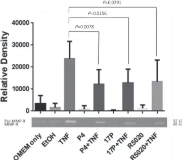
P=0.0391 P=0.0156 P=0.0078
Fig. 2. Effects of progesterone and TNF-α on MMP-9 activity in chorio- decidual membranes. Choriodecidual membranes were isolated, cul- tured, and pretreated with P4, 17P, R5020, or an EtOH as a vehicle for 24 hours, and treated with TNF-α for 48 hours. Densitometric analysis of zymography of MMP-9 (top). Representative zymography (bottom).
Shown are mean±standard deviation (n=8). MMP, matrix metallopro- teinase; OMEM, Opti-Minimum Essential Media; EtOH, ethanol; TNF, tumor necrosis factor; P4, progesterone; 17P, 17α-hydroxyprogesterone caproate; R5020, promegestone.
that of vehicle alone (P<0.01).
In this tissue culture study, progesterone did not decrease basal MMP-9 activity. There were no differences in MMP-9 protein expression among P4, 17P or R5020 treated tissue and vehicle only samples (P=0.56, P=0.68, and P=0.84, respec- tively). Zymography also did not identify any differences in MMP-9 activity in media among progesterone pre-treated and vehicle treated samples (P=0.64, P=0.05, and P>0.99, respec- tively).
Zymography showed that pre-treatment with P4, 17P, and R5020 significantly diminished TNF-α induced MMP-9 acti- vity when compared to TNF-α alone (51%, 54%, and 56%, respectively) (Fig. 1). In Western blot analysis (n=6), TNF-α induced MMP-9 expression was decreased by P4, 17P, and R5020 pretreatment when compared with the stimulated control (31%, 14%, and 21%, respectively). There were no differences in MMP-9 activity among the P4, 17P, and R5020 pretreated group in Western blot (P=0.25), and zymography analysis (P=0.36).
Discussion
Progesterone and 17P are currently being used in the clinical setting for the prevention of preterm birth in high risk groups.7,8 The exact molecular mechanism of progesterones effect has not been clearly described. The current study demonstrated that progestins reduce TNF-α mediated MMP-9 activity and expression in human choriodecidual membranes culture.
Infection and/or inflammation is now widely accepted to play a pivotal role in preterm parturition.9-11 Intrauterine infection induces secretion of proinflammatory cytokines such as TNF- α.12 In an LPS-induced murine model, the pre-treatment of anti-TNF-α decreases preterm delivery.13 TNF-α signaling, mediated through TNF receptor (TNFR)1, forms the TNF- TNFR1 complex and leads to activation of nuclear factor-κB (NF-κB). NF-κB promotes induction of MMP-9 expression and activity, ultimately leading to inflammation and potential weakening of the fetal membranes. The TNF-TNFR2 complex directly interacts with TNF receptor associated factor-2, which results in prostaglandin production and promotes uterine contractions.4
Increased activity of MMPs has been documented in fetal membranes from women with PPROM.14 Fetal membranes are composed of several layers and each layer has a distinct extracellular matrix (ECM) composition.15 MMPs hydrolyze ECM, resulting in cervical ripening and rupture of the fetal membranes.16 The fibrillar collagens have different suscepti- bility to cleavage by MMPs.17 MMP-9 degrade type IV and V collagen as well as basement membrane components, which maybe important for the maintenance of the amnion epithelial cells. MMP-9 expression has been found to be increased in term rupture as well as PPROM17-20 and is associated with fetal membrane weakening in vitro.19
Fetal membranes at the rupture site have been shown to have disruption in connective tissue and thinning of the chorion layer and increased MMP-9 activity including the zone over- lying the cervix.17 In this study we intentionally sampled mem- branes distant from the edge to avoid the weak zones.
Several putative mechanisms by which progesterone is able to prevent preterm birth have been proposed, including anti-inflammatory action, interfering with cortisol-mediated regulation of placental gene expression, and inhibiting oxy- tocin binding.21 Progesterone has also been shown to inhibit apoptosis and secretion of proinflammatory modulators induced by TNF-α in fetal membranes.22-24 It has also been proposed that binding TNF-α to TNF-R1 in the fetal membranes in- duces both the apoptotic process and MMP-9 expression.25 In this study we showed the ability of progesterones to inhibit TNF-α induced MMP-9 expression and this could prevent the associated ECM that lead to membrane weakening.
In summary, the current study shows that progesteone inhi- bits TNF-α mediated MMP-9 activity in human choriodecidual membranes. These findings may provide further support for the role of progesterone in preventing preterm birth.
Conflict of Interest
No potential conflict of interest relevant to this article was re ported.
Acknowledgments
This research was supported by Basic Science Research Program through the National Research Foundation of Korea (NRF) by the Ministry of Education, Science and Technology (2010-0005271).
References
1) Beck S, Wojdyla D, Say L, Betran AP, Merialdi M, Requejo JH, et al. The worldwide incidence of preterm birth: a systematic review of maternal mortality and morbidity. Bull World Health Organ 2010;88:31-8.
2) Martin JA, Hamilton BE, Sutton PD, Ventura SJ, Mathews TJ, Kirmeyer S, et al. Births: final data for 2007. Natl Vital Stat Rep 2010;58:1-85.
3) Maymon E, Ghezzi F, Edwin SS, Mazor M, Yoon BH, Gomez R, et al. The tumor necrosis factor alpha and its soluble receptor profile in term and preterm parturition. Am J Obstet Gynecol 1999;181(5 Pt 1):1142-8.
4) Fortunato SJ, Menon R, Lombardi SJ. Role of tumor necrosis factor- alpha in the premature rupture of membranes and preterm labor pathways. Am J Obstet Gynecol 2002;187:1159-62.
5) Mackenzie AP, Schatz F, Krikun G, Funai EF, Kadner S, Lockwood CJ.
Mechanisms of abruption-induced premature rupture of the fetal membranes: Thrombin enhanced decidual matrix metalloproteinase-3 (stromelysin-1) expression. Am J Obstet Gynecol 2004;191:1996-2001.
6) Allen TK, Feng L, Grotegut CA, Murtha AP. Progesterone receptor membrane component 1 as the mediator of the inhibitory effect of progestins on cytokine-induced matrix metalloproteinase 9 activity in vitro. Reprod Sci 2014;21:260-8.
7) Hassan SS, Romero R, Vidyadhari D, Fusey S, Baxter JK, Khandelwal M, et al. Vaginal progesterone reduces the rate of preterm birth in women with a sonographic short cervix: a multicenter, randomized, double- blind, placebo-controlled trial. Ultrasound Obstet Gynecol 2011;38:18- 31.
8) Meis PJ, Klebanoff M, Thom E, Dombrowski MP, Sibai B, Moawad AH, et al. Prevention of recurrent preterm delivery by 17 alpha-hydroxyproge- sterone caproate. N Engl J Med 2003;348:2379-85.
9) Goldenberg RL, Hauth JC, Andrews WW. Intrauterine infection and preterm delivery. N Engl J Med 2000;342:1500-7.
10) Gonçalves LF, Chaiworapongsa T, Romero R. Intrauterine infection and prematurity. Ment Retard Dev Disabil Res Rev 2002;8:3-13.
11) Romero R, Espinoza J, Goncalves LF, Kusanovic JP, Friel L, Hassan S. The role of inflammation and infection in preterm birth. Semin Reprod Med 2007;25:21-39.
12) Romero R, Espinoza J, Kusanovic JP, Gotsch F, Hassan S, Erez O, et al. The preterm parturition syndrome. BJOG 2006;113 Suppl 3:17-42.
13) Holmgren C, Esplin MS, Hamblin S, Molenda M, Simonsen S, Silver R.
Evaluation of the use of anti-TNF-alpha in an LPS-induced murine model. J Reprod Immunol 2008;78:134-9.
14) Draper D, McGregor J, Hall J, Jones W, Beutz M, Heine RP, et al. Elevated protease activities in human amnion and chorion correlate with pre- term premature rupture of membranes. Am J Obstet Gynecol 1995;173:
1506-12.
15) Parry S, Strauss JF 3rd. Premature rupture of the fetal membranes. N Engl J Med 1998;338:663-70.
16) Cockle JV, Gopichandran N, Walker JJ, Levene MI, Orsi NM. Matrix metalloproteinases and their tissue inhibitors in preterm perinatal com plications. Reprod Sci 2007;14:629-45.
17) Strauss JF 3rd. Extracellular matrix dynamics and fetal membrane rup- ture. Reprod Sci 2013;20:140-53.
18) Goldman S, Weiss A, Eyali V, Shalev E. Differential activity of the gelati- nases (matrix metalloproteinases 2 and 9) in the fetal membranes and decidua, associated with labour. Mol Hum Reprod 2003;9:367-73.
19) Kumar D, Fung W, Moore RM, Pandey V, Fox J, Stetzer B, et al. Proinflam- matory cytokines found in amniotic fluid induce collagen remodeling, apoptosis, and biophysical weakening of cultured human fetal mem- branes. Biol Reprod 2006;74:29-34.
20) McLaren J, Taylor DJ, Bell SC. Increased concentration of pro-matrix metalloproteinase 9 in term fetal membranes overlying the cervix before labor: implications for membrane remodeling and rupture. Am J Obstet Gynecol 2000;182:409-16.
21) Sfakianaki AK, Norwitz ER. Mechanisms of progesterone action in inhibiting prematurity. J Matern Fetal Neonatal Med 2006;19:763-72.
22) Luo G, Abrahams VM, Tadesse S, Funai EF, Hodgson EJ, Gao J, et al.
Progesterone inhibits basal and TNF-alpha-induced apoptosis in fetal membranes: a novel mechanism to explain progesterone-mediated prevention of preterm birth. Reprod Sci 2010;17:532-9.
23) Kumar D, Springel E, Moore RM, Mercer BM, Philipson E, Mansour JM, et al. Progesterone inhibits in vitro fetal membrane weakening. Am J Obstet Gynecol 2015;213:520.e1-9.
24) Pineda-Torres M, Flores-Espinosa P, Espejel-Nunez A, Estrada-Gutierrez G, Flores-Pliego A, Maida-Claros R, et al. Evidence of an immunosuppres- sive effect of progesterone upon in vitro secretion of proinflammatory and prodegradative factors in a model of choriodecidual infection.
BJOG 2015;122:1798-807.
25) Arechavaleta-Velasco F, Mayon-Gonzalez J, Gonzalez-Jimenez M, Her- nandez-Guerrero C, Vadillo-Ortega F. Association of type II apoptosis and 92-kDa type IV collagenase expression in human amniochorion in prematurely ruptured membranes with tumor necrosis factor recep- tor-1 expression. J Soc Gynecol Investig 2002;9:60-7.