Yonsei Med J http://www.eymj.org Volume 53 Number 5 September 2012 1045
Brief Communication
http://dx.doi.org/10.3349/ymj.2012.53.5.1045pISSN: 0513-5796, eISSN: 1976-2437 Yonsei Med J 53(5):1045-1048, 2012
Serum Amyloid A Circulating Levels and Disease Activity in Patients with Juvenile Idiopathic Arthritis
Luca Cantarini,
1Teresa Giani,
2Antonella Fioravanti,
1Francesca Iacoponi,
3Gabriele Simonini,
2Ilaria Pagnini,
2Adriano Spreafico,
1Federico Chellini,
1Mauro Galeazzi,
1and Rolando Cimaz
21Rheumatology Unit, Department of Clinical Medicine and Immunologic Sciences, University of Siena, Siena;
2Department of Pediatrics, Rheumatology Unit, Anna Meyer Children’s Hospital and University of Florence, Florence;
3Department of Biomedical Sciences, Applied Biology Section, University of Siena, Siena, Italy.
Received: October 27, 2011 Revised: December 6, 2011 Accepted: December 6, 2011
Corresponding author: Dr. Luca Cantarini, Rheumatology Unit, Department of Clinical Medicine and Immunologic Sciences, University of Siena, Viale Bracci 1, 53100 Siena, Italy.
Tel: 39 347 9385457, Fax: 39 (0) 577 40450 E-mail: [email protected]
∙ The authors have no financial conflicts of interest.
© Copyright:
Yonsei University College of Medicine 2012 This is an Open Access article distributed under the terms of the Creative Commons Attribution Non- Commercial License (http://creativecommons.org/
licenses/by-nc/3.0) which permits unrestricted non- commercial use, distribution, and reproduction in any medium, provided the original work is properly cited.
The aim of our study was to evaluate the association between circulating levels of serum amyloid A protein (SAA) and disease activity in patients with juvenile idio- pathic arthritis (JIA). Our study group included 41 JIA patients (9 male, 32 fe- male), classified according to the International League of Associations for Rheu- matology (ILAR) criteria (5); 16 had polyarticular onset disease and 25 had oligoarticular onset disease. Among 25 patients with oligoarticular disease, three had extended oligoarthritis. Serum amyloid A (SAA), erythrocyte sedimentation rate (ESR) and C-reactive protein (CRP) were measured in both patients and 26 healthy controls. SAA levels were higher in JIA patients versus healthy controls (p<0.001). Significant positive correlations were found between SAA and the presence of active joints (rho=0.363, p<0.05), the number of active joints (rho=0.418, p<0.05), ESR (R=0.702, p<0.05) and CRP (R=0.827, p<0.05). No significant correlations between ESR and the presence of active joints (rho=0.221, p=0.225) or between ESR and the number of active joints (rho=0.118, p=0.520) were demonstrated in JIA patients. No significant correlations were obtained be- tween CRP and the presence of active joints (rho=0.034, p=0.855) or between CRP and the number of active joints (rho=0.033, p=0.859). We discovered a sig- nificant increase in SAA levels in JIA patients, compared to controls, and a strong positive correlation between SAA level and JIA disease activity. We also discerned SAA to be a more sensitive laboratory marker than ESR and CRP for evaluating the presence and number of active joints. We suggest that SAA can be used as an additional indicator of disease activity in JIA.
Key Words: Serum amyloid A, juvenile idiopathic arthritis, inflammatory mark- ers, disease activity
In addition to erythrocyte sedimentation rate (ESR) and C-reactive protein (CRP), there are other known inflammatory markers, such as serum amyloid A protein (SAA).1 SAA is an acute-phase reactant transported mainly as an apolipoprotein in high-density lipoprotein, and is primarily synthesized in the liver by activated mono-
Luca Cantarini, et al.
Yonsei Med J http://www.eymj.org Volume 53 Number 5 September 2012 1046
Data are summarized as means and standard deviations or as medians and interquartile ranges, depending on the variable distribution. Comparisons between the control group and JIA group were performed using Student’s t-test for continuous variables (age and SAA) or the chi-square test for qualitative variables (gender). In the JIA group, bi- variate correlations were calculated using the Pearson corre- lation coefficient (R) or Spearman’s rho coefficient (rho).
Comparisons between examined variables were performed using the Mann Whitney U-test.
To evaluate the predictive role of variables with a correla- tion coefficient greater than 0.5 on dependent variable SAA, linear regression analysis was performed. Estimate coeffi- cients (slope) with a 95% confidence interval (CI 95%) were reported. p-values <0.05 (two-tailed) were considered statisti- cally significant. All analyses were performed using SPSS sta- tistical package, version 13 (SPSS Inc., Chicago, IL, USA).
Clinical and demographic characteristics were compara- ble between patients and controls, except for gender distri- bution (p<0.02) (Table 1). SAA levels were significantly higher in JIA patients versus healthy controls (p<0.001).
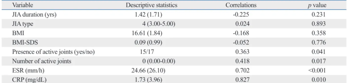
Descriptive statistics and correlation coefficients of all vari- ables in comparison with SAA are reported in Table 2. Sig- nificant positive correlations were found between SAA and the presence of active joints (rho=0.363, p<0.05), the num- ber of active joints (rho=0.418, p<0.05), ESR (R=0.702, p<0.05) and CRP (R=0.827, p<0.05). No significant corre- lations were obtained between SAA and JIA duration (R=
-0.225, p=0.231), JIA type (rho=0.024, p=0.893), BMI (R=
-0.168, p=0.358) and BMI-SDS (R=-0.052, p=0.776). Also, no significant correlations between ESR and the presence of active joints (rho=0.221, p=0.225) or between ESR and number of active joints (rho=0.118, p=0.520) were demon- strated in JIA patients. Similarly, no significant correlations were obtained between CRP and the presence of active joints (rho=0.034, p=0.855) or between CRP and the number of active joints (rho=0.033, p=0.859).
Good bivariate correlations between SAA versus ESR and CRP variables allowed us to construct a simple linear regression model in which SAA was the dependent variable and ESR or CRP was the independent variable, in order to predict SAA based on any ESR or CRP value. The estimate regression coefficients of ESR (slope=6.79, CI 95%: 4.35- 9.23) and CRP (slope=87.56, CI 95%: 66.45-108.66) were positive and significant (p<0.001); the regression model trend is shown in Fig. 1.
In vivo SAA serum concentrations may increase dramati- cytes and macrophages in response to proinflammatory cyto-
kines. Several cytokines, principally interleukin-6 (IL-6), IL-1 and tumor necrosis factor (TNF), are involved in the in- duction of SAA synthesis in hepatocytes.2
SAA has been shown to correlate with disease activity in ankylosing spondylitis, and the superiority of SAA to ESR and CRP in this regard has recently been suggested.3,4
The aim of our study was to evaluate the association be- tween circulating levels of SAA and disease activity in pa- tients with juvenile idiopathic arthritis (JIA).
Our study group included 41 children (9 males, 32 fe- males) with JIA, classified according to the International League of Associations for Rheumatology (ILAR) criteria.5 Sixteen had polyarticular onset disease and 25 had oligoar- ticular onset disease. Among 25 patients with oligoarticular disease, three had extended oligoarthritis. No patient had systemic onset disease. Eleven patients with polyarticular and seven with the oligoarticular disease were treated with oral methotrexate; 2 patients with oligoarticular disease were receiving oral sulphasalazine. The remaining patients were treated with nonsteroidal anti-inflammatory drugs only.
Twenty-six healthy children (14 male, 12 female) attending our outpatient clinic (Department of Paediatrics, Rheuma- tology Unit, Anna Meyer Children’s Hospital, University of Florence, Florence, Italy) for musculoskeletal pain and without signs of inflammation were enrolled as controls.
Table 1 summarizes the baseline demographic characteris- tics and acute phase reactant circulating levels in the JIA patients and healthy controls. We excluded subjects who had a medical or surgical condition such as recent infection, trauma, and/or a neoplastic disease, in order to remove con- founding factors affecting SAA levels.
SAA serum concentration was determined by a commer- cial solid phase sandwich Enzyme linked-immuno-sorbent assay (Human SAA; BioSource Europe S.A., Nivelles, Bel- gium). The assay sensitivity was <4 ng/mL. The normal value of SAA was <10.0 mg/L.
ESR was measured using the Westergren method, and expressed in mm/hour. An ESR of <15 mm/hour was con- sidered to be normal for males and an ESR of <20 mm/
hour was considered to be normal for females. Serum CRP concentrations were measured using a nephelometric im- munoassay, and expressed in mg/dL. A CRP of <0.5 mg/dL was considered to be normal. All the patients were evaluat- ed for disease duration, different onset types of JIA, body mass index (BMI), BMI-standard deviation score (SDS), the presence of active joints and number of active joints.
Juvenile Idiopathic Arthritis, Serum Amyloid A and Disease Activity
Yonsei Med J http://www.eymj.org Volume 53 Number 5 September 2012 1047
The present study attempted to correlate SAA levels with disease activity in JIA in comparison to ESR and CRP. Our data demonstrated that SAA levels are elevated in JIA pa- tients, and that they correlate well with disease activity, which has only once been previously demonstrated.9 Ac- cording to Scheinberg, et al.,9 higher SAA levels in children cally during acute inflammatory conditions in response to
inflammatory stimuli, such as IL-1, IL-6 and TNF-α. Sever- al studies regarding the relationship between SAA level and disease activity have been reported in rheumatic diseases such as rheumatoid arthritis, polymyalgia rheumatica and ankylosing spondylitis.3,4,6-8
Table 1. Clinical and Demographic Characteristics of Patients
Characteristics JIA patients Controls p value
No of subjects 41 26
Sex (female/male) 34/7 12/14 0.002
Age (yrs) 6.81 (3.84) 8.84 (4.67) 0.563
BMI (kg/m2) 16.76 (1.82) 17.04 (1.96) 0.554
BMI-SDS 0.08 (0.15) 0.09 (0.21) 0.821
NSAIDs 21 - -
MTX 18 - -
SSZ 2 - -
SAA levels (mg/L) 175.34 (243.83) 42.62 (38.79) <0.001
CRP levels (mg/dL) 1.73 (3.96) 0.37 (0.21) <0.001
ESR levels (mm/h) 24.65 (26.10) 8.37 (5.46) <0.001
JIA, juvenile idiopathic arthritis; BMI, body mass index; SDS, standard deviation score; NSAIDs, nonsteroidal anti-inflammatory drugs; MTX, methotrexate;
SSZ, sulfasalazine; SAA, serum amyloid A protein; CRP, C-reactive protein; ESR, erythrocyte sedimentation rate.
Data are summarized as means and standard deviations or as medians and interquartile ranges, depending on the variable distribution.
Table 2. Descriptive Statistics and Correlation Coefficients of All Variables versus SAA
Variable Descriptive statistics Correlations p value
JIA duration (yrs) 1.42 (1.71) -0.225 0.231
JIA type 4 (3.00-5.00) 0.024 0.893
BMI 16.61 (1.84) -0.168 0.358
BMI-SDS 0.09 (0.99) -0.052 0.776
Presence of active joints (yes/no) 15/17 0.363 0.041
Number of active joints 0 (0.00-0.00) 0.418 0.017
ESR (mm/h) 24.66 (26.10) 0.702 <0.001
CRP (mg/dL) 1.73 (3.96) 0.827 0.010
SAA, serum amyloid A; JIA, juvenile idiopathic arthritis; BMI, body mass index; SDS, standard deviation score; ESR, erythrocyte sedimentation rate; CRP, C-reactive protein.
Data are summarized as means and standard deviations or as medians and interquartile ranges, depending on the variable distribution.
Fig. 1. Plots with regression lines between SAA and ESR (A) and CRP (B) variables. SAA, serum amyloid A; ESR, erythrocyte sedimentation rate; CRP, C-reactive protein.
0 0
200 200
400 400
600 600
800 800
1000 1000
1200 1200
SAA (mg/L) SAA (mg/L)
0 1 2 3 4 5 6 7 8 9 10 0 20 40 60 80 100 120
CRP (mg/dL) ESR (mm/hour)
A B
Luca Cantarini, et al.
Yonsei Med J http://www.eymj.org Volume 53 Number 5 September 2012 1048
loid A (SAA): biochemistry, genetics and the pathogenesis of AA amyloidosis. Amyloid 1994;1:119-37.
2. Uhlar CM, Whitehead AS. Serum amyloid A, the major vertebrate acute-phase reactant. Eur J Biochem 1999;265:501-23.
3. Jung SY, Park MC, Park YB, Lee SK. Serum amyloid A as a use- ful indicator of disease activity in patients with ankylosing spon- dylitis. Yonsei Med J 2007;48:218-24.
4. Lange U, Boss B, Teichmann J, Klör HU, Neeck G. Serum amy- loid A--an indicator of inflammation in ankylosing spondylitis.
Rheumatol Int 2000;19:119-22.
5. Cleary AG, Sills JA, Davidson JE. Revision of the proposed clas- sification criteria for juvenile idiopathic arthritis: Durban, 1997. J Rheumatol 2000;27:1568.
6. O’Hara R, Murphy EP, Whitehead AS, FitzGerald O, Bresnihan B.
Acute-phase serum amyloid A production by rheumatoid arthritis synovial tissue. Arthritis Res 2000;2:142-4.
7. Shimojima Y, Matsuda M, Gono T, Ishii W, Ikeda S. Serum amy- loid A as a potent therapeutic marker in a refractory patient with polymyalgia rheumatica. Intern Med 2005;44:1009-12.
8. de Vries MK, van Eijk IC, van der Horst-Bruinsma IE, Peters MJ, Nurmohamed MT, Dijkmans BA, et al. Erythrocyte sedimentation rate, C-reactive protein level, and serum amyloid a protein for pa- tient selection and monitoring of anti-tumor necrosis factor treat- ment in ankylosing spondylitis. Arthritis Rheum 2009;61:1484- 9. Scheinberg MA, Hubscher O, Morteo OG, Benson MD. Serum 90.
amyloid protein levels in south american children with rheumatoid arthritis: a co-operative study. Ann Rheum Dis 1980;39:228-30.
10. Hachulla E, Saile R, Parra HJ, Hatron PY, Gosset D, Fruchart JC, et al. Serum amyloid A concentrations in giant-cell arteritis and polymyalgia rheumatica: a useful test in the management of the disease. Clin Exp Rheumatol 1991;9:157-63.
11. Hagihara K, Nishikawa T, Isobe T, Song J, Sugamata Y, Yoshizaki K. IL-6 plays a critical role in the synergistic induction of human serum amyloid A (SAA) gene when stimulated with proinflamma- tory cytokines as analyzed with an SAA isoform real-time quanti- tative RT-PCR assay system. Biochem Biophys Res Commun 2004;314:363-9.
12. Yamada T. Serum amyloid A (SAA): a concise review of biology, assay methods and clinical usefulness. Clin Chem Lab Med 1999;37:381-8.
with juvenile rheumatoid arthritis were associated with the polyarticular and systemic forms of the disease. In addition, SAA levels were correlated with disease activity, increasing during acute exacerbations and decreasing during remis- sion, in patients undergoing prednisone therapy.
According to several reports concerning clinical markers in rheumatic disorders, SAA was shown to be superior to CRP and ESR with regard to sensitivity and specificity, respective- ly, and serum levels of SAA are known to reliably indicate disease activity.7,10 Included among disease activity scores of both clinical and biological parameters, both ESR and CRP are now used to evaluate response criteria. In this regard, SAA could also prove useful, upon validation by others.
Both SAA and CRP are produced in the liver, but serum levels of the former depend on a larger number of inflam- matory cytokines, such as IL-1 and IL-6, than those of the latter.11,12
The discrepancy between CRP and SAA in JIA patients may be explained by the types and amounts of cytokines involved.
In conclusion, we discovered a significant increase in SAA levels in JIA patients, compared to controls, and a strong positive correlation between SAA level and JIA dis- ease activity. We also discerned SAA to be a more sensitive laboratory marker than ESR and CRP for evaluating the presence and number of active joints. Accordingly, we sug- gest that SAA can be used as an additional indicator of dis- ease activity in JIA.
REFERENCES
1. Husby G, Marhaug G, Dowtor B, Sletten K, Sipe JD. Serum amy-