22(4) : 246-251 (2016)
https://doi.org/10.20307/nps.2016.22.4.246
246
Effects of (−)-Sesamin on Memory Deficits in MPTP-lesioned Mouse Model of Parkinson’s Disease
Ting Ting Zhao, Keon Sung Shin, and Myung Koo Lee*
1Department of Pharmacy and Research Center for Bioresource and Health, College of Pharmacy, Chungbuk National University, 1, Chungdae-ro, Seowon-gu, Cheongju 28644, Korea
Abstract – This study investigated the effects of (−)-sesamin on memory deficits in 1-methyl-4-phenyl-1,2,3,6- tetrahydropyridine (MPTP)-lesioned mouse model of Parkinson’s disease (PD). MPTP lesion (30 mg/kg/day, 5 days) in mice showed memory deficits including habit learning memory and spatial memory. However, treatment with (−)-sesamin (25 and 50 mg/kg) for 21 days ameliorated memory deficits in MPTP-lesioned mouse model of PD: (−)-sesamin at both doses improved decreases in the retention latency time of the passive avoidance test and the levels of dopamine, norepinephrine, 3,4-dihydroxyphenylacetic acid, and homovanillic acid, improved the decreased transfer latency time of the elevated plus-maze test, reduced the increased expression of N-methyl-D- aspartate (NMDA) receptor, and increased the reduced phosphorylation of extracellular signal-regulated kinase (ERK1/2) and cyclic AMP-response element binding protein (CREB). These results suggest that (−)-sesamin has protective effects on both habit learning memory and spatial memory deficits via the dopaminergic neurons and NMDA receptor-ERK1/2-CREB system in MPTP-lesioned mouse model of PD, respectively. Therefore, (−)- sesamin may serve as an adjuvant phytonutrient for memory deficits in PD patients.
Keywords – (−)-Sesamin, MPTP-lesioned mouse model of Parkinson’s disease, Habit learning memory, Spatial memory, Dopaminergic neuron, NMDA receptor
Introduction
Parkinson’s disease (PD) is one of the common neurodegenerative disorders that caused by the loss of dopaminergic neurons in the substantia nigra,
1and many PD patients suffer from learning and memory impairment problems, including habit learning memory and spatial memory deficits during the PD process.
2,3In the memory systems of CNS, the nigro-striatal system is involved in habit learning memory via dopaminergic neurons and the hippocampal system is mainly involved in spatial memory via N-methyl-D-aspartate (NMDA) receptor.
4,5( −)-Sesamin is a major lignan constituent of Asiasari Radix (Asiasarum heterotropoides F. Maekawa var. mands- huricum F. Maekawa, Aristolochiaceae). (−)-Sesamin has ameliorative effects on lung cancer, nitric oxide production, and cholesterol and triglycerides.
6-8( −)-Sesamin also enhances dopamine biosynthesis and reduces 6-hydroxydopamine (6-OHDA)-induced cytotoxicity in dopaminergic neuronal
and PC12 cells.
9In addition, (+)-sesamin is a lignan compound in Sesamum indicum DC (Sesame seeds) and is epimeric isomer lignans with ( −)-sesamin. (+)-Sesamin has preventive effects on the loss of dopaminergic neuronal cells induced by rotenone.
10(+)-Sesamin also induces dopamine biosynthesis and reduces L-3,4-dihydroxypheny- lalanine (L-DOPA)-induced cytotoxicity in PC12 cells.
11These results indicate that ( −)-sesamin shows protective effects on nigro-striatal dopaminergic neurons.
In this study, therefore, the effects of ( −)-sesamin on memory deficits in 1-methyl-4-phenyl-1,2,3,6-tetrahy- dropyridine (MPTP)-lesioned mouse model of PD were investigated in order to confirm the neuropharmacological functions of ( −)-sesamin.
Experimental
Materials – (−)-Sesamin was isolated from A. heterotropoides and identified previously described.
9,12A voucher specimen was deposited in the herbarium of College of Pharmacy, Chungbuk National University.
Dopamine, norepinephrine, 3,4-dihydroxyphenylacetic acid (DOPAC), homovanillic acid (HVA), isoproterenol,
*Author for correspondence
Myung Koo Lee, College of Pharmacy and Research Center for Bioresource and Health, Chungbuk National University, 1, Chun- gdae-ro, Seowon-gu, Cheongju 28644, Korea
Tel: +82-43-261-2822; E-mail: [email protected]
MPTP, and phospho-NMDA receptor (type 1) (Ser 897) were purchased from Sigma-Aldrich (St. Louis, MO, USA). Extracellular signal-regulated kinase (ERK1/2), phospho-ERK1/2 (Thr 202/Tyr 204), cyclic AMP (cAMP)- response element binding protein (CREB), phospho- CREB (Ser 133), and β-actin were purchased from Cell Signaling Tech (Beverly, MA, USA). Tyrosine hydroxylase (TH) antibody was obtained from Chemicon International (Temecula, CA, USA). Vectasatin diaminobenzidine (DAB), avidin/biotinylated enzyme complex (ABC) kits, and anti- mouse IgG were purchased from Vector Laboratories (Burlingame, CA). All other chemicals were of analytical grade.
Experimental design – Mice (C57BL/6, male, 20 – 25 g) were obtained from Samtako Co. (Osan, Korea).
Animals were in a temperature (23 ± 2
oC) and humidity (60 ± 2%) under a 12-h light/dark cycle, with ad libitum access to water and standard diet. All procedures were approved by the Animal Ethics Committee of Chungbuk National University (Approval No., CBNUA-716-14-01).
Mice were randomly divided into 4 groups and each group contained 8 – 10 animals (Fig. 1). The control group received 0.9% saline. The MPTP-lesioned group (MPTP in the Figures) was injected with MPTP (30 mg/kg, daily for 5 days, i.p.).
13( −)-Sesamin (25 and 50 mg/kg) were treated for 21 days after the 5 days of MPTP injections (MPTP + ( −)-Sesamin in the Figures). After the final treatments, all mice were subjected to behavioral tests.
The mice were then anaesthetized (Zoletil 50,100 mg/kg, i.p., Virbac, Carros, France) and sacrificed to obtain brain
tissues for biochemical, western blotting and immuno- histochemical analyses.
Latency time in the step-through passive avoidance test – The apparatus consisted of an illuminated chamber connected to dark chamber by a guillotine door (Med Associates Inc., Vermont, USA). Each mouse was placed in the illuminated chamber. After the habituation period, the guillotine door was opened. If the mice entering the dark chamber, the guillotine door was closed and delivered an inescapable electric shock (0.5 mA, 3 s). For the initial trial, the initial latency time of entrance into the dark chamber was recorded. Twenty-four hours later, the retention trial was carried out.
14TH-immunohistochemistry – Mice were intracardially perfused with a paraformaldehyde solution (4% in 0.1 M phosphate buffered saline, pH 7.4). The brains were removed and preserved in 30% sucrose buffer. Coronal brain sections (30 μm) were made through the cell bodies of dopaminergic neurons in the substantia nigra (Vibratome, Leica Microsystems GmbH, Wetzelar, Germany), and the sections were processed using an anti-TH primary antibody (1:200, in 0.3% Triton X-100) overnight at 4
oC, a biotinylated goat anti-rabbit secondary antibody (1:250) and ABC kit procedure with DAB kit as chromogens according to protocols. The TH-immunopositive cells were counting using Axiovision software (Carl Zeiss MicroImaging, GmbH, Jena, Germany) with an Axiophot microscope (100x magnification) (Zeiss Axiophot, Carl Zeiss MicroImaging).
Determination of dopamine, norepinephrine, DOPAC, and HVA levels – The levels of dopamine, norepinephrine, DOPAC, and HVA in the striatum were determined by an HPLC method, as described previously with a slight modification.
15The striatum was dissected out immediately and frozen in −70
oC until analysis. The tissue samples were homogenized in 300 µl of 0.1 M perchloric acid containing 100 mM EDTA and 10 μM isoproterenol, and then centrifuged (13,000 × g, 4
oC, 15 min). The supernatant was passed a filter (Millex-GV, 0.45 μm, Waters) and the filtrate (100 μl) was injected into an HPLC system, which consisted of a solvent delivery pump (Model 1525, Waters, Milford, MA, USA), an electrochemical detector (+0.85 V, Ag/AgCl reference electrode; Model 2465;
Waters), and a Waters 120 ODS-BP column (5 μm, 50 × 4.6 mm).
16All data were expressed were expressed as percentage of the control group.
Transfer latency time in the elevated plus-maze test – The elevated plus-maze apparatus consists of two open and two closed arms (each size, 30 cm × 5 cm), with 16- cm-high black walls elevated 45 cm. Each mouse was placed on the open arm facing outwards. The time entered
Fig. 1. Experimental design. Mice (C57BL/6, male, 20 − 25 g)were divided into 4 groups (8 – 10 animals and lesions) and PD models were established with by MPTP injection (30 mg/kg, daily for 5 days, i.p.). Five days after lesion formation, MPTP- lesioned groups were treated with (−)-sesamin (25 and 50 mg/kg) orally for 21 days. After the final treatments, all mice were subjected to behavioral tests. The mice were then sacrificed to obtain brain tissues for biochemical, western blotting and immune histochemical analyses.
the closed arm in the first trial was noted as initial transfer latency time. After 24-h, retention transfer latency time was noted. The retention transfer latency time was expressed as percentage of the initial transfer latency time (%ITL).
17Western blot analysis – The hippocampal regions were dissected and immediately incubated in 1 ml of RIPA lysis buffer containing 1% protease inhibitor and 1% phosphatase inhibitor, and sonicated to obtain homogenates. Proteins in samples (30 μg in each lane) were electrophoresed in 8% or 10% sodium dodecyl sulfate-polyacrylamide gels and transferred to a polyvinylidene difluoride membrane. The blots were blocked with 5%
bovine serum albumin (BSA) in Tris-buffered saline (TBS-T), and incubated overnight using primary antibodies (dilutions, 1:1,000 using TBS-T containing 5% BSA) at 4
oC and secondary antibodies (dilutions, 1:5,000 using TBS-T containing 5% BSA) for 1 h at room temperature.
The blots were washed with TBS-T and the transferred proteins were incubated with ECL substrate solution (Amersham Pharmacia Biotech, Inc., Piscataway, NJ, USA), and then visualized with radiographic film.
Statistical analysis – The results were analyzed by one- way analysis of variance (ANOVA) followed by Tukey’s test and are expressed as the means ± S.E.M., with a P value < 0.05 being considered statistically significant.
Result and Discussion
The habit learning memory has been examined by the retention latency time in the step-through passive avoidance test,
14which was shown in Table 1. On the first day, all groups showed slightly different initial latency times, but it was not significant (average of initial latency time from each group, 19.9 s). The change of latency time between the initial latency time and retention latency time significantly increased to 108.8 s ( ± 21.0) after 24-h in the control group. However, the changes between the initial latency time and retention latency time in the MPTP- lesioned group decreased to 60.4 s ( ± 18.4) (P < 0.01), compared with the control group. This change by MPTP lesion was reversed to 91.3 s ( ± 21.3) (P < 0.05) and 95.9 s ( ± 19.3) (P < 0.05) by treatment with (−)-sesamin (25 and 50 mg/kg), respectively, compared with the MPTP- lesioned group.
Representative photomicrographs showed that TH- immunopositive neuronal cells were reduced by MPTP lesion in the substantia nigra, which were then recovered by treatment with ( −)-sesamin (25 and 50 mg/kg) (Fig.
2A). In addition, the number of TH-immunopositive
Table 1. Effects of (−)-sesamin on latency time in the passive avoidance test
Latency time (s) Initial trial Retention trial Control 21.6± 1.68 130.4 ± 19.3
MPTP 17.9± 1.97 578.3± 16.4**
MPTP + (−)-Sesamin
(25 mg/kg) 18.5± 2.76 109.8 ± 18.5 MPTP + (−)-Sesamin
(50 mg/kg) 21.7± 3.01 117.6 ± 16.3# Mice (C57BL/6, male, 20 – 25 g) were orally treated with (−)- sesamin (25 or 50 mg/kg) for 21 days after the 5 days of MPTP injections (30 mg/kg, i.p.). The control group was treated with 0.9% saline. After the final treatments, all mice were subjected to the passive avoidance test. The results are expressed as the means± S.E.M. (8 – 10 animals per group). **P < 0.01 compared with the control group, #P < 0.05 compared with the MPTP- lesioned group (one-way ANOVA followed by Tukey’s test).
Fig. 2. Representative photographs illustrating the effects of (−)- sesamin on TH-immunohistochemistry (A) and the number of TH-immunopositive cells (B) in the substantia nigra. Mice (C57BL/
6, male, 20 – 25 g) were orally treated with (−)-sesamin (25 or 50 mg/kg) for 21 days after the 5 days of MPTP injections (30 mg/
kg, i.p.). The control group was treated with 0.9% saline. (A) Immunoblots of lysates from the brain were probed with TH antibodies, and the TH-immunopositive neuronal cells were measured as described under the Experimental section. (B) The number of TH-immunopositive cells was counted in the sub- stantia nigra and was expressed as a percentage of the control groups. The results are expressed as the means± S.E.M. (8 − 10 animals per group). **P < 0.01 compared with the control group;
#P < 0.05 compared with the MPTP-lesioned group (one-way ANOVA followed by Tukey’s test).
neuronal cells was significantly reduced to 44.7% (P <
0.01) in the MPTP-lesioned group, compared with the control group (Fig. 2B). However, the number of TH- immunopositive neuronal cells also increased to 67.4%
(P < 0.05), and 76.5% (P < 0.05) by treatment with ( −)- sesamin (25 and 50 mg/kg), respectively, compared with the MPTP-lesioned group (Fig. 2B).
In addition, the levels of dopamine, norepinephrine, DOPAC, and HVA in the striatum were shown in (Figs.
3A, B, C and D). The levels of dopamine, norepinephrine, DOPAC and, HVA significantly decreased to 47.5%
(P < 0.01), 67.4% (P < 0.01), 68.6% (P < 0.01), and 70.8%
(P < 0.01) in the MPTP-lesioned group, respectively, compared with the control group. However, the decreased levels of dopamine, norepinephrine, DOPAC, and HVA by MPTP lesion increased to 75.3% and 81.6% (both, P < 0.05), 81.7%, and 86.5% (both, P < 0.05), 82.4% and 87.3% (both, P < 0.05), and 84.2% and 87.1% (both, P < 0.05) by treatment with ( −)-sesamin (25 and 50 mg/kg), respectively, compared with the MPTP-lesioned group.
The lesion of substantia nigra and striatum regions may
induce learning and memory impairments.
4,18It has also been reported that MPTP causes dopaminergic neuronal degeneration, that may induce learning and memory impairments.
3,19These results show that treatment with ( −)-sesamin can improve MPTP-induced habit learning memory deficits by protecting the dopaminergic neuronal cell death.
Next, in order to explore whether MPTP induces spatial memory, the transfer latency time in the elevated plus- maze test was examined by the ratio of retention transfer latency time to initial transfer latency time (%ITL), which is popular to determine spatial memory.
17The retention transfer latency time (%ITL) in the MPTP-lesioned group significantly increased to 144.2% (P < 0.01), compared with the control group (Fig. 4). However, the %ITL in the MPTP-lesioned group significantly decreased to 125.4%
(P < 0.05) and 116.7% (P < 0.05) by treatment with ( −)- sesamin (25 and 50 mg/kg), respectively, compared with the MPTP-lesioned group (Fig. 4).
In addition, MPTP lesion significantly induced the
expression of NMDA receptor (type 1) phosphorylation
Fig. 3. Effects of (−)-sesamin on dopamine (A), norepinephrine (B), DOPAC (C) and HVA (D) levels in the striatum. Mice (C57BL/6, male, 20 – 25 g) were orally treated with (25 or 50 mg/kg) for 21 days after the 5 days of MPTP injections (30 mg/kg, i.p.). The control group was treated with 0.9% saline. The control levels (ng/mg tissue) of dopamine (A), norepinephrine (B), DOPAC (C) and HVA (D) were 8.01± 0.71, 5.34 ± 0.61, 3.15 ± 0.49, and 1.96 ± 0.52, which were determined by HPLC analysis. The results are expressed as the means± S.E.M. (8 − 10 animals per group). **P < 0.01compared with the control group; #P < 0.05 compared with the MPTP-lesioned group (one-way ANOVA followed by Tukey’s test).to 1.61-fold (P < 0.01), compared with the control group (Fig. 5). However, the expression of NMDAR1 pho-
sphorylation induced by MPTP lesion decreased to 1.48- fold (P < 0.05) and 1.47-fold (P < 0.05) by treatment with ( −)-sesamin (25 and 50 mg/kg), respectively (Fig. 5).
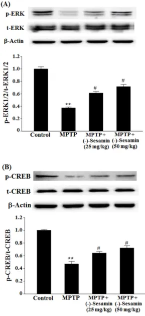
By contrast, the phosphorylation of ERK1/2 and CREB was reduced by MPTP lesion to 0.37-fold (P < 0.01) and 0.47-fold (P < 0.01), respectively, compared with the control
Fig. 4. Effects of (−)-sesamin on transfer latency time (%ITL) inthe elevate plus-maze test. Mice (C57BL/6, male, 20 – 25 g) were orally treated with (−)-sesamin (25 or 50 mg/kg) for 21 days after the 5 days of MPTP injections (30 mg/kg, i.p.). The control group was treated with 0.9% saline. After the 24-h of initial transfer latency test, the retention transfer latency time was noted and expressed as percentage of the initial transfer latency time (%ITL). The results are expressed as the means± S.E.M.
(8− 10 animals per group). **P < 0.01 compared with the control group; #P < 0.05 compared with the MPTP-lesioned group (one- way ANOVA followed by Tukey’s test).
Fig. 5. Effects of (−)-sesamin on phosphorylation of NMDAR1 in the hippocampus. Mice (C57BL/6, male, 20 – 25 g) were orally treated with (−)-sesamin (25 or 50 mg/kg) for 21 days after the 5 days of MPTP injections (30 mg/kg, i.p.). The control group was treated with 0.9% saline. Immunoblots of lysates from the mice were probed with phospho-NMDAR1 antibody, and the phosphorylation of NMDAR1 (Ser 897) (p-NMDAR1) and β- actin were analyzed by western blot analysis. The value of the relative density ratio of p-NMDAR1/β-actin is expressed in arbitrary units. The results are expressed as the means± S.E.M.
(8− 10 animals per group). **P < 0.01 compared with the control group; #P < 0.05 compared with the MPTP group (one-way ANOVA followed by Tukey’s test).
Fig. 6. Effects of (−)-sesamin on phosphorylation of ERK1/2 (A) and CREB (B) in the hippocampus. Mice (C57BL/6, male, 20 – 25 g) were orally treated with (−)-sesamin (25 or 50 mg/kg) for 21 days after the 5 days of MPTP injections (30 mg/kg, i.p.). The control group was treated with 0.9% saline. Immunoblots of lysates from the mice were probed with phospho-ERK1/2 antibody, and the phosphorylation of ERK1/2 (Thr 202/Tyr 204) (p-ERK1/2), total ERK1/2 (t-ERK1/2) and β-actin were analyzed by western blot analysis. The value of the relative density ratio of p-ERK1/2/
t-ERK1/2 is expressed in arbitrary units. The results are expressed as the means± S.E.M. (8 − 10 animals per group). **P < 0.01 compared with control group; #P < 0.05 compared with MPTP group (one-way ANOVA followed by Tukey’s test).
group (Figs. 6A and B). However, treatment with ( −)- sesamin (25 and 50 mg/kg) in the MPTP-lesioned group increased the phosphorylation of ERK1/2 and CREB to 0.61-fold and 0.71 (both P < 0.05), and 0.64 and 0.72-fold (both P < 0.05), respectively (Figs. 6A and B).
The function of NMDA receptor in the hippocampus is essential for the learning and memory processes,
20however, the overexpression of NMDA receptor in the hippocampus can damage memory.
5The phosphorylation of ERK1/2 may be an essential component of NMDA receptor pathway for the learning and memory processes via NMDA receptor (type 2B).
21The deficiency of ERK1/2 and CREB phosphorylation in the hippocampus can also induce the learning and memory impairments.
21,22In this study, the MPTP lesion enhances NMDAR1 phosphorylation in the hippocampus. These results suggest that MPTP lesion induces the spatial memory deficits, and the MPTP- induced spatial memory deficits is recovered by treatment with ( −)-sesamin (25 and 50 mg/kg) by regulating the functions of NMDA receptor, ERK1/2 and CREB.
MPTP can pass brain-blood barrier and spread in the brain, which can induce the nigro-striatal dopaminergic neuronal cell death,
23and then may also damage the hippocampal region. MPTP lesion may reduce the movement of the mice. In this experiment, MPTP lesion induced both habit learning memory and spatial memory deficits and ( −)-sesamin improved the MPTP-lesioned memory deficits measured by passive avoidance and elevated plus-maze. These experiments are also influenced by locomotor activity.
In conclusion, this study suggest that ( −)-sesamin has protective effects on memory deficits of both habit learning memory and spatial memory via the dopaminergic neurons and NMDA receptor-ERK1/2-CREB system in MPTP- lesioned mouse model of PD. ( −)-Sesamin may serve as an adjuvant phytonutrient for the memory deficits of PD patients.
Acknowledgements
This work was supported by a research grant from Chungbuk National University (2015).
References
(1) Mayeux, R. Annu. Rev. Neurosci. 2003, 26, 81-104.
(2) Buter, T. C.; van den Hout, A.; Matthews, F. E.; Larsen, J. P.;
Brayne, C.; Aarsland, D. Neurology 2008, 70, 1017-1022.
(3) Williams-Gray, C. H.; Foltynie, T.; Brayne, C. E.; Robbins, T. W.;
Barker, R. A. Brain 2007, 130, 1787-1798.
(4) Da Cunha, C.; Angelucci, M. E. M.; Canteras, N. S.; Wonnacott, S.;
Takahashi, R. N. Cell. Mol. Neurobiol. 2002, 22, 227-237.
(5) Shi, L.; Adams, M. M.; Long, A.; Carter, C. C.; Bennett, C.;
Sonntag, W. E.; Nicolle, M. M.; Robbins, M.; D’Agostino, R.; Brunso- Bechtolda, J. K. Radiat. Res. 2006, 166, 892-899.
(6) Wang, H. M.; Cheng, K. C.; Lin, C. J.; Hsu, S. W.; Fang, W. C.;
Hsu, T. F.; Chiu, C. C.; Chang, H. W.; Hsu, C. H.; Lee, A. Y. Cancer Sci.
2010, 101, 2612-2620.
(7) Han, A. R.; Kim, H. J.; Shin, M.; Hong, M.; Kim, Y. S.; Bae, H.
Chem. Biodivers. 2008, 5, 346-351.
(8) Peñalvo, J. L.; Hopia, A.; Adlercreutz, H. Eur. J. Nutr. 2006, 45, 439-444.
(9) Park, H. J.; Zhao, T. T.; Lee, K. S.; Lee, S. H.; Shin, K. S.; Park, K.
H.; Choi, H. S.; Lee, M. K. Neurochem. Int. 2015, 83-84, 19-27.
(10) Fujikawa, T.; Kanada, N.; Shimada, A.; Ogata, M.; Suzuki, I.;
Hayashi, I.; Nakashima, K. Biol. Pharm. Bull. 2005, 28, 169-172.
(11) Zhang, M.; Lee, H. J.; Park, K. H.; Park, H. J.; Choi, H. S.; Lim, S.
C.; Lee, M. K. Neuropharmacology 2012, 62, 2219-2226.
(12) Li, C. Y.; Chow, T. J.; Wu, T. S. J. Nat. Prod. 2005, 68, 1622-1624.
(13) Schober, A. Cell Tissue Res. 2004, 318, 215-224.
(14) Roghani, M.; Baluchnejadnojarad, T. Basic Clin. Neurosci. 2010, 1, 52-55.
(15) Izurieta-Sánchez, P.; Sarre, S.; Ebinger, G.; Michotte, Y. Eur. J.
Pharmacol. 1998, 353, 33-42.
(16) Shin, K. S.; Choi, H. S.; Zhao, T. T.; Suh, K. H.; Kwon, I. H.; Choi, S. O.; Lee, M. K. Arch. Pharm. Res. 2013, 36, 759-767.
(17) Kumar, B.; Kuhad, A.; Chopra, K. Psychopharmacology 2011, 214, 819-828.
(18) El Massioui, N.; Delatour, B. V. Neurosci. Res. Comm. 1997, 21, 103-111.
(19) Matsumoto, N.; Hanakawa, T.; Maki, S.; Graybiel, A. M.; Kimura, M. J. Neurophysiol. 1999, 82, 978-98.
(20) Bannerman, D. M.; Good, M. A.; Butcher, S. P., Ramsay, M.;
Morris, R. G. Nature 1995, 378, 182-186.
(21) Krapivinsky, G.; Krapivinsky, L.; Manasian, L.; Ivanov, A.; Tyzio, R.; Pellegrino, C.; Ben-Ari, Y.; Clapham, D. E.; Medina, I. Neuron 2003, 40, 775-784.
(22) Carlezon Jr, W. A.; Duman, R. S.; Nestler, E. J. Trends Neurosci.
2005, 28, 436-445.
(23) Heikkila, R. E.; Hess, A.; Duvoisin, R. C. Science 1984, 224, 1451-1453.
Received March 3, 2016 Revised June 10, 2016 Accepted June 10, 2016