pISSN 2289-0203ㆍeISSN 2383-7101 https://doi.org/10.15747/jcn.2021.13.1.17
생체 전기 임피던스 분석으로 측정한 내장 지방 단면적이 대장 수술의 임상병리학적 결과에 미치는 영향
김경의
1,2, 송우진
1,2, 석민지
3, 배성욱
1,4, 정운경
1, 백성규
11계명대학교 의과대학 외과학교실, 2계명대학교 일반대학원, 3계명대학교 동산병원 간호부, 4계명대학교 동산병원 영양집중지원팀
Impact of Visceral Fat Area Measured by Bioelectrical Impedance Analysis on Clinico-Pathologic Outcomes of Colorectal Surgery
Kyeong Eui Kim
1,2, Woo Jin Song
1,2, Minji Seok
3, Sung Uk Bae
1,4, Woon Kyung Jeong
1, Seong Kyu Baek
11Department of Surgery, Keimyung University School of Medicine, 2Keimyung University Graduate School, 3Department of Nursing, Keimyung University Dongsan Hospital, 4Nutrition Support Team, Keimyung University Dongsan Medical Center, Daegu, Korea
Purpose: This study investigated the relationship between the visceral fat area (VFA) and clinico-pathological outcomes in patients with colorectal cancer (CRC).
Methods: This retrospective study included 204 patients who underwent anthropometric measurement by bioelectrical impedance analysis (BIA) before surgical treatment for CRC between January 2016 and June 2020.
Results: According to the average value of the visceral fat area, 119 (58.3%) patients had a low visceral fat area, and 85 (59.1%) patients had a high visceral fat area. Patients with visceral obesity showed a higher BMI compared to patients without visceral obesity, (21.8±1.9 vs. 25.7±2.5, P<0.001). There was no significant difference in the overall perioperative outcomes including total operation time, time to gas out, sips of water, soft diet, hospital stay, and morbidity between patients in the low and high VFA groups. We divided patients into two subgroups according to the degree of cancer progression and more advanced cases with low VFA showed significantly more total and positive retrieved lymph nodes (LNs) (20.9±10.3 vs. 16.1±7.1, P=0.021 and 3.3±2.9 vs. 2.2±2.3, P=0.019, respectively) and a higher proportion of more than 12 retrieved LNs compared to patients with a high VFA (95.1% vs. 90.0%, P=0.047). Body composition analysis showed that phase angle, muscle composition, and body fluid composition were not statistically different between the two groups. However, body fat mass was statistically higher in the high VFA group (22.0±4.6 vs. 12.8±3.1, P<0.001).
Conclusion: Visceral obesity measured by BIA showed lower total and positive retrieved LNs and was not associated with adverse peri-operative outcomes, inflammatory and nutritional, and pathologic outcomes for CRC.
Key Words: Colorectal neoplasm, Nutrition assessment, Body composition, Electric impedance, Prognosis
Received June 24, 2021; Accepted June 26, 2021
Correspondence to Sung Uk Bae https://orcid.org/0000-0002-7876-4196 Department of Surgery, Keimyung University School of Medicine, 1035 Dal- gubeol-daero, Dalseo-gu, Daegu 42601, Korea
Tel: +82-53-258-4708, Fax: +82-53-258-4710, E-mail: [email protected] Conflict of interest: None.
This is an Open Access article distributed under the terms of the Creative Commons Attribution Non-Commercial License (http://creativecommons.org/licenses/by-nc/4.0) which permits unrestricted non-commercial use, distribution, and reproduction in any medium, provided the original work is properly cited.
INTRODUCTION
Colorectal cancer (CRC) is the third most frequently diag- nosed cancer and second most mortality in worldwide.
Obesity is a global health growing problem. According to World Health Organization, 39% of adults aged 18 years and over were overweight, and 13% of adults were obese. The re-
lationship between body weight and several cancers is now well recognized obesity is now a well-established risk factor for development of CRC and is also associated with in- creased mortality from CRC.1-3 In clinical setting, body mass index has been used to one of the most reliable anthro- pometric methods to check obesity, however it doesn’t reflect the accumulation of adipose tissue, especially intra-abdomi- nal or visceral fat tissue.4,5
Some studies showed that increase of visceral fat was asso- ciated with post-operatively clinical outcomes and oncologic outcomes. A systemic review demonstrated that visceral obe- sity, especially, is associated with an increased risk of longer hospital stay, higher morbidity, and longer operative time af- ter colon surgery and that obese patients had lower chances of survival and more aggressive biological tumor features.6 However, a study reported that patients with visceral obesity tended to have significantly better overall survival than pa- tients with non-visceral obesity and controversies exist re- garding the correlation between visceral obesity and the out- come of colon cancer.
Bioelectrical impedance analysis (BIA) is a non-invasive technique that requires a low cost equipment available at many health care services for routine nutritional assessment describes the percentages of fat, protein, minerals in human bodies. Recently, several studies have established a relation- ship between some parameters of body composition such as skeletal muscle mass index, the index of sarcopenia or phase angle and clinical and oncologic outcomes of CRC.7-9 However, to our knowledge, there were no studies about us- ing BIA to find the effects of visceral fat on outcomes of CRC. Therefore, our study aimed to compare the effects of visceral obesity measuring by bioelectrical impedance analy- sis using Inbody 770 (Biospace, Seoul, Korea) on clinical and pathologic outcomes to patients who was treated with sur- gery for CRC.
MATERIALS AND METHODS
1. Patients and data collection
The study group included 204 patients who underwent lap- aroscopic surgery for colorectal adenocarcinoma between January 2016 and June 2020. The patients were divided into
low and high groups according to visceral fat area (VFA) measured by BIA. The exclusion criteria included synchro- nous or previous malignancies, malignancies other than ad- enocarcinoma, and familial adenomatous polyposis or heredi- tary nonpolyposis colorectal cancer. This study protocol was approved by the Institutional Review Board of the Dongsan Medical Center, and informed consent was obtained from all patients.
Data on patient demographics, including age, sex, pre- operative carcinoembryonic antigen, body mass index (BMI), and location of the tumor, platelet-lymphocyte ratio (PLR), neutrophil-lymphocyte ratio (NLR) and platelet-neutrophil index (PNI) were collected retrospectively. Perioperative out- comes included operation time, time to gas out, sips of water, and soft diet, hospital stay, morbidity within 30 days and Clavien-Dindo classification. Pathologic outcomes included tumor, node, metastasis (TNM) stage, histology, number of harvested lymph nodes and positive lymph nodes, metastatic lymph node ratio, tumor size, lymphovascular invasion, peri- neural invasion, and extranodal tumor deposits.
2. Bioimpedance analysis
BIA was performed using Inbody 770 (Biospace) to esti- mate patient’s body composition at their first visit. Among various parameters of BIA, we categorized variables as body composition and metabolic index, fat index, muscle index, obesity index, and phase angle. We used the average value of the VFA as the cut-off level, because there have been no pre- vious studies on the cut-off value for VFA using BIA. Skeletal muscle index (SMI) was calulated using Baumgartner’s defi- nition (appendicular skeletal muscle mass/height2).
3. Preoperative evaluation and surgical treatment
All of the patients underwent preoperative evaluation in- cluding colonoscopy, computed tomography scan of chest and abdomen, and magnetic resonance imaging of the pelvis.Some patients underwent positron emission tomography scans for check distant metastasis. We followed the general principles of complete mesocolic or mesorectal excision and central vascular ligation for CRC. The primary tumor was re- sected by sharp dissection of the visceral plane from the pari- etal fascia layer along with the entire regional mesocolon in
Table 1. Patient characteristics Variables Low VFA
(n=119)
High VFA
(n=85) P-value
Age (y) 66.4±9.9 65.5±10.1 0.544
Sex 0.053
Male 88 (73.9) 52 (61.2)
Female 31 (26.1) 33 (38.8)
Preoperative CEA (ng/mL)
8.0±23.2 3.4±4.8 0.073
Preoperative CRP 0.74±1.6 0.35±0.5 0.060
ASA score 0.872
I 34 (28.6) 25 (29.4)
II 68 (57.1) 50 (58.8)
III 17 (14.3) 10 (11.8)
BMI (kg/m2) 21.8±1.9 25.7±2.5 <0.001
Location of tumor 0.256
Right-sided 32 (26.9) 17 (20.0) Left-sided 87 (73.1) 68 (80.0)
PLR 181.7±110.7 190.7±102.9 0.548
NLR 3.3±3.4 3.1±2.6 0.700
PNI 67.0±27.0 72.6±32.6 0.199
Values are presented as mean±standard deviation or number (%).
VFA = visceral fat area; CEA = carcinoembryonic antigen; CRP = C-reactive protein; ASA = American Society of Anesthesiologists;
BMI = body mass index; PLR = platelet-to-lymphocyte ratio; NLR
= neutrophil to lymphocyte ratio; PNI = prognostic nutritional index.
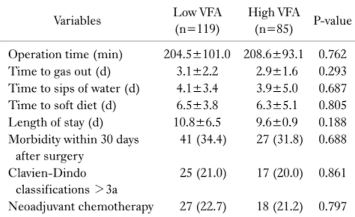
Table 2. Perioperative clinical outcomes
Variables Low VFA
(n=119)
High VFA (n=85) P-value Operation time (min) 204.5±101.0 208.6±93.1 0.762 Time to gas out (d) 3.1±2.2 2.9±1.6 0.293 Time to sips of water (d) 4.1±3.4 3.9±5.0 0.687 Time to soft diet (d) 6.5±3.8 6.3±5.1 0.805 Length of stay (d) 10.8±6.5 9.6±0.9 0.188 Morbidity within 30 days
after surgery
41 (34.4) 27 (31.8) 0.688
Clavien-Dindo classifications >3a
25 (21.0) 17 (20.0) 0.861
Neoadjuvant chemotherapy 27 (22.7) 18 (21.2) 0.797 Values are presented as mean±standard deviation or number (%).
VFA = visceral fat area.
an intact package. For right-sided colon cancer, radical lym- phadenectomy including D2 or D3 dissection along the pri- mary feeding vessels along a vertical line to expose the supe- rior mesenteric vein was performed. For left-sided colon or rectal cancer, high ligation or selectively low ligation of the inferior mesenteric artery with lymph node dissection ac- cording to the tumor location was performed. Tumor stages were classified in accordance with the American Joint Committee on Cancer 8th Edition staging system.
4. Statistical analysis
The results are presented as medians with ranges for con- tinuous outcomes and as frequencies with percentages for categorical outcomes. Categorical variables were analyzed using chi-square and Fisher’s exact tests. Continuous varia- bles were analyzed with independent t-test and Mann-Whitney test. A P-value <0.05 was considered to indicate statistical significance. The statistical analyses were performed with IBM SPSS Statistics version 25 (IBM Corp., Armonk, NY, USA).
RESULTS
1. Patient characteristic
According to the average value of VFA, 119 (58.3%) pa- tients had low VFA and 85 (59.1%) patients had high VFA.
Patients’ characteristics which were divided into amount of visceral fat are summarized in Table 1. Preoperative carci- noembryonic antigen and C-reactive protein tended to high- er in non-visceral obesity (8.0±23.2 vs. 3.4±4.8, P=0.073 and 0.74±1.6 vs. 0.35±0.5, P=0.060, respectively). Patients with high VFA showed significantly higher BMI than patients with low VFA (21.8±1.9 vs. 25.7±2.5, P<0.001). All in- flammation indexes including PLR, NLR, PNI had no stat- istical differences between two groups.
2. Perioperatively clinical outcomes
Table 2 showed no significant difference in the overall per- ioperative outcomes including total operation time, time to gas out, sips of water, soft diet, and hospital stay between pa- tients with low and high VFA groups. Also, there were no sig- nificant difference in morbidity within 30 days after surgery, the proportion of Clavien-Dindo classification >3a, and neo- adjuvant chemotherapy.
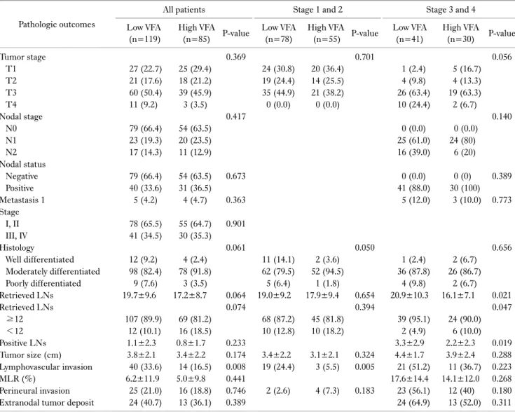
3. Postoperatively pathologic outcomes
Comparing to non-visceral obesity, there were no sig- nificant difference in overall TNM stage, differentiation, the number of total lymph node harvested and pathologically
Table 3. Postoperative pathologic outcomes Pathologic outcomes
All patients Stage 1 and 2 Stage 3 and 4
Low VFA (n=119)
High VFA
(n=85) P-value Low VFA (n=78)
High VFA
(n=55) P-value Low VFA (n=41)
High VFA
(n=30) P-value
Tumor stage 0.369 0.701 0.056
T1 27 (22.7) 25 (29.4) 24 (30.8) 20 (36.4) 1 (2.4) 5 (16.7)
T2 21 (17.6) 18 (21.2) 19 (24.4) 14 (25.5) 4 (9.8) 4 (13.3)
T3 60 (50.4) 39 (45.9) 35 (44.9) 21 (38.2) 26 (63.4) 19 (63.3)
T4 11 (9.2) 3 (3.5) 0 (0.0) 0 (0.0) 10 (24.4) 2 (6.7)
Nodal stage 0.417 0.140
N0 79 (66.4) 54 (63.5) 0 (0.0) 0 (0.0)
N1 23 (19.3) 20 (23.5) 25 (61.0) 24 (80)
N2 17 (14.3) 11 (12.9) 16 (39.0) 6 (20)
Nodal status
Negative 79 (66.4) 54 (63.5) 0.673 0 (0.0) 0 (0) 0.389
Positive 40 (33.6) 31 (36.5) 41 (88.0) 30 (100)
Metastasis 1 5 (4.2) 4 (4.7) 0.363 5 (12.0) 3 (10.0) 0.773
Stage
I, II 78 (65.5) 55 (64.7) 0.901
III, IV 41 (34.5) 30 (35.3)
Histology 0.061 0.050 0.656
Well differentiated 12 (9.2) 4 (2.4) 11 (14.1) 2 (3.6) 1 (2.4) 2 (6.7)
Moderately differentiated 98 (82.4) 78 (91.8) 62 (79.5) 52 (94.5) 36 (87.8) 26 (86.7)
Poorly differentiated 9 (7.6) 3 (3.5) 5 (6.4) 1 (1.8) 4 (9.8) 2 (6.7)
Retrieved LNs 19.7±9.6 17.2±8.7 0.064 19.0±9.2 17.9±9.4 0.654 20.9±10.3 16.1±7.1 0.021
Retrieved LNs 0.074 0.394 0.047
≥12 107 (89.9) 69 (81.2) 68 (87.2) 45 (81.8) 39 (95.1) 24 (90.0)
<12 12 (10.1) 16 (18.5) 10 (12.8) 10 (18.2) 2 (4.9) 6 (10.0)
Positive LNs 1.1±2.3 0.8±1.7 0.233 3.3±2.9 2.2±2.3 0.019
Tumor size (cm) 3.8±2.1 3.4±2.2 0.174 3.4±2.2 3.1±2.1 0.324 4.4±1.7 3.9±2.4 0.288
Lymphovascular invasion 40 (33.6) 14 (16.5) 0.008 19 (24.4) 3 (5.5) 0.005 21 (51.2) 11 (36.7) 0.223
MLR (%) 6.2±11.9 5.0±9.8 0.441 17.6±14.4 14.1±12.0 0.268
Perineural invasion 25 (21.0) 16 (18.8) 0.746 2 (2.6) 4 (7.3) 0.183 23 (56.1) 12 (40) 0.180
Extranodal tumor deposit 24 (40.7) 13 (36.1) 0.389 24 (64.9) 13 (52.0) 0.311
Values are presented as mean±standard deviation or number (%).
VFA = visceral fat area; LN = lymph node; MLR = metastatic lymph nodes ratio.
positive lymph node, tumor side, tumor size, lymphovascular invasion, perineural invasion, and extranodal tumor deposit between two groups, except there were more lymphovascular invasion in patients with low VFA (33.6% vs. 16.5%, P=
0.008) (Table 3).
To investigate the impact of VFA in nodal disease, we div- ided into two subgroups including stage one and two CRC and stage three and four CRC. In earlier CRC, there were no significant difference in tumor stage and patients with low FVA showed more poorly differentiated tumor histology (6.4% vs. 1.8%, P=0.050). The mean number of total and positive retrieved lymph nodes, the proportion of more than 12 lymph nodes harvested, and perineural invasion were not
significantly different, however low VFA group had more lymphovascular invasion than high VFA group (24.4% vs.
5.5%, P=0.005).
In more advanced CRC, patients with low VFA showed sig- nificantly more total and positive retrieved lymph nodes (20.9±10.3 vs. 16.1±7.1, P=0.021 and 3.3±2.9 vs. 2.2±2.3, P=0.019, respectively) and higher proportion of more than 12 retrieved lymph nodes compared to patients with high VFA (95.1% vs. 90.0%, P=0.047). Tumor sizes, lymphovas- cular invasion, metastatic lymph nodes ratio, perineural in- vasion, and extranodal tumor deposits were not significantly different between two groups.
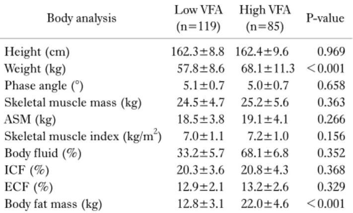
Table 4. Inbody 770 body composition analysis of patients Body analysis Low VFA
(n=119)
High VFA
(n=85) P-value
Height (cm) 162.3±8.8 162.4±9.6 0.969
Weight (kg) 57.8±8.6 68.1±11.3 <0.001
Phase angle (°) 5.1±0.7 5.0±0.7 0.658
Skeletal muscle mass (kg) 24.5±4.7 25.2±5.6 0.363
ASM (kg) 18.5±3.8 19.1±4.1 0.266
Skeletal muscle index (kg/m2) 7.0±1.1 7.2±1.0 0.156
Body fluid (%) 33.2±5.7 68.1±6.8 0.352
ICF (%) 20.3±3.6 20.8±4.3 0.368
ECF (%) 12.9±2.1 13.2±2.6 0.329
Body fat mass (kg) 12.8±3.1 22.0±4.6 <0.001 Values are presented as mean±standard deviation.
VFA = visceral fat area; ASM = appendicular skeletal muscle mass;
ICF = intracellular fluid; ECF = extracellular fluid.
4. Inbody 770 body composition analysis of patients
Table 4 showed the body composition analysis of patients between non-visceral obesity and visceral obesity patients us- ing Inbody 770. Patients with high VFA had higher weight compared to patients with low VFA. Phase angle, muscle compositions including skeletal muscle mass, appendiceal skeletal muscle mass and SMI were not statistically different between two groups. Body fluid, intracellular fluid composi- tion, and extracellular fluid composition showed no sig- nificant differences between two groups, however body fat mass was statistically higher in high VFA group (22.0±4.6 vs.12.8±3.1, P<0.001).
DISCUSSION
In this study, we investigated the surgical outcomes and short-term oncologic outcomes for viscerally obese patients with CRC. To our knowledge, this study is the first report to evaluate the effects of visceral obesity on CRC using BIA.
The present study shows that among CRC patients, VFA measured by BIA was not associated with peri-operative out- comes, inflammatory and nutritional, and pathologic out- comes after colorectal surgery. However, patients with low VFA showed more total and positive retrieved lymph nodes and the proportion of more than 12 retrieved lymph nodes compared to patients with high VFA.
Traditionally, body fat composition The WHO BMI defi- nition of obesity ≥30 kg/m2 was adopted, but we also in-
cluded studies in which BMI was defined as ≥25 kg/m2 for Asian populations. Visceral fat tissue has been acknowledged to be more pathogenic than BMI and visceral adipose tissue could be quantified by computerized tomography, and has been identified as a risk factor for colon cancer.10,11 Com- pared to subcutaneous adipose tissue, visceral revealed high levels of markers of inflammatory lipid metabolism and some of them associated with cancer stage.10 Gao et al.12 reported that VFA measured by BIA showed satisfactory reliability with that measured by CT and suggested specific cut-off val- ue for VFA by BIA in diagnosing visceral obesity for patients with gastric cancer in the Chinese population. Our study showed positive relationships between BMI and body fat mass and visceral fat have positive relationships. We think that VFA measured by BIA can be an index as surrogates of visceral obesity, although we could not compare the accuracy of BIA in estimating VFA with other index such as BMI, waist circumference, waist-to-hip ratio, or VFA measured by CT scan.
Some studies showed that obese patients have a significant risk of overall postoperative complications, surgical site in- fection, anastomotic leakage and colostomy complications.
Kang et al.13 divided into the obese group and the non-obese group who underwent laparoscopic surgery for rectal cancer according to BMI and VFA measured by abdominal CT and demonstrated that VFA was more reliable predictive in- dicator than BMI in estimating early surgical outcomes for patients who underwent rectal cancer surgery. Yu et al.14 in- vestigated VFA and general obesity and to compare visceral and general obesity as predictors of surgical outcomes of a CRC resection and described that there was no differences in morbidity, mortality, postoperative bowel recovery, and re- admission rate after surgery between the visceral obesity and visceral non-obesity groups. In the current study, visceral obesity has no influence on intraoperative difficulties, post- operative complications, and postoperative recovery in pa- tients with CRC. Prospective studies with more sample-size are needed.
Some studies evaluated the importance of lymph node metastasis in colon cancer and found that visceral obesity was associated with a lower likelihood of metastatic lymph node involvement.15,16 Park et al.17 showed that a larger num-
ber of lymph nodes removed in patients without obesity than in patients with BMI=25.0~29.9 kg/m2, but no differences compared with patients with higher BMI (>30.0 kg/m2). A study that evaluated the impact of visceral obesity on lymph node metastasis and overall survival in colon cancer reported that metastatic lymph node ratio was significantly associated only with lower VFA to total fat area ratio.18 Meanwhile, cur- rent guidelines for CRC treatment suggest that a minimum 12 lymph nodes need to be examined to establish nodal stage.
Those guidelines recommend that less than 12 lymph nodes retrieved constitute the high-risk factors for recurrence and adjuvant chemotherapy is beneficial to those patients. In our study, non-visceral obesity patients showed more total and pathological lymph nodes harvested than patients with vis- ceral obesity. We think that surgeon may have more difficulty to perform a radical lymphadenectomy in the excess fat tis- sue around major vessels in patients with visceral obesity.
And identification of lymph nodes were difficult for pa- thologists.19
A recent study showed that sarcopenia had negatively im- pact on overall survival, disease-free survival, recurrence-free survival, and cancer-specific survival in patients with non- metastatic and metastatic CRC.20 Phase angle that is defined as the ratio of resistance (intracellular and extracellular re- sistance) to reactance (cell membrane-specific resistance) expressed as an angle and is considered an indicator of cell membrane function. There were few studies about relation phase angle and other gastrointestinal cancers that low phase angle showed bad clinical and pathological outcomes.21,22 We tried to find the association between VFO and other nutri- tional index measured by BIA such as SMI and phase angle, however there was no statistical relationship between visceral obesity and those parameters.
Some previous studies reported the PLR are associated with fat respectively. Bahadır et al.23 reported that lympho- cyte count significantly was higher while increasing BMI and Samocha-Bonet et al.24 found that platelet count had positive relation to BMI only in females. Because female had high body fat mass and excessive adipose tissue was shown to in- duce systemic and chronic inflammation through the release of inflammatory cytokines including interleukin-6 (IL-6).
Yudkin et al.25 have demonstrated an association between
obesity and IL-6 levels. IL-6 is inflammatory cytokines that plays a crucial role in increasing platelet count. However, in- flammation factors including PLR, PNI, NLR showed no re- markable differences in non-visceral obesity patients to vis- ceral obesity patients in this study.
The limitations of this study include its retrospective de- sign, which is subject to incomplete data and potential se- lection bias in single institution. Secondly, our study in- cluded only small number of patients and didn’t include sur- vival data. Thirdly, the cut-off value of visceral obesity was the average value of the patients included in our study.
Further prospective study with receiver operating character- istic curve to determine the cut-off value of visceral obesity measured by BIA is needed. In conclusion, visceral obesity measured by BIA showed lower total and positive retrieved lymph nodes and was no associated with peri-operative out- comes, inflammatory and nutritional, and pathologic out- comes for CRC.
ORCID
Kyeong Eui Kim, https://orcid.org/0000-0001-9984-9976 Woo Jin Song, https://orcid.org/0000-0002-9045-3657 Minji Seok, https://orcid.org/0000-0002-4159-8665 Sung Uk Bae, https://orcid.org/0000-0002-7876-4196 Woon Kyung Jeong, https://orcid.org/0000-0001-8421-218X Seong Kyu Baek, https://orcid.org/0000-0001-6427-8675
REFERENCES
1. Renehan AG, Tyson M, Egger M, Heller RF, Zwahlen M.
Body-mass index and incidence of cancer: a systematic review and meta-analysis of prospective observational studies. Lancet 2008;371(9612):569-78.
2. Calle EE, Kaaks R. Overweight, obesity and cancer: epidemio- logical evidence and proposed mechanisms. Nat Rev Cancer 2004;4(8):579-91.
3. Murphy TK, Calle EE, Rodriguez C, Kahn HS, Thun MJ. Body mass index and colon cancer mortality in a large prospective study. Am J Epidemiol 2000;152(9):847-54.
4. WHO Expert Consultation. Appropriate body-mass index for Asian populations and its implications for policy and inter- vention strategies. Lancet 2004;363(9403):157-63.
5. Examination Committee of Criteria for ‘Obesity Disease’ in
Japan; Japan Society for the Study of Obesity. New criteria for
‘obesity disease’ in Japan. Circ J 2002;66(11):987-92.
6. Cakir H, Heus C, van der Ploeg TJ, Houdijk AP. Visceral obesity determined by CT scan and outcomes after colorectal surgery; a systematic review and meta-analysis. Int J Colorectal Dis 2015;
30(7):875-82.
7. Song WJ, Kim KE, Bae SU, Jeong WK, Baek SK. Association between body composition measured by bioelectrical im- pedance analysis and platelet-to-lymphocyte ratio in colorectal cancer. Korean J Clin Oncol 2019;15(1):7-14.
8. Oh RK, Ko HM, Lee JE, Lee KH, Kim JY, Kim JS. Clinical impact of sarcopenia in patients with colon cancer undergoing laparoscopic surgery. Ann Surg Treat Res 2020;99(3):153-60.
9. Gupta D, Lis CG, Dahlk SL, King J, Vashi PG, Grutsch JF, et al.
The relationship between bioelectrical impedance phase angle and subjective global assessment in advanced colorectal cancer.
Nutr J 2008;7:19.
10. Liesenfeld DB, Grapov D, Fahrmann JF, Salou M, Scherer D, Toth R, et al. Metabolomics and transcriptomics identify pathway differences between visceral and subcutaneous adipose tissue in colorectal cancer patients: the ColoCare study. Am J Clin Nutr 2015;102(2):433-43.
11. Suzuki S, Goto A, Nakatochi M, Narita A, Yamaji T, Sawada N, et al. Body mass index and colorectal cancer risk: a Mendelian randomization study. Cancer Sci 2021;112(4):1579-88.
12. Gao B, Liu Y, Ding C, Liu S, Chen X, Bian X. Comparison of visceral fat area measured by CT and bioelectrical impedance analysis in Chinese patients with gastric cancer: a cross-sec- tional study. BMJ Open 2020;10(7):e036335.
13. Kang J, Baek SE, Kim T, Hur H, Min BS, Lim JS, et al. Impact of fat obesity on laparoscopic total mesorectal excision: more reliable indicator than body mass index. Int J Colorectal Dis 2012;27(4):497-505.
14. Yu H, Joh YG, Son GM, Kim HS, Jo HJ, Kim HY. Distribution and impact of the visceral fat area in patients with colorectal cancer. Ann Coloproctol 2016;32(1):20-6.
15. Watanabe J, Tatsumi K, Ota M, Suwa Y, Suzuki S, Watanabe A, et al. The impact of visceral obesity on surgical outcomes of laparoscopic surgery for colon cancer. Int J Colorectal Dis 2014;
29(3):343-51.
16. Görög D, Nagy P, Péter A, Perner F. Influence of obesity on lymph node recovery from rectal resection specimens. Pathol Oncol Res 2003;9(3):180-3.
17. Park JW, Lim SW, Choi HS, Jeong SY, Oh JH, Lim SB. The impact of obesity on outcomes of laparoscopic surgery for colorectal cancer in Asians. Surg Endosc 2010;24(7):1679-85.
18. Park SW, Lee HL, Doo EY, Lee KN, Jun DW, Lee OY, et al.
Visceral obesity predicts fewer lymph node metastases and better overall survival in colon cancer. J Gastrointest Surg 2015;
19(8):1513-21.
19. Yang T, Wei M, He Y, Deng X, Wang Z. Impact of visceral obesity on outcomes of laparoscopic colorectal surgery: a meta-analysis.
ANZ J Surg 2015;85(7-8):507-13.
20. Vergara-Fernandez O, Trejo-Avila M, Salgado-Nesme N. Sarco- penia in patients with colorectal cancer: a comprehensive review.
World J Clin Cases 2020;8(7):1188-202.
21. Yu B, Park KB, Park JY, Lee SS, Kwon OK, Chung HY.
Bioelectrical impedance analysis for prediction of early com- plications after gastrectomy in elderly patients with gastric cancer: the phase angle measured using bioelectrical impedance analysis. J Gastric Cancer 2019;19(3):278-89.
22. Hui D, Moore J, Park M, Liu D, Bruera E. Phase angle and the diagnosis of impending death in patients with advanced cancer:
preliminary findings. Oncologist 2019;24(6):e365-73.
23. Bahadır A, Baltacı D, Türker Y, Türker Y, Iliev D, Öztürk S, et al.
Is the neutrophil-to-lymphocyte ratio indicative of inflammatory state in patients with obesity and metabolic syndrome? Anatol J Cardiol 2015;15(10):816-22.
24. Samocha-Bonet D, Justo D, Rogowski O, Saar N, Abu-Abeid S, Shenkerman G, et al. Platelet counts and platelet activation markers in obese subjects. Mediators Inflamm 2008;2008:
834153.
25. Yudkin JS, Stehouwer CD, Emeis JJ, Coppack SW. C-reactive protein in healthy subjects: associations with obesity, insulin resistance, and endothelial dysfunction: a potential role for cytokines originating from adipose tissue? Arterioscler Thromb Vasc Biol 1999;19(4):972-8.