179
Two Cases of Successful Primary Percutaneous Coronary
Intervention in Patients With an Anomalous Right Coronary Artery Arising From the Left Coronary Cusp
Jong Yeon Kim, MD1, Sang Goo Yoon, MD1, Joon Hyung Doh, MD1,2, Hyun Min Choe, MD1, Sung Uk Kwon, MD1, June Namgung, MD1, Sung Yun Lee, MD1 and Won Ro Lee, MD1
1Department of Internal Medicine, Vision 21 Cardiac and Vascular Center,
2Clinical Research Center, Ilsan-Paik Hospital, Inje University College of Medicine, Goyang, Korea
ABSTRACT
An anomalous origin of the right coronary artery (RCA) from the left coronary cusp is a rare congenital anomaly.
Because of the unusual location and the noncircular luminal orifice of this anomaly, cannulation of this artery during coronary angiography and percutaneous coronary intervention (PCI) poses significant technical difficulties when using the currently available guiding catheters. Primary PCI should be performed as quickly as possible when a patient displays this condition. When we face the situation of an anomalous artery during primary PCI, it takes a much longer time to open the occluded artery. We report here on two cases of successful primary PCI with using manually manipulated catheters and Ikari type guiding catheters in 2 patients who both had an anomalous RCA arising from the left coronary cusp. (Korean Circ J 2008;38:179-183)
KEY WORDS: Coronary vessel anomalies; Percutaneous transluminal coronary angioplasty; Tomography, X-ray computed.
Introduction
Isolated coronary artery anomalies have been described in up to 1% of all patients who have undergone cardiac catheterization.1) An anomalous right coronary artery originating from the left coronary cusp is uncommon, and this represents 8 to 16% of all coronary artery ano- malies.2) Because of the unusual location and the non- circular luminal orifice of this anomaly, selective cathe- terization of an anomalous RCA can be difficult and time-consuming. When this anomaly is encountered with coexistent atherosclerotic coronary disease, performing percutaneous coronary intervention presents significant technical challenges. Primary PCI should be performed as quickly as possible. It takes much longer time to open the occluded artery when operators face the situation of an anomalous artery during primary PCI. In this situation, if operators have never experienced such types of coro-
nary anomalies, then they might also waste time in trying different guiding catheters and techniques. We report here two cases of successful primary PCI for which we used specialized guiding catheters and techniques in patients with an anomalous RCA arising from the left coronary cusp.
Case
Case 1
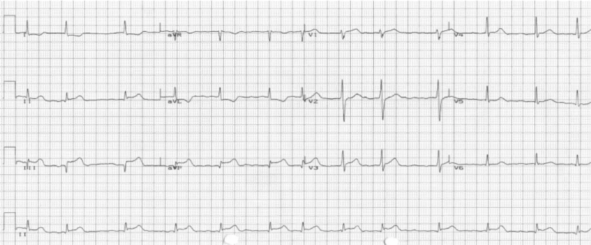
A 49-year-old male visited the emergency room and presented with chest pain of 2 hours duration. He was a smoker and had no medical history of hypertension or diabetes. At the time of admission, his blood pressure and pulse rate were 130/80 mmHg and 88 beats per minute, respectively. He showed clear consciousness. The physical examination showed normal findings. The elec- trocardiogram at admission revealed ST segment ele- vation in leads II, III and aVF (Fig. 1). Based on the results of the examinations, he was diagnosed as having an acute inferior myocardial infarction. He was referred to the cardiac catheterization laboratory for emergency coronary angiography. The CAG showed two signifi- cant stenoses of the left coronary system. Because of the anomalous position of the RCA ostium originating from
Received: September 12, 2007 Accepted: October 11, 2007
Correspondence: Joon Hyung Doh, MD, Department of Internal Medicine, Vision 21 Cardiac and Vascular Center Clinical Research center, Ilsan-Paik Hospital, Inje University College of Medicine, 2240 Daehwa-dong, Ilsanseo- gu, Goyang 411-706, Korea
Tel: 82-31-910-7830, Fax: 82-31-910-7829 E-mail: [email protected]
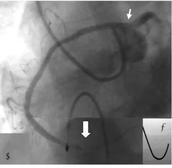
the left coronary cusp, we could not easily engage the RCA with using several guiding catheters such as a 6 French (Fr) sized Judkin 4, a 5 right (JR, Cordis, USA), an Amplatz 1 and 2 left (AL, Cordis, USA) and a La- uncher extra-backup 3.5 (EBU, Medtronic, USA). We successfully engaged the RCA using a smaller sized 5 Fr Launcher EBU 4 guiding catheter (Medtronic, USA) and the RCA showed a subtotal occlusion in its mid- portion (Fig. 2). In order to successfully engage the RCA, the tip of the guiding catheter was manipulated manually after it was heated using a hair dryer (Fig. 3). We then inserted a Whisper guide wire (Guidant, USA) across the lesion and dilated the artery using a 2.0×20 mm sized Art balloon (AMG Co. Germany). After balloon dilation, we implanted a 3.0×28 mm sized Taxus stent (Boston Scientific Corporation, USA) at 18 atmospheres. The final angiogram after stenting showed successful restoration of coronary blood flow (Fig. 4). We finished the proce- dure without any peri-procedural complications. Second stage PCI was done at the left circumflex artery 5 days
later and he was discharged 2 days later. He has been followed-up at the out patient clinic without any adverse cardiovascular events. Because of the great technical diffi- culties in performing CAG, we decided to verify the stent
Fig. 1. An electrocardiogram showed ST segment elevation in the II, III and aVF leads.
Fig. 2. Baseline coronary angiogram showed an anomalous right coronary artery (RCA) ostium (small arrow) and subtotal occlusion of the mid-RCA (large arrow).
Before After
Fig. 3. 5 French launcher EBU4 guiding catheter. Before: the natu- ral shape of the EBU4 guiding catheter. After: the manually ma- nipulated EBU4 guiding catheter (arrow) using a hair dryer. EBU:
extra-backup.
Fig. 4. The final coronary angiogram showed a successfully implan- ted stent at the mid right coronary artery (arrow).
patency at 9 months via 64 slice multi-detector computed tomography (MDCT, Aquillon, Toshiba, Japan). The MDCT showed an anomalous origin of the RCA from the left coronary cusp and a patent stent lumen without restenosis (Fig. 5).
Case 2
A 79-year-old female visited the emergency room and was presented with chest pain of 5 hours duration. She had no history of hypertension. Upon admission, the blood pressure was lower than 90/60 mmHg and the pulse rate was 75 beats per minute. She showed clear consciousness. The physical examination showed an ele- vated jugular venous pressure. The ECG at admission revealed ST segment elevation in leads II, III and aVF (Fig. 6). She was diagnosed as having an acute inferior myocardial infarction with right ventricular infarction.
She was referred to the cardiac catheterization laboratory for emergency CAG. A temporary pacemaker and an in- traaortic balloon pump were inserted for treating her hy- potension and bradycardia. A CAG showed a normal left
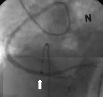
coronary system. Because of the anomalous position of the RCA ostium originating from the left coronary cusp, we could not easily engage the RCA using several guiding catheters such as a JR, an AL and an EBU guiding cathe- ter. Using a 6 Fr Heartrail Ikari right 1.5 guiding catheter (Terumo, Japan), we could selectively engage the RCA and the RCA showed total occlusion at its distal portion (Fig. 7). We next started the primary PCI. To overcome the instability and poor guiding support, we inserted two BMW guide wires (Guidant, USA) across the lesion.
The distal RCA lesion was dilated using a 2.5×20 mm Yangtze balloon (Minvasys, France) and then we implan- ted a 3.5×21 mm Premier stent (Minvasys, France) at 18 atmospheres.
We dilated the stent using a 4.0×15 mm Ryujin bal- loon (Terumo, Japan). A final angiogram showed succe- ssful revascularization of the distal RCA (Fig. 8). The patient’s clinical condition was stabilized after the proce- dure and she was discharged 1 week later. One year later, a MDCT showed that the blood flow through the stent was normal without restenosis (Fig. 9).
Fig. 6. An electrocardiogram showed ST segment elevation in the II, III and aVF leads.
Fig. 5. The 64 slice multi-detector computed tomography (MDCT) showed an anomalous right coronary artery ostium (small arrow) and patent stent lumen (large arrow).
B
A
Fig. 7. A: baseline coronary angiogram using a 6 French Heartrail Ikari right 1.5 guiding catheter showed an anomalous right coronary artery (RCA) ostium (small arrow) and total occlusion of the distal RCA (large arrow). B: 6 French Heartrail Ikari right 1.5 guiding catheter.
Discussion
An anomalous RCA originating from the left coronary cusp is uncommon, and this represents 8 to 16% of all coronary artery anomalies.2) There is controversy over whether this anomaly may cause myocardial ischemia or even sudden cardiac death, and the anatomical and the clinical characteristics of this anomaly have not been established.3-7) Selective catheterization of an anomalous RCA may be difficult because of the unusual location and the noncircular luminal orifice of this anomaly. Because there are no standardized guidelines to select a catheter
for an anomalous coronary artery, the anatomy of this anomaly and the operator’s preferences are key factors for successfully performing CAG.8)9) The operators are occasionally faced with multiple technical challenges that required frequent catheter exchanges to find the best fitting catheter. Non-selective cannulation can lead to misdiagnosis during CAG and a lack of proper guide sup- port often prolongs the interventional procedures and increases the procedural risk.10) A primary PCI should be performed as quickly as possible. When we face the situ- ation of an anomalous artery during a primary PCI, it takes much longer to open the occluded artery.
A PCI of an anomalous coronary artery requires pro- per angiographic recognition of the anatomic details. An anomalous RCA arising from the left coronary cusp is characterized by the anterior location of the ostium, the tortuous proximal portion and the initial anterior-caudal/
rightward course.3) Knowledge of these unique anatomic features can help to select the appropriate catheters and provide excellent backup support. Successful cannulation and optimal guiding catheter support depends on stable coaxial alignment, and the guiding catheter tip should be able to directly access the right anterior at a 45 to 90 degree angle.11) There have been several case reports of successful cannulation of an anomalous RCA with using AL and Judkins 5 Left guiding catheters and manually manipulated guiding catheters such as a Leya catheter.11-14) As in our two cases, we also could successfully cannulate the anomalous RCA with using a manually manipulated EBU guiding catheter and the recently developed Hear- trail Ikari right 1.5 guiding catheter.15) When performing primary PCI, the operators and catheterization laborato- ries have to be aware of this anomaly and prepare spe- cialized guiding catheters to facilitate PCI.
Because of the technical difficulties during performing CAG, we performed 64 slice MDCT to confirm the stent patency after stent implantation. The recently developed 64 slice MDCT showed excellent visualization of the coronary artery and intra-stent lumen compared to the previous versions and it helped us recognize the anatomical characteristics of the anomalous coronary artery.16) The MDCT images of our cases also showed the clear lumen and the anatomy of the anomalous RCA.
In the future, we can get more useful information about coronary artery anomalies with using MDCT. We report here two cases of successful primary PCI with using spe- cialized guiding catheters and techniques in 2 patients who both had an anomalous RCA arising from the left coronary cusp, and we include a review of the relevant literature.
REFERENCES
1) Kimbris D, Iskanderian AS, Segal BL, Bemis CE. Anomalous aor- tic origin of coronary arteries. Circulation 1978;58:606-15.
2) Yamanaka O, Hobbs RE. Coronary artery anomalies in 126,595 Fig. 8. The final coronary angiogram showed a successfully implan-
ted stent at the distal right coronary artery (arrow).
Fig. 9. The 64 slice multi-detector computed tomography showed a patent stent lumen (arrows).
patients undergoing coronary angiography. Cathet Cardiovasc Diagn 1990;21:28-40.
3) Rigatelli G, Docali G, Rossi P, et al. Congenital coronary artery anomalies angiographic classification revisited. Int J Cardiovasc Imaging 2003;19:361-6.
4) Garcia-Rinaldi R, Sosa J, Olmeda S, Cruz H, Carballido J, Quintana C. Surgical treatment of right coronary arteries with anomalous origin and slit ostium. Ann Thorac Surg 2004;77:1525-9.
5) Benge W, Martins J, Funk DC. Morbidity associated with ano- malous origin of the right coronary artery from the left sinus of val- salva. Am Heart J 1980;99:96-100.
6) Isner JM, Shen EM, Martin ET, Fortin RV. Sudden unexpected death as a result of anomalous origin of the right coronary artery from the left sinus of valsalva. Am J Med 1984;76:155-8.
7) Spargias K, Kariofyllis P, Mavrogeni S. Percutaneous coronary intervention in anomalous right coronary arteries arising from the left sinus of valsalva: a report of two cases and observations on the pattern of atherosclerosis. J Invasive Cardiol 2006;18:E78-81.
8) Choi KL, Kwon JI, Jung WH, et al. Stenting of an anomalous coro- nary artery in acute myocardial infarction. Korean Circ J 1998;
28:1378-81.
9) Cha KS, Keum DJ, Park HR, et al. Transradial stenting of an ano- malous right coronary artery originating from the left sinus of valsalva. Korean Circ J 1998;28:2056-60.
10) Topaz O, DiSciascio G, Gondreau E, et al. Coronary angioplasty of anomalous of coronary arteries: notes on technical aspects.
Cathet Cardiovasc Diagn 1990;21:106-11.
11) Qayyum U, Leya F, Steen L, et al. New catheter design for cannu- lation of the anomalous right coronary artery arising from the left sinus of valsalva. Catheter Cardiovasc Interv 2003;60:382-8.
12) Cohen MG, Tolleson TR, Peter RH, Harrison JK, Sketch MH Jr.
Successful percutaneous coronary intervention with stent implan- tation in anomalous right coronary arteries arising from the left sinus of valsalva: a report of two cases. Catheter Cardiovasc Interv 2002;55:105-8.
13) Oral D, Dagalp Z, Pamir G, et al. Percutaneous transluminal coro- nary angioplasty of anomalous coronary arteries: case reports.
Angiology 1996;47:77-82.
14) Lorin JD, Robin B, Lochow P, Lorenzo A, Sedlis SP. The right radial approach for stenting of lesions in the right coronary artery with anomalous take-off from the left sinus of valsalva. J Invasive Cardiol 2000;12:478-80.
15) Ikari Y, Nakajima H, Iijima R, et al. Initial characterization of Ikari Guide catheter for transradial coronary intervention. J Inva- sive Cardiol 2004;16:65-8.
16) Sato Y, Ichikawa M, Masubuchi M, et al. MDCT of the anomalous origin of the right coronary artery from the left sinus of valsalva as a single coronary artery. Int J Cardiol 2006;109:125-6.