5S와 45S rDNA 유전자를 이용한 제주도산 애기더덕 ( Codonopsis minima )과 더덕 ( C. lanceolata )의 FISH 패턴 분석
김수영*†·김찬수**
*국립생물자원관 야생생물유전자원센터, **국립산림과학원 난대산림연구소
Analysis of FISH patterns using 5S and 45S rDNAs in Codonopsis minima and C. lanceolata from Jeju Island
Soo Young Kim
*†and Chan Soo Kim
***
Wildlife Genetic Resources Center, National Institute of Biological Resources, Incheon 404-708, Korea.
**
Warm-Temperate Forest Research Center, Korea Forest Research Institute, Seogwipo 697-050, Korea.
ABSTRACT : The chromosome number was identified and fluorescence in situ hybridization(FISH) mapping of 5S and 45S rDNAs were conducted for C. minima and C. lanceolata in the genus Codonopsis from Jeju island. In this study, we have con- firmed that the somatic metaphase chromosome number determined as 2n = 2x = 16 was the same as the findings from the previous studies. While the conventional staining method makes it rather difficult to distinguish satellite chromosomes due to high degree of variability, FISH analysis produced the exact number and location of 5S and 45S rDNAs. Both species in the genus Codonopsis have a pair of 5S rDNA and their gene loci were observed on chromosome 3. Although two pairs of 45S rDNAs (one on chromosome 1 and the other on chromosome 8) were identified in both species, the 45S rDNA signals on chromosome 8 in C. minima were significantly weaker than those on chromosome 1. In addition, the 45S rDNA signals on chromosome 1 in C. lanceolata showed that the chromosome is non-homologus. In this study, we have determined cytoge- netic characteristics of C. minima and C. lanceolata according to their gene replication patterns.
Key Words : Chromosome, FISH, rDNAs, C. minima, C. lanceolata , Satellite
서 언
더덕속 식물은 동아시아에
59
종이 분포하며(Lammers, 2007),
중국에39
종(Fu et al ., 2004),
일본에4
종(Shimizu, 1993)
이분포하는것으로알려져있다.
우리나라에는4
종이분 포하는데거의대부분이약용,
식용,
관상용으로이용되고있다(Fu et al ., 2004; Mabberley, 1990).
그 중 더덕{ Codonopsis lanceolata (Siebold & Zucc.) Trautv.}
은 우리나라를 비롯하 여중국,
일본,
러시아등동아시아에널리분포하는종으로서(Yoo, 2007), phytoderin, leoithin, pentosane, saponin
을 함유하고 있으며
(Song et al . 1990),
해독(
解毒),
거담(
去痰),
최유
(
催乳)
에 유용한 약재료 알려져 있다(Hotta et al ., 1989).
애기더덕( C. minima Nakai)
은유연관계가 깊은여타 의한국산 더덕속 식물들과 유사하지만줄기가 보다 가늘며 꽃과잎이2~3
배작고,
잎의양면에털이있는점에서구별된 다고하여설정된종으로한라산해발1100~1300 m
에드물게분포하는국내고유종이다
(Lee, 1996; Lee, 2005; Yoo, 2007;
Kim, 2009; Koh, 2010).
더덕속( Codonopsis Wall.)
은 드물게직립하는종도 있지만대부분다른물체를감아오르거나 기어오르는 다년생 초본으로흔히 스컹크같은 향을 가지고 있다
.
뿌리는대형으로괴경 모양이며,
잎은마주나거나모여 나고,
잎자루는있거나거의없는경우도있다.
꽃은대형이고 대부분아래를향하지만드물게곧추서는경우도있으며,
소 화경이있고,
단생으로정생 또는액생한다.
열매는건과로서3~5
실로되어있고,
종자에는날개가있다.
더덕속은이와같은특성으로초롱꽃과
(Campanulaceae)
의다른속들과구분이된다
.
더덕에 관한 대다수의 연구는 성분 분석과 관련이 있으며 한국산더덕속에대한외부형태
,
식생조사,
해부학적형질,
염 색체수및화분학적형질에따른분류학적연구결과가보고 된바있다(Yoo and Lee, 1989).
또한분자생물학적 방법인RAPD
를이용하여지역적인유연관계를보고한바있다(Doo
†
Corresponding author: (Phone) +82-32-590-7111 (E-mail) [email protected]
Received 2010 April 26 / 1st Revised 2010 May 25 / 2nd Revised 2010 June 11 / 3rd Revised 2010 June 14 / Accepted 2010 june 14
et al ., 2002).
더덕의 염색체수는Krasnoborov
등(1980)
과Nishikawa (1985)
에 의해 처음으로 보고되었고 국내에서는Yoo
와Lee (1989)
를통해 연구되었으며 지역에 따른 핵형학 적차이를 보고한바있다(Yoon et al ., 1992).
더덕의다양 한연구 결과에비해애기더덕에관한 연구는염색체수를포 함한 분류학적 형질에 관한 연구만 보고되었다(Yoo and Lee., 1989).
애기더덕은외부형태학적특성이다른종과는뚜 렷하게차이를나타내고있으며,
한라산의고지대에희소하게분포하는점으로볼때약학적가치와함께경제성향상을위 한육종재료로서도활용 가능성이높다고할수있다
.
그러 므로이러한두분류군에대하여분류학적으로도유용한형질 일뿐만아니라앞으로자원개발에있어서도필수적인정보라 고할수있는염색체의특성을규명하는것은필수적이다.
분자세포유전학적기술인
fluorescence in situ hybridization
(FISH)
는특정 유전자를염색체 상에 직접 확인 할수있는방법으로국내자생하는약용식물인시호
,
지모,
깽깽이풀,
황기속 식물 등을 대상으로
5S
와45S rDNA
를이용하여물리 지도(physical mapping)
를 작성한 결과가 보고된 바 있다(Koo et al ., 2003a; Kim et al ., 2004, 2005, 2006).
따라서 본연구는제주지역에분포하는더덕과제주도특산식물이면 서희귀식물인애기더덕의세포학적연구를통해rDNA
유전 자의 물리지도를 작성하여더덕속 식물의 유전체 구조 분석 및세포유전학적연구에기초자료로활용하고자수행되었다.
재료 및 방법
1. 식물재료
본연구에사용한더덕과애기더덕은제주도한라산에자생 하고 있는 개체
(
난대산림연구소,
확증표본Jul. 24, 2007, Moon M.O., s.n.)
를 온실에 이식하여 새로운 뿌리를 유도한후염색체분석용재료로사용하였다
.
2. 체세포 염색체 관찰 및 핵형분석
채취된근단은 중기염색체상을 얻기위하여 증류수
(4
℃)
에 담가
24
시간 동안 저온처리 한 다음, Carnoy's solution (glacial acetic acid : ethanol = 1 : 3, v/v)
에담가4
℃에 보관하면서 재료로이용하였다
.
염색체 관찰을위해 고정한 근단을1N HCl (60
℃)
에서5
분간연화 한다음,
증류수로 수세하고Feulgen
용액에서 염색한 후, 1% aceto-carmine
을 이용하여 압착법으로프레파라트를만들어염색체를관찰하였다.
3. FISH 슬라이드 제작
FISH
실험을위한슬라이드제작을위해고정된근단을증류수로 수세한후 효소 혼합용액
(2% cellulase Onozuka R- 10, 1.5% macerozyme R-10, 1% pectolyase Y-23, 0.5 mM
EDTA, pH 4.2)
에 담가30
분간 처리(37
℃)
후, Carnoy's
solution
을이용하여가는핀셋으로슬라이드글라스위에서염색체를전개 한뒤상온에서
2~3
일간건조시켰다.
위상차현 미경하에서분열상이양호한슬라이드를선발하여FISH
에이 용하였다.
4. 탐침의 준비와 bicolor-FISH
FISH
를 위한 탐침으로는biotin-16-dUTP
로 표지된45S rDNA
와digoxigenin-11-dUTP
로 표지된5S rDNA
를 이용하였으며
bicolor-FISH
는Kim
등(2006)
의방법을변용하여사 용하였다.
슬라이드상의 염색체는70% formamide/2xSSC
용 액(
−70
℃)
에서2
분간 변성시키고70%
에탄올(
−20
℃)
에서 급냉 후, 95%
와99%
에탄올에서각각5
분씩 탈수하여상온 에서30
분 정도 건조시켰다.
탐침 혼합액(biotin-16-dUTP
와digoxigenin-11-dUTP
로 각각 표지된100 ng
의probe DNA, 50% formamide, 2X SSC, 10% dextran sulfate, 10 ng ssDNA)
은90
℃에서10
분간 변성시킨후,
급냉시켜준비하였 다.
건조된 슬라이드상에20
㎕의 탐침혼합액을 가한다음,
커버글라스를덮고
paper bond
로봉하여, 37
℃에서16
시간이 상혼성화(hybridization)
하였다.
혼성화 시킨 슬라이드는
40
℃의2xSSC, 50% formamide/
2xSSC, 2xSSC, 4xSSC
용액에서각각5
분씩 수세하였다.
탐침의 비 특이적인결합을 막기 위해 염색체 슬라이드를
5%
BSA/BT (1M NaHCO
3+ 0.5% Tween-20, pH 8.3)
완충액으 로37
℃에서5
분간blocking
한 후, 1%
의avidin-FITC (fluorescein isothiocyanate)
와anti-digoxigenin rhodamine
이 포함된100
㎕의1% BSA/4XSSC
의혼합액을슬라이드에가 한다음37
℃에서30
분동안반응시켜biotin
과digoxigenin
으로 표지된
DNA
탐침을 동시에 검출하였다. 4xSSC/0.2%
tween-20
완충액으로37
℃에서5
분씩3
번 수세 후, 1
㎍/
㎖DAPI (4,6-diamidine-2-phenylindole dihydrochloride)
용액을 포함한Vectashield (Vector Lab.) 15
㎖를 도포하여 커버를 덮은 후cooled CCD
카메라(Cool SNAP, Photometrics)
와 형광현미경을이용하여signal
을관찰하고사진을촬영하였다.
확인된
signal
들은Meta Imaging Serries TM 4.6 (Universal Imaging Corporation)
소프트웨어를사용하여합성하였으며실험의정확성및재현성을높이기위해종당
10
개체이상을분 석하였다.
결과 및 고찰
제주도에 서식하는 더덕과 애기더덕의 체세포 염색체수는
2n = 2x = 16
으로 관찰되었다(Fig. 1).
본연구결과와 기존에보고된 더덕 및 애기더덕의 염색체수
(2n = 16)
를 통하여(Krasnoborov et al ., 1980; Nishikawa, 1985; Yoo and Lee,
1989; Yoon et al ., 1992)
더덕속의 염색체수가 안정적임을알수있었다
.
염색체핵형분석결과가더덕의품질을직접적으로반영하는형태나약효성분은아니지만핵형학적차이가 형태나품질의차이를나타낼수있는일종의지표로이용가 능하다는보고
(Yoon et al ., 1992)
가있으므로본연구에서도 특정염색체에존재하는satellite
의유무와hetero
로형태를갖는 염색체를 구분하여 핵형학적 차이를 구분하였다
.
그러나aceto-carmine
과orceine
등을이용한일반염색법으로염색체를관찰할경우
,
동일한슬라이드의세포에서도satellite
가관찰 되는경우와그렇지않은양상을쉽게관찰할수있고,
염색 체의 응축 정도에따라서도변이가 심하기때문에satellite
를보다 확실하게구분하고관찰 할수있는
FISH
방법이 정확하다고할수있다
.
5S
와45S rDNA
는리보소옴의구성성분으로, 5S rDNA
의경우
120 bp
의conserved coding sequence
를 포함한200~
500 bp
의 반복적인 배열을 하고 있으며,
하나의loci
에서rDNA
복제수의변화가 심하여 수의변이와 위치 분포가식물종간의관계와진화에유용하게사용되고있다
(Mukai et
al. , 1991; Maluszynska and Heslop-Harrison, 1993; Castilho and Heslop-Harrison, 1995). 45S rDNA
유전자는모든 식물 종의 염색체 상에서1
쌍 이상이존재하고(Maluszynska and Heslop-Harrison, 1991),
인형성부위를포함하고있는부수체염색체에서 관찰되며
(Leitch and Heslop-Harrison, 1992),
5S rDNA
와동일한 위치에서 나타나지않는다.
따라서 일반염색법으로 핵형분석이어렵거나
,
동일한 속내에서의종간의 차이를구분할수있는유용한유전자로사용된다.
더덕과애기더덕의염색체상에서의
5S
와45S rDNA
유전자 를이용한물리지도작성을위해digoxigenin-16dUTP
로표지한
5S rDNA
는rhodamine
으로, biotin-11dUTP
로 표지한45S rDNA
는FITC
로 검출하였다.
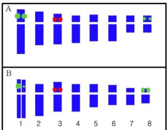
애기더덕에서1
쌍의5S rDNA
유전자가3
번염색체의동원체주위에서확인되었고, 2
쌍의
45S rDNA
유전자는1
번과8
번 염색체의단완 말단 부 위에서 각각 관찰되었다.
특이한 점은2
쌍의45S rDNA signal
가운데1
번염색체에 있는1
쌍의signal
이8
번염색체 에서 관찰되는 것보다 강하게 관찰되었다(Fig. 3 and Fig.
5A).
반면 더덕은5S rDNA
와45S rDNA
의signal
수와 위치가각각
3
번염색체에1
쌍, 1
번과8
번염색체에2
쌍으로애기 더덕과 동일하였으나1
번 염색체에서 관찰되는1
쌍의45S
Fig. 1.
Somatic metaphase chromosomes of Codonopsis minima and C. lancelolata (2n = 2x = 16). A, C. minima ; B, C.
lanceolata . Bar, 5
㎛.
Fig. 2.
An idiogram of a metaphase chromosome. The arrowheads indicate the satellite and Nucleolus organizer region (NOR).
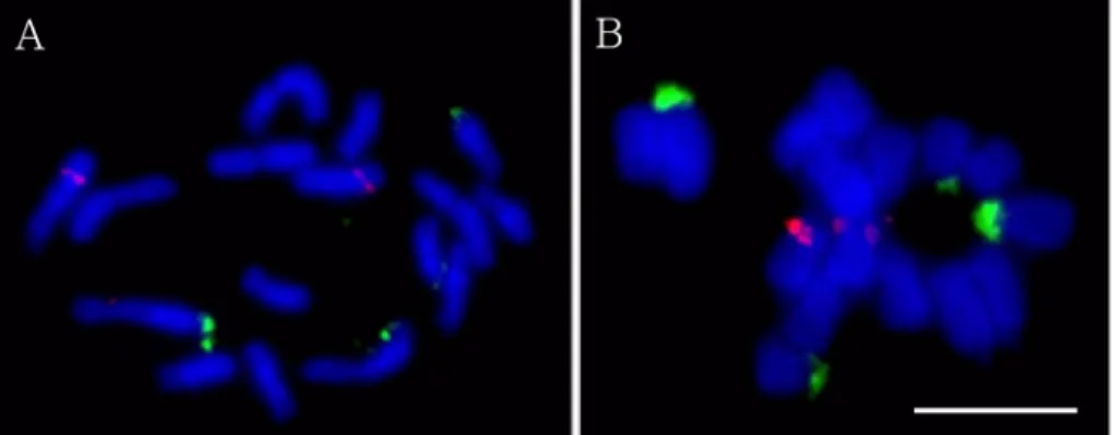
Fig. 3.
Bicolor FISH pattern of the metaphase chromosomes of C. minima using both 5S (red) and 45S (green) rDNA genes. A, early-
metaphase chromosome; B, metaphase chromosome . Bar, 5
㎛.
rDNA signal
중하나는 강하고다른 또하나는아주 약하게 관찰되는양상을보였다(Fig. 4 and Fig 5B).
애기더덕과더 덕의 물리지도 작성의 큰 차이는1
번과8
번에 위치한45S rDNA
의염색체상에서의signal
크기라고할수있다.
이러한결과는 상동염색체간의
45S rDNA
복제수가 서로 다르다는 것을의미하지만실험조건이나염색체의응축정도에따라변 이를 보일수도있다(Koo et al ., 2003b).
따라서FISH
결과 에따른애기더덕과더덕의세포유전학적특징은애기더덕의 경우2
쌍의동일한45S rDNA
를갖고있다하더라도1
번염 색체에위치하는유전자가8
번염색체에위치하는것보다복제수가많고더덕은
1
번염색체에위치한45S rDNA
복제수가비상동적인것으로구분할수있다는것이다
.
이러한실험 결과는한종당10
개체이상의반복적실험을통하여정확도 를높이고재현성을고려하였다.
본연구에서수행한rDNAs
의물리지도작성결과는제주지역에서식하는애기더덕과더 덕의 게놈 구조를이해하는데필수적이며
,
특정 유전자의물리적위치를탐색하는기초연구로서더덕속식물의염색체지 도구축에필요한중요한자료로이용될수있을것으로사료 된다
.
감사의 글
이연구논문은
2010
년국립생물자원관(NIBR)
일반연구과제
-‘
멸종위기및주요생물자원의염색체도감발간’
과제의연 구비를지원받아수행된연구결과로이에감사드립니다.
LITERATURE CITED
Castilho A. and Heslop-Harrison JS. (1995). Physical mapping of 5S and 18S-25S rDNA and repetitive DNA sequences in
Aegilops umbellulata. Genome. 38:91-96.
Doo HS, Ryu JH, Lee KS, Li HL and Liu XH. (2002). Analysis of genetic relationship by RAPD technique for
Codonopsis lanceolatatrauty collected from Baekdoo mountain and Korea.
Korean Journal of Medicinal Crop Science. 10:194-199.
Fukui K, Ohmido N and Khush GS. (1994). Variability in rDNA loci in the genus
Oryzadetected through fluorescence
in situhybridization. Theoretical and Applied Genetics. 87:893-899.
Fu LT, Chen K, Lang T, Hong QL and Li R. (2004). Higher Plants of China (10). Qingdao Publishing House. Qingdao, China. p. 455-466.
Hotta M, Ogata K, Nitta A, Hosikawa K, Yanagi M and Yamazaki K. (1989). Useful Plant of The World. Heibonsha LTD Publishers. Tokyo, Japan. p. 296-297.
Kim CS. (2009). Vascular plant diversity of Jeju Island, Korea.
Korean Journal of Plant Research. 22:558-570.
Kim SY, Choi HW and Bang JW. (2004). Physical mapping of rDNAs using McFISH in
Anemarrhena asphodeloidesBunge.
Korean Journal of Medicinal Crop Science. 12:515-518.
Kim SY, Choi HW, Koo DH, Kim CS and Bang JW. (2005).
Karyotype analysis and physical mapping of rDNAs using McFISH in
Jeffersonia dubiaBenth. Korean Journal of Medicinal Crop Science. 13:48-51.
Kim SY, Choi HW, Kim CS, Sung JS, Lee JK and Bang JW . (2006). Cytogenetic analysis of
Astragalusspecies. Korean Journal of Medicinal Crop Science. 14:250-254.
Koh JG . (2010). Endemic plants in Jeju island. Institute of Environmental Resource Research, Jeju Special Self-Governing Province. Jeju, Korea. p. 90-91.
Koo DH, Seong NS, Seong JS, Bang KH and Bang JW.
(2003a). Karyotype analysis and physical mapping of rDNAs in
Bupleurum longeradiatum. Korean Journal of Medicinal Crop Science. 11:402-407.
Koo DH, Kim SY, Bang KH, Seong NS and Bang JW. (2003b).
Cytogeneic analysis of
Angelicaplants using Feulgen staining and multicolor fluorescence
in situhybridization. Korean Journal of Plant Biotechnology. 30:123-127.
Fig. 4.
Bicolor FISH pattern of the metaphase chromosomes of C. lanceolata using both 5S (red) and 45S (green) rDNA genes. Bar, 5
㎛.
Fig. 5.