http://dx.doi.org/10.14316/pmp.2014.25.3.157
This work was supported by the research grant of Chungbuk National University in 2012.
Received 25 August 2014, Revised 4 September 2014, Accepted 11 September 2014
Correspondence: Yong Sook Goo ([email protected]) Tel: 82-43-261-2870, Fax: 82-43-272-1603 cc This is an Open-Access article distributed under the terms of the Creative Commons Attribution Non-Commercial License (http://creativecommons.org/licenses/by-nc/3.0) which permits unrestricted non-commercial use, distribution, and reproduction in any medium, provided the original work is properly cited.
Effect of ATP on Calcium Channel Modulation in Rat Adrenal Chromaffin Cells
Kyung Ah Kim*, Yong Sook Goo†
Departments of *Biomedical Engineering and †Physiology, Chungbuk National University School of Medicine, Cheongju, Korea
ATP in quantity co-stored with neurotransmitters in the secretory vesicles of neurons, by being co-released with the neurotransmitters, takes an important role to modulate the stimulus-secretion response of neurotransmitters.
Here, in this study, the modulatory effect of ATP was studied in Ca2+ channels of cultured rat adrenal chromaffin cells to investigate the physiological role of ATP in neurons. The Ca2+ channel current was recorded in a whole-cell patch clamp configuration, which was modulated by ATP. In 10 mM Ba2+ bath solution, ATP treatment (0.1 mM) decreased the Ba2+ current by an average of 36±6% (n=8), showing a dose-dependency within the range of 10−4∼10−1 mM. The current was recovered by ATP washout, demonstrating its reversible pattern. This current blockade effect of ATP was disinhibited by a large prepulse up to +80 mV, since the Ba2+ current increment was larger when treated with ATP (37±5%, n=11) compared to the control (25±3%, n=12, without ATP). The Ba2+ current was recorded with GTPγS, the non-hydrolyzable GTP analogue, to determine if the blocking effect of ATP was mediated by G-protein. The Ba2+ current decreased down to 45% of control with GTPγS. With a large prepulse (+80 mV), the current increment was 34±4% (n=19), which 25±3% (n=12) under control condition (without GTPγS). The Ba2+ current waveform was well fitted to a single-exponential curve for the control, while a double-exponential curve best fitted the current signal with ATP or GTPγS. In other words, a slow activation component appeared with ATP or GTPγS, which suggested that both ATP and GTPγS caused slower activation of Ca2+ channels via the same mechanism. The results suggest that ATP may block the Ca2+
channels by G-protein and this Ca2+ channel blocking effect of ATP is important in autocrine (or paracrine) inhibition of adrenaline secretion in chromaffin cell.
Key Words: Adrenal chromaffin cell, Ca2+ channel, ATP, G-protein, Autocrine inhibition
서 론
부신수질의 크로마핀(chromaffin) 세포는 내장신경(splanchnic nerve) 말단에서 유리되는 아세틸콜린에 반응하여 신경전 달물질인 카테콜아민(catecholamine)을 분비하는 세포로써 형태학적으로는 내분비세포에 해당하고 발생학적으로는 교감신경의 절전세포와 기원이 동일한 신경-내분비 세포
(neuro-endocrine cell)이다.
카테콜아민 분비(exocytosis) 과정에 Ca2+이 중요한 작용 을 함은 이미 잘 밝혀져 있다.1) 내장신경 말단에서 분비된 아세틸콜린이 세포막의 Na+투과성을 증가시켜 세포가 저 분극되면, 칼슘통로가 열려 세포 내 Ca2+ 농도가 증가한다.
세포 내로 유입된 Ca2+은 카테콜아민이 포함되어 있는 소 포(vesicle)에 작용하여 소포가 세포막에 쉽게 접합(fusion) 되도록 하여 카테콜아민의 세포외분비(exocytosis)를 자극한 다. 이 현상에 기여하는 칼슘통로는 주로 막전압 의존성 (voltage-dependant) 칼슘통로라고 생각된다.2) 신경세포의 막 전압의존성 칼슘통로의 아형(subtype)은 L-, N-, P/Q-type 등 으로 구분되며3) 이들 각각의 칼슘통로를 조절함으로써 Ca2+
의 시냅스전뉴론으로의 유입과 신경전달물질의 분비가 조 절된다.
부신수질 크로마핀 세포에서는 카테콜아민과 함께 ATP
(Adenosine 5’-triphosphate)가 공동저장(co-store), 공동분비 (co-release) 된다.4) 부신수질 크로마핀 세포의 과립(granule) 에는 카테콜아민과 ATP가 약 5:1의 비율로 포함되어 있 고, 그 밖에 ADP (Adenosine 5’-diphosphate), AMP (Adenosine 5’-monophosphate), GTP (Guanosine triphosphate), UTP (Uracil triphosphate) 등의 뉴클레오티드는 ATP에 비해 소량 포함 되어 있는 것으로 알려져 있다.4)
Dunlap과 Fischbach 는 노아드레날린(noradrenaline)이 세 포에서 기능적으로 작용을 나타내는 칼슘통로의 수를 감소 시킬 것이라고 처음 발표하였고,5) 이 후 여러 신경전달물 질들(noradrenaline, GABA)의 이온 통로 조절 기전에 대해 많은 연구가 진행되어 이들 신경전달물질들에 의해 신경 세포의 막전압 의존성 칼슘통로가 조절된다고 보고되었
다.6-10) 다른 여러 신경전달물질들의 역할로 미루어 ATP도
이와 유사한 기능을 가질 것으로 생각되며, 위의 신경전달 물질들의 작용과 유사하게 micromole (μM) 농도의 ATP를 세포막의 외부에 적용시켰을 때 여러 종류의 세포에서 이 온 통로와 생물학적 신호를 변화시킴이 알려져 있다.11, 12) Ca2+ 전류에 대한 ATP 조절 작용은 Diverse-Pierlussi 등13)에 의해 직접적인 증거가 보고되었고, 그들은 ATP가 Pertussis toxin (PTX)-sensitive pathway를 통해서 Ca2+ 전류의 억제나 증가를 유도한다고 추론하였다. Holz 등은 신경전달물질에 의한 칼슘통로의 억제가 GTP-binding protein에 의한 현상이 라고 처음 발표하였으며,14) 칼슘통로 조절에 있어서 G-pro- tein의 역할에 대한 연구가 진행되고 있다.15)
그러나 현재까지 진행된 연구에서 ATP를 처치하였을 때 칼슘통로의 활성화 변화를 kinetic study로 관찰한 연구는 드물기 때문에 본 연구에서는 흰쥐 부신수질 크로마핀 세 포를 대상으로 1. 칼슘통로를 통한 whole-cell Ba2+ 전류를 기록하고, 2. 칼슘통로에 대한 ATP와 GTPγS의 효과를 보 고, 3. 아주 큰 저분극 prepulse를 주어 facilitation (소통) 유 도 시에 이들 약물의 효과가 어떻게 나타나는지를 보았고, 4. Kinetics study를 병용하여 칼슘통로에 미치는 ATP의 효 과가 G-protein을 매개로 하는 지를 규명함으로써 ATP의 칼슘통로 조절 기전을 밝히고자 하였다.
재료 및 방법
1. 세포 분리(Adrenal chromaffin cell preparation) 부신 수질로부터의 단일 크로마핀 세포 분리는 Akaike 등의 방법16)을 개선하여 시행하였다. 체중 200∼250 gm되 는 성숙한 흰쥐(Sprague-Dawley rat)를 암수 구분 없이 사용
하였다. 목뼈를 탈구(cervical dislocation)하여 희생시킨 즉시 가능한 무균상태에서 부신을 적출하였다. 적출한 부신을 생리적 완충 용액[(mM) ; 145 NaCl, 5.4 KCl, 1 NaH2PO4, 11.2 glucose, 15 HEPES (pH 7.4 by NaOH)]에 옮긴 후, 현미 경 시야에서 캡슐과 피질(cortex)을 제거하여 수질(medulla) 만을 분리한 후 작은 조각(1 mm3)으로 절단하였다.
부신 조각을 생리적 완충 용액으로 세 번 세척한 후 소화 용액[(0.2% collagenase (Sigma, type IA), 0.1% hyaluronidase (Sigma, type IS)]을 처치하여 항온수조(Jeiotek, Korea)에서 30분 동안 진탕 처리하였다(80 rpm). 소화된 조직을 생리적 완충 용액으로 세 번 씻은 후, 0.1% bovine serum albmin (BSA, Sigma)을 포함한 생리적 완충 용액 내에서 약 15분 간 조심스럽게 pipetting하여 단일세포로 떨어져 나올 수 있 도록 하였다. 분리된 세포를 원심분리(100 G force, 10 min) 하고 상층액을 버린 후 배양액[Dulbecco’s modified Eagle medium (DMEM) with 10% fetal bovine serum]에 넣어 최종 부유시켰다.
Polyethylene imine (PEI, Sigma)을 전처치한 커버슬립(2×7 mm) 위에 세포를 분주하고 CO2 incubator (37°C, 5% CO2
phase)에 넣어 보관하였다. 세포배양 후 1∼3 일까지의 세 포를 사용하여 실험하였다.
2. Ba2+ 전류의 기록
부신수질 크로마핀 세포가 붙어 있는 커버슬립을 세포외 용액으로 채운 기록 체임버로 옮겨 실험에 사용하였다. 크 로마핀 세포를 역전도 현미경(inverted microscope, Diaphot, Nikon, Japan) 시야에서 선택한 후 저항이 2∼4 MΩ 정도 되도록 본 실험실에서 제작한 유리미세전극을 3-D micro- manipulator MX-1 (Narishige, Japan)로 조작하여 대상 세포 의 세포막에 접근시킨 후 세포 내 공간과 유리미세전극이 전기적으로 등가회로를 형성하도록 whole-cell configuration patch clamp technique17)을 사용하여 칼슘전류를 기록하였다.
유리미세전극은 실험실에서 Kimax-51 borosilicate capil- lary tubes (Kimble, USA)를 사용하여 microelectrode puller pp-83 (Narishige, Japan)로 뽑은(pulling) 후 저항이 2∼4 MΩ 정도 되는 전극만을 선택하여 사용하였다.
막전류의 기록을 위하여 전압자극은 pCLAMP5.5 Soft- ware (Axon Instrument, USA)를 사용하여 필요한 자극 프로 토콜을 만든 후 세포에 인가하였다. 이때 기록된 전류데이 터는 EPC-7 amplifier (List-electronics, Germany)를 거쳐 12 bit A/D conver (Lab master, Warner Instrument, USA)로 디지 털신호화한 후 PC에 저장하였다. 모든 실험은 실온(20∼
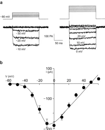
Fig. 1. Peak current (IBa)-voltage (Vm) relationship recorded in adrenal chromaffin cell held at −80 mV activated by 150 ms depolarizing pulses applied in 10 mV steps. (a) Activation of Ba2+
currents. Upper and lower traces are voltage and current traces, respectively. (b) Voltage-current relationship of Ba2+ current from 25 cells.
25°C)에서 수행하였다.
3. 용액조성(Solutions)
사용한 용액의 조성은 다음과 같다. 세포외용액(단위 mM):140 NaCl, 5.4 KCl, 0.5 MgCl2, 10 HEPES, 5 glucose, 10 BaCl2 (pH 7.3 by NaOH). 전극속 용액(electrode-filling solution)의 조성(단위 mM):120 CsCl, 20 TEACl, 11 EGTA, 1 CaCl2, 2MgCl2, 10 HEPES, 2 MgATP (pH 7.4 by CsOH).
Na+-전류는 세포외용액에 0.5 μM TTX (tetrodotoxin, Sigma)을 추가하여 완전히 차단하였고, 전극속 용액에 120 CsCl와 20 TEACl (tetraethylammonium chloride)를 넣음으로 써 K+-전류를 차단하여 오직 Ca2+-전류만이 기록될 수 있 도록 하였다.
4. 실험순서
- 전극이 세포외용액에 담가지기 시작하여 whole-cell patch를 형성할 때까지의 과정을 모니터링 하기 위해 10 mV, 100 Hz의 test pulse를 −80 mV (Vh, holding potential)에 인 가하였다.
- Test pulse의 상승기와 하강기에서 시스템의 캐패시턴 스(capacitance) 성분에 의한 전류를 관찰할 수 있으며 EPC- 7의 보상 회로를 사용하여 이를 제거하였다.
- 캐패시턴스를 보상한 후 Ba2+ 전류가 안정적으로 일정 하게 기록될 때까지 −80 mV (Vh) → 10 mV (Vt)의 pulse protocol을 10초에 한 번씩 수행하였다.
- Facilitation protocol:0 mV의 test pulse 주기 직전에 − 80 mV (Vh) → +80 mV (Vt)의 높은 전압의 저분극 pre- pulse를 주었다.
5. 데이터분석
1) Curve fitting: PC로부터 전류 데이터를 재생한 후 Igorpro software (Wavemetrics, USA)를 사용하여 분석하였 고 칼슘전류 활성화과정을 curve fitting 하기 위하여 Marquardt-Levenberg algorithm을 사용하여 전류기록을 가장 잘 반영하는 이중지수함수(I(t)=A[1−exp(−t/τ1)]+B[1−exp (−t/τ2)])를 도출하였다. Goodness of fit을 판단하는 지표로 R-square 값이 0.95 이상일 경우만 데이터에 포함시켜 τ1, τ2 값을 읽었다.
2) 통계처리(Statistics): 통계자료의 평균값은 평균 ± 표준오차(SEM)로 표시하였다. 측정한 값들 사이의 통계적 인 의미는 student’s t-test를 실시하여 확인하였으며 p-value
가 0.05 미만일 때를 유의한 차이가 있다고 판정하였다.
결 과
1. Ba2+ current kinetics
칼슘통로의 Charge carrier로는 Ca2+ 대신 10 mM Ba2+을 사용하였다. Ba2+ 전류의 전류-전압 곡선(current-voltage re- lationship, I-V curve)을 얻기 위해 150 ms의 test pulse를
−60 mV부터 +60 mV에 걸쳐 10 mV 간격으로 증가시켰 다. 이때 holding potential은 −80 mV로 하였다. Fig. 1a의 전류기록을 얻은 세포에서는 150 ms 이내에는 Ba2+ 전류 가 비활성화 되지 않았으나 세포마다 비활성화 정도는 다 양한 양상을 보였다(Fig. 1a). 총 25개의 크로마핀 세포에서 얻은 값의 I-V curve에서 Ba2+ 전류는 −40 mV부터 활성화 되기 시작하여 0 mV에서 최대값(−283.5±23.3 mV, n=25)에 이른 후 +50 mV에서 역전되었다(Fig. 1b).
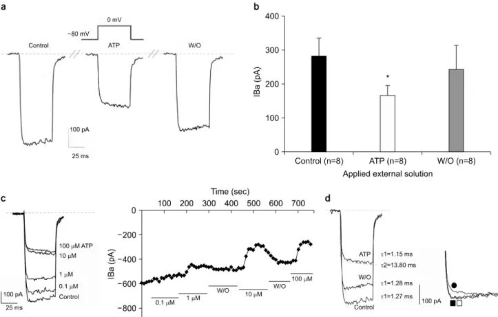
Fig. 2. Effect of ATP on Barium current (IBa). (a) Upper trace is voltage protocol. Lower traces are control Ba2+ current trace (left), current trace when the external solution is switched to ATP-containing solution (middle), and current trace after wash-out of ATP (right), respectively. (b) Effect of ATP on Ba2+ current. Peak amplitude of control Ba2+ current was 281.37±53.53 pA while that with ATP was 168.05±28.6 pA, with wash-out of ATP was 243.45±70.7 pA (mean±S.E.M., n=8). *p-value<0.05. (c) Left: Current traces show Ba2+ currents recorded and concentration of ATP in one cell. Right: Time course of inhibitory effects of ATP on IBa. IBa was activated by 50 ms depolarization pulses from -80 mV to 0 mV applied at a frequency of 0.1 Hz. As indicated by the bars, ATP (0.1, 1, 10, 100 μM) was added to or removed (W/O, washed out) from the external solution. Concentration was successively changed from 0.1 μM to 100 μM. (d) Left: Current traces of control, and with and after washout of ATP. Curve fitting was performed and activation time constant (τ) was calculated in each trace. Right: Normalized current : normalization of raw current with respect to the final point. Upper trace (●) is current during ATP, middle (□), current after washout of ATP, and lower trace (■), control current.
2. 칼슘통로에 미치는 ATP의 효과
크로마핀 세포를 관류시키는 세포외용액(재료 및 방법의 용액조성 부분 참조)에 ATP를 첨가하여 최종 ATP 농도가 micromole (μM) 농도가 되도록 만든 세포외용액을 관류시 켰을 때 적용시킨 ATP는 Ca2+ 통로를 통한 Ba2+ 전류의 크기를 감소시켰다. ATP 효과는 가역적인 억제현상으로 ATP 가 첨가되지 않은 세포외용액으로 다시 관류시켜주자 -즉 ATP를 wash-out 해주자 전류 크기는 약 control current 에 비해 80% 내외로 회복되었다(Fig. 2a). 100 μM ATP는 control current의 크기(281.37±53.53 pA)를 168.05±28.6 pA로
평균 36±6% (n=8) 감소시켰으며 ATP를 wash-out 해주자 243.45±70.7 pA 수준으로 회복하였다. Wash-out 후 Control current의 크기 수준으로 완전히 회복하지 않은 것은 동일 한 크로마핀 세포를 상대로 연속적으로 control−ATP 처치
−ATP wash out 실험을 연속적으로 진행하다 보니 시간이 경과됨에 따라 whole-cell mode 에서 필연적으로 발생하는 전류의 run-down 현상으로 해석된다(p-value<0.05)(Fig. 2b).
동일 크로마핀 세포의 whole cell patch 상태에서 세포외 용액에 0.1, 1, 10, 100 μM (10−4∼10−1 mM)로 각기 다른 ATP의 농도가 되도록 ATP를 점차 증가시키며 적용한 후, Ba2+ 전류를 기록하고 다시 대조 세포외용액으로 관류하기
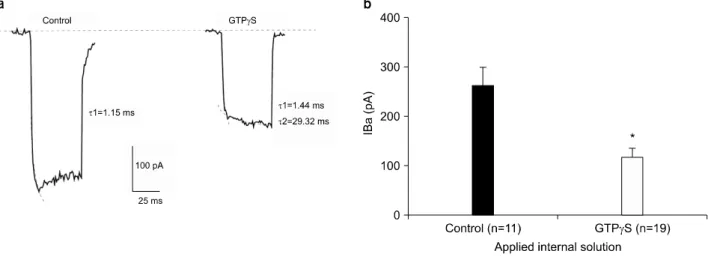
Fig. 3. Effect of GTPγS (50 μM) on Ba2+ current. (a) Effect of GTPγS on gating kinetics of Ca2+ channel. Current trace of control condition vs intracellular GTPγS. Curve fitting was performed and activation time constant was calculated for each trace as shown.
(b) Averaged results are shown (mean±S.E.M.) with the number of cells given in parentheses. Peak amplitude of IBa was 261.65±38.84 pA (n=11) while that with GTPγS was 118.78±17.59 pA (n=19). *p-value<0.05.
Table 1. Activation time constants of Ca2+ channel when external solutions were switched to different condition on the same chromaffin cells. Each value is presented as mean±SEM.
Condition of External Solution (N=8)
Activation Time Constant
τ1 (ms) τ2 (ms)
Control 1.24±0.15
ATP added 1.17±0.22 13.5±2.75
ATP wash-out 1.26±0.17
를 반복하면서 ATP의 농도에 따른 전류억제 관계를 시간 에 따라 나타내었을 때 ATP는 농도 의존적으로 Ba2+ 전류 의 크기를 억제하였으며 10 μM 이상의 농도에서 ATP의 전류억제 효과가 최대임을 관찰하였다(Fig. 2c).
ATP의 전류 억제가 단순히 크기만을 억제하는 것인지 칼슘통로의 개폐과정에도 영향을 나타내는지를 알아보고 자, 대조 세포외용액 상태(control)와 ATP를 대조 세포외용 액에 추가한 후, 그리고 ATP를 wash-out 후 기록한 current trace의 활성화(activation) 과정만을 지수함수(아래 (1)과 (2))로 fitting한 후 이 때 얻은 각각의 시정수(time constant, τ) 값을 비교하였다.
I(t)=A[1−exp(−t/τ1)] (1) I(t)=A[1−exp(−t/τ1)]+B[1−exp(−t/τ2)] (2)
Control current나 wash-out 후에 기록한 Ba2+ 전류는 sin- gle-exponential curve (공식 (1))로 fitting 되는 데 반해서, ATP 처치 후의 Ba2+ 전류는 double-exponential curve (공식 (2))로 fitting이 잘 이루어졌다(Fig. 2d). Control current의 ac- tivation time constant (τ)는 1.27 ms로 비교적 빠른 성분인 반면, ATP 처치시 control current의 τ 값과 유사한 1.15 ms 의 빠른 활성을 가지는 성분과, control에서는 보이지 않던 13.80 ms의 느린 활성을 가지는 성분이 나타났다. Wash-out 후의 current는, τ 값이 1.28 ms로써 control의 τ 값과 아주 유사한 값을 가진다(Fig. 2d). Control−ATP 처치 후−ATP
wash out 후 time constant의 변화를 Table 1에 정리하였다.
이 결과는 ATP에 의해 칼슘통로의 activation kinetics 자체 가 느리게 변화하였음을 시사한다.
3. GTPγS의 적용
ATP의 Ba2+ 전류 억제 효과가 G-protein (GTP-binding protein)에 의해 매개되는 것인지 알아보기 위하여, non-hy- drolyzable GTP analogue인 GTPγS를 사용하였고, GTPγS 는 극한농도라고 알려진 100 μM 의 반인 50 μM 을 사용 하였다.18)
GTPγS로 세포를 dialysis 시킨 후와 control condition에서 기록된 전류 기록의 활성화 부분을 fitting하여 activation time constant를 얻었다. GTPγS는 세포 내 용액에 첨가하 여야 하므로 하나의 세포에서 GTPγS 처치 전후를 동시에 비교하는 것은 불가능하여 같은 날 측정한 세포군에서 기
Table 2. Activation time constants of Ca2+ channel when different internal solutions were dialyzed. Each value is presented as mean±SEM.
Condition of Internal Solution Activation time constant τ1 (ms) τ2 (ms)
Control (N=11) 1.22±0.20
GTPγS added (N=19) 1.35±0.27 25.4±5.56
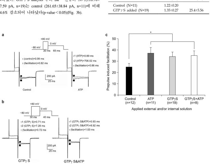
Fig. 4. Prepulse induced facilitation recorded in different dialyzing conditions. (a) Left: Ba2+ currents were recorded without (●) and with (■) prepulse of 20 ms to +80 mV from Vh=−80 mV(upper trace) in control condition. Right: Records were obtained using the same pulse protocol but with extracellular ATP. Notice the pronounced inactivation on the facilitated currents that slowly relaxes to its control level. (b) Left: Ba2+ currents were recorded without (●) and with (■) prepulse of 30 ms to +80 mV from Vh=−80 mV(upper trace) in the application of intracellular GTPγS. Right: Records were obtained using same pulse protocol but with the application of intracellular GTPγS and additional extracellular ATP. There was no additional facilitation increment with GTPγS and ATP in comparison with GTPγS alone. (c) Prepulse induced facilitation in control condition was 25±3% (n=12) increase while that with extracellular ATP, with intracellular GTPγS, with intracellular GTPγS and extracellular ATP was 37±5% (n=11) increase, was 34±4% (n=19) increase, and 35±4% (n=8) increase, respectively. There was significant difference between control group and ATP- and or GTPγS- treated group. *p-value<0.05.
록된 전류를 비교하였다. 정상 환경 하에서의 activation time constant는 1.15 ms인 반면, GTPγS 처리후의 time con- stant는 1.44 ms의 fast component (τ1)와 29.32 ms의 slow component (τ2), 두 component가 나타났다(Fig. 3a). Control
−GTPγS 처치 후 time constant의 변화를 Table 2에 정리 하였다. ATP와 유사하게 GTPγS가 Ca2+ 통로의 활성화 과 정을 느리게 조절함을 알 수 있었다.
GTPγS 처치 후의 Ba2+ 전류 크기를 control current와 비 교하였다. GTPγS dialysis 후의 Ba2+ 전류의 크기(118.78±
17.59 pA, n=19)는 control (261.65±38.84 pA, n=11)에 비해 54.6% 감소되어 나타났다(p-value<0.05)(Fig. 3b).
4. Facilitation에 대한 ATP, GTPγS의 효과
여러 신경전달물질들의 칼슘통로에 대한 억제 현상이 매
우 높은 저분극의 conditioning pulse를 주었을 때 역전 (reverse)된다는 보고가 있어서,19,20) Ba2+ 전류에 대한 APT 의 억제 효과를 확인한 후, 0 mV의 test pulse를 주기 직전 에 2.5∼30 ms의 기간동안 +80 mV의 큰 prepulse를 주는 실험을 수행하였다. 이때의 holding potential은 −80 mV이 었다. 본 실험에서 Prepulse의 기간이 20∼30 ms인 구간에 서는 facilitation 정도에는 차이가 없었다.
Prepulse 이후 기록된 current를 control과 비교하기 위하여 kinetics study를 실시하였다(Fig. 4a, 4b). Control에서 pre- pulse를 주기 이전에 얻은 current의 활성화 부분의 time constant (τ)는 0.99 ms이었고 prepulse 이후 current의 time constant는 0.82 ms이었다. 모두 single-exponential component 로 fitting되었다(Fig. 4a left). 반면 ATP 적용 후 기록된 전 류는 double-exponential로 fitting되어 fast component (τ1)가 0.89 ms, slow component (τ2)가 706.02 ms로 나타났고, ATP를 적용시킨 상태에서 large prepulse 직후에 기록된 전 류는 0.86 ms의 time constant를 가지는 single-exponential curve로 fitting이 되었다(Fig. 4a Right). Control 조건에서는 25±3%의 전류 크기 증가(facilitation)가 있은 데 반해 ATP 처치 후 37±5%의 증가가 있었다(p-value<0.05)(Fig. 4c).
GTPγS 처치시, facilitation이 있기 전의 time constant는 0.71 ms (τ1)와 7.28 ms (τ2)의 double-exponential을 나타났 고, facilitation 이후의 time constant는 0.72 ms로 나타났다 (Fig. 4b left). Fast activation component는 facilitation 전후에 변화가 없었고, 높은 전압을 인가함으로써 GTPγS에 의해 나타났던 slow activation component가 사라졌다. GTPγS를 처치한 상태에서 추가로 ATP가 첨가된 세포외용액을 관 류시키며 facilitation을 유도하여 kinetic study를 실시해 보 면, 2개의 time constant (τ1=0.93 ms, τ2=8.92 ms) 중에서 slow activation component (τ2)가 높은 저분극 pulse에 의해 사라지는 것을 볼 수 있었다(Fig. 4b Right).
GTPγS가 있는 상태에서 높은 저분극 pulse를 걸어준 후 test pulse를 주었을 때, control에서 facilitation이 25±3% (n=12) 인데 비해, GTPγS에 의한 facilitation은 34±4% (n=19)이었 으며 GTPγS와 ATP가 모두 처치되었을 때의 facilitation 은 35±4% (n=8)이었다. Control과 약물처치군에서 유의한 차이 가 있었다(p-value<0.05)(Fig. 4c).
고 찰
본 연구에서 ATP는 막전압 의존적으로 작용하는 칼슘통 로를 통한 Ba2+ 전류의 크기를 감소시켰고(Fig. 2c), 또한
칼슘통로의 activation kinetics를 느리게 하였다(Fig. 2d).
Dunlap 등은 노르아드레날린에 의해 칼슘전류가 감소하는 것은 functional 칼슘통로의 수를 줄이기 때문이라고 발표하 였으나,5) 본 연구에서는 거의 모든 통로가 약물 처치 후에 도 여전히 기능을 하고 최대 저분극 자극에 의해 milli- second (ms) 이내에 열릴 수 있는 능력이 있음을 확인하였 다(Fig. 4a, 4b).
본 연구에서는 선택적인 칼슘통로 억제제를 사용하지 않 았기 때문에 ATP에 의한 칼슘통로의 조절이 L, N, P/Q-type 칼슘통로 중 어느 type 에 의해 매개되었는 지를 확실히 구 분하기는 어려우나 ATP의 칼슘통로 억제 효과는 어느 한 type에만 한정된 것처럼 보이지는 않는다. 왜냐하면 ATP 적용시 transient N-type 뿐만 아니라 sustained L-type 부분도 함께 감소되어 나타났기 때문이다(Fig. 2d).
본 실험에서 GTPγS를 세포 내에 주었을 때 ATP와 마 찬가지로 Ba2+ 전류를 감소시켰고, activation kinetics를 느 리게 변화시켰다(Fig. 3). GTPγS의 효과 또한 ATP의 효과 와 마찬가지로 L-type이나 N-type 어느 한 type에 국한된 효 과가 아닌 것으로 해석된다.
ATP가 membrane의 Ca2+ 투과성(permeability)을 높여 Ca2+- induced inactivation mechanism을 통하여 칼슘통로를 억제하 리라는 주장이 있지만,21) 본 실험에서는 11 mM EGTA를 사용하여 세포 내 Ca2+ 농도가 심각하게 높아지는 것을 막 았으므로 이 주장 역시 배제할 수 있다.
크로마핀 세포에서 ATP는 P2Y 수용체에 결합한 후 칼슘 통로를 억제함으로써 카테콜아민 분비를 억제시킨다는 보 고가 있다.11,22) 본 실험에서는 ATP가 결합하는 purinergic receptor의 아형을 확인하기 위해 purinergic agonist와 antag- onist를 사용하지는 않았으므로 추후 ATP가 작용하는 수용 체를 확인하기 위하여 suramin, PPADS (pyridoxalphosphate- 6-azophenyl 1-2’,4’-disulphonic acid) 등의 P2 receptor antago- nist와 ADPβS, 2-MeSADP (2-methylthio ADP), AMP-PCP (β,γ-methylene ATP), AMP-CPP (α,β-methylene ATP) 등 의 agonist23) 실험이 수반되어야 할 것이다.
Intracellular GTPγS에 의한 칼슘통로 억제 효과도 NA (noradrenalin), GABA (γ-aminobutyric acid)6-10) 등에 의해 보 고된 것과 유사하게 나타났으며 ATP에 의한 효과와도 유 사하게 나타났다. 본 실험에서 사용한 electrode-filling sol- ution 성분 중 2 mM MgATP는 GTPγS에 의한 current 감소 (run-down)를 70% 정도 차단한다.18) 그러나 GTPγS는 0 mV (Vt)에서 전류 크기의 54.6%를 감소 시켰고 electrode 내 에 50 μM 농도의 GTPγS를 포함시켰을 때는 2개의 ex-
Fig. 5. Schematic diagram of chromaffin cell stimulus-secretion coupling. The splanchnic nerve that innervates chromaffin cell release acetylcholine (Ach) that activates the nicotinic acety- lcholine receptor (nAChR) that gates Na+, causing the mem- brane to depolarize (ΔV) sufficiently to trigger the action potentials to activate voltage-operated Ca2+ channels (VOCC).
Entry of external Ca2+ increases the intracellular Ca2+ con- centration ([Ca2+i]) that triggers exocytosis and the release of chromaffin granule contents that contain adrenaline and ATP.
The ATP may feedback to activate receptors that use the G protein to inhibit the VOCC. (Broken arrow represents the possibility).
ponential component의 합으로 전류기록이 잘 fitting되었다.
I(t)=A[1−exp(−t/τ1)]+B[1−exp(−t/τ2)]
Fast component의 time constant (τ1=1.44 ms)는 control의 time course와 유사했으나 slow component의 time constant (τ2=29.32 ms)는 τ1보다 훨씬 길어졌다(Fig. 3a). 이는 ATP 를 세포외부에 주었을 때와 같은 결과이며 GTPγS와 ATP 를 함께 처치하였을 때도 double-exponential curve로 fitting 되었다(Fig. 4b).
ATP를 주었을 때와 마찬가지로 GTPγS를 electrode 내에 포함시켰을 때 Ba2+전류는 slow activation을 보이며, 동시 에 억제된 Ba2+ 전류는 높은 저분극 자극에 의해 역전되었 다(37% facilitation). 그러나 facilitation 이후에 GTPγS나 ATP에 의해 나타났던 slow activation 성분이 사라졌다(Fig.
4a, 4b). 즉, ATP와 GTPγS에 의한 칼슘통로의 억제가 높 은 prepulse에 의해 풀렸고, facilitation 이후의 전류는 ATP 와 GTPγS가 없을 때의 control 전류와 유사한 τ값을 가지 는 single-exponential curve로 fitting되어 1 개의 fast compo- nent만을 가진다(I(t)=A[1−exp(−t/τ1)]).
보다 큰 facilitation 효과는 ATP 존재 시와 GTPγS가 있 을 때 나타났고 이는 regulatory G-protein의 disinhibition에 의한 것으로 사료된다. 즉, ATP와 GTPγS가 regulatory G-protein을 활성화시켜 Ca2+ 통로를 억제하고 이 통로의 activation kinetics를 변화시켰던 것을 높은 양(positive) 전압을 걸어줌으로써 G-protein에 의한 억제가 풀려 ATP, GTPγS 가 없을 때와 동일한−즉 control current와 같은 kinetics를 보이는 것이라고 해석된다.
G-protein의 channel modulation 기전은 크게, protein phos- phorylation과 연관된 Protein Kinase를 경유할 것이라는 의 견,24) 그리고 G-protein이 직접 channel에 binding하여 조절 하는 ‘direct modulation’이라는 의견14)으로 나뉘어 있다.
ATP에 의한 칼슘통로의 조절은 direct G-protein modu- lation으로 사료된다. 왜냐하면 ATP 또는 GTPγS 처치로 G-protein activation으로 인한 전류 감소 후 facilitation proto- col을 수행했을 때, channel의 state가 매우 빠른 시간 안에 (∼ms 이내) 회복되는 것은 large depolarization에 의해 G-protein이 channel 단백질 subunits의 G-protein binding site 로부터 떨어지게(release) 될 것이기 때문이다. 그러나 본 실 험에서는 G-protein coupled receptor (GPCR) blocker와 G-protein 아래 단계에서 작용하는 각종 signaling pathway 에 관여하는 blocker 를 처치하지 않았으므로 본 연구에서
확인한 ATP의 칼슘통로 억제효과는 G-protein과의 관련성 을 직접적이 아니라 간접적으로 보여준다.
결론적으로 ATP는 칼슘통로를 down-regulation하는 G- protein을 활성화시킴으로써 전류의 억제와 채널 활성화를 느리게 유도하는 것으로 사료된다. 그러나 약물 처치 이후 facilitation을 수행하여도 원래의 current 크기로는 회복되지 않는 결과로 미루어 current reducing과 slow activation이 같 은 pathway를 가지는지의 문제가 남아있고 이를 해결하기 위하여 이후에 GDPβS나 다른 ATP analog를 이용한 실험 을 더 수행하고자 한다.
결 론
본 실험 결과의 생리학적 의미는 부신수질 크로마핀 과 립에 함께 저장되어있던 여러 신경전달물질들이 카테콜아 민과 함께 분비되는 과정에서 찾을 수 있다. 부신수질 크로 마핀 세포의 경우 아드레날린을 함유하는 adrenergic secre- tory vesicles에 함께 포함된 adenosine nucleotide compound 중에서는 ATP가 가장 많은 분율을 차지한다(약 90% 정도).
이렇게 부신수질 크로마핀 세포의 분비과립에 아드레날린
과 함께 다량 포함되어 있던 ATP가 아드레날린과 함께 방 출된 후 칼슘통로를 억제함으로써, 시냅스 전 억제(auto- crine inhibition)를 하거나 혹은 주변의 크로마핀 세포에 작 용하여(paracrine inhibition) 카테콜아민 분비 과정을 국소적 으로 조율할 것으로 사료된다(Fig. 5).
References
1. Augustine GJ, Neher E: Calcium requirments for secretion in bovine chromaffin cells. J Physiol 450:247-271 (1992) 2. Boarder MR, Marriot D, Adams M: Stimulus secretion cou-
pling in cultured chromaffin cells. Biochem Pharm 56(1):163-167 (1987)
3. Albillos A, Artalejo AR, Lopez MG, Gandia L, Garcia AG, Carbone E: Calcium channel subtypes in cat chromaffin cells. J Physiol 477:197-213 (1994)
4. Zimmermann H: Signalling via ATP in the nervous system.
Trends Neurosci 17:420-426 (1994)
5. Dunlap K, Fischbach GD: Neurotransmitters decrease the calcium conductance activated by depolarization of embryonic chick sensory neurons. J Physiol 317:519-535 (1981) 6. Forscher P, Oxford GS: Modulation of calcium channels by
norepinephrine in internally dialyzed avian sensory neurons. J Gen Physiol 85:743-763 (1985)
7. Galvan M and Adams PR: Control of calcium current in rat sympathetic neurons by norepinephrine. Brain Res 244:135-144 (1982)
8. Dolphin AC and Scott RH: Calcium channel currents and their inhibition by (-)-baclofen in rat sensory neurons: modu- lation by guanine nucleotides. J Physiol 386:1-17 (1987) 9. Grassi F and Lux HD: Voltage-dependent GABA-induced
modulation of calcium currents in chick sensory neurons.
Neurosci Lett 1015:113-119 (1989)
10. Menon-Johansson AS, Berrow N, Dolphin AC: G(o) transduces GABAB-receptor modulation by N-type calcium channels in cultured dorsal root ganglion neurons. Pflugers Arch 425:335-343 (1993)
11. Harkins AB and Fox AP: Activation of purinergic receptors by ATP inhibits secretion in bovine adrenal chromaffin cells.
Brain Res 885:231-239 (2000)
12. Carbone E, Carabelli V, Casetti T, Baldelli P, Hernandez- Guijo JM, Giusta L: G-protein and cAMP-dependent L- channel gating mechanism: a manifold system to control calcium entry in neurosecretory cells. Pflugers Arch 442(6):801-813 (2001) 13. Diverse-Pierluissi M, Dunlap K, Westhead EW: Multiple actions of extracellular ATP on calcium currents in cultured bo- vine chromaffin cells. Proc Nat Acad Sci USA 88:1261-1265 (1991)
14. Holz GG IV, Rane SG, Dunlap K: GTP-binding proteins mediate transmitter inhibition of voltage-dependent calcium channels. Nature 319:670-672 (1986)
15. Kajikawa Y, Saitoh N, Takahashi T: GTP-binding protein beta gamma subunits mediate presynaptic calcium current in- hibition by GABA(B) receptor. Proc Nat Acad Sci USA 98:
8054-8058 (2001)
16. Akaike A, Mine Y, Sasa M, Takaori S: Voltage and current clamp studies of M- and Nicotinic excitation of the rat chromaffin cells. J Pharmacol Exp Ther 255:333-339 (1990)
17. Hamil OP, Marty A, Neher E, Sakmann B, Sigworth FJ:
Improved patch-clamp techniques for high resolution current re- cording from cells and cell free membrane patches. Pflugers Arch 391:85-100 (1981)
18. Marchetti C, Robello M: Guanosine-5’-O-(3-thiotriphosphate) modifies kinetics of voltage-dependent calcium current in chick sensory neurons. Biophys J 56:1267-1272 (1989)
19. Artalejo CR, Rossie S, Perlman RL, Fox AP: Voltage dependent phosphorylation may recruit Ca2+ current facilitation in chromaffin cells. Nature 358:63-66 (1992)
20. Dolphin AC: Facilitation of Ca2+ current in excitable cells.
Trends Neurosci 19(1):35-43 (1996)
21. R. Eckert and J.E. Chad: Inactivation of calcium channels:
Prog. Biophysi. Mol. Biol., 44:215-267 (1984)
22. Powell AD, Teschemacher AG, Seward EP: P2Y purino- ceptors inhibit exocytosis in adrenal chromaffin cells via modu- lation of voltage-operated calcium channels. J Neurosci 20(2):
606-616 (2000)
23. Dubyak GR, el-Moatassim C: Signal transduction via P2- purinergic receptors for extracellular ATP and other nucleotides.
Am J Physiol 265:C577-606 (1993)
24. Carabelli V, Hernández-Guijo JM, Baldelli P, Carbone E: Direct autocrine inhibition and cAMP-dependent potentiation of single L-type Ca2+ channels in bovine chromaffin cells. J Physiol 532(Pt 1):73-90 (2001)
흰쥐 부신 크로마핀 세포 칼슘통로 조절에 미치는 ATP의 효과
충북대학교 의과대학 *의공학교실, †생리학교실
김경아*⋅구용숙†
뉴론에서 ATP는 분비 과립내에 신경전달물질과 함께 다량 저장되어 있다가 신경전달물질과 함께 분비되는 것으로 알려 져 있으므로 신경전달물질의 자극-분비(stimulus-secretion) coupling 과정에 있어 중요한 조절작용을 할 것으로 기대된다.
그러므로 본 연구에서는 뉴론과 그 발생학적 기원이 동일한 부신수질 세포(adrenal chromaffin cell)를 대상으로 하여 세포 막 칼슘통로를 통한 세포막 전류에 미치는 ATP의 영향을 측정함으로써 신경전달물질이 자극-분비 coupling 과정에 작용 하는 ATP의 조절 작용을 알아보고자 하였다. 부신수질 세포의 칼슘통로를 통한 세포막 전류는 패치클램프 테크닉으로 기록하였다. 10 mM Ba2+을 포함한 세포 외 용액에서, Ba2+ current는 0.1 mM ATP를 세포외부에 처치했을 때, 평균 36±6% (n=6) 감소되어 나타났고 ATP를 씻어준 후 전류는 다시 회복되는 가역적 반응을 보였다. ATP의 전류 억제 기전 을 알아보고자 칼슘통로에서 관찰되는 현상 중의 하나인 소통(facilitation)현상을 기록하였다. +80 mV의 큰 prepulse를 준 후 바로 테스트 펄스를 주며 측정한 전류는 큰 prepulse에 의해 억제효과가 풀리는(disinhibition) 현상을 나타내었다. ATP 처치 후 큰 자극을 주어 37±5% (n=11)의 Ba2+ 전류 증가가 있었고 이는 ATP가 없는 상태에서 순수하게 큰 자극에 의해 소통되는 25±3% (n=12)과 유의한 차이를 보였다(p<0.05). ATP의 억제 기전이 G-protein을 매개로 한 것인지를 알아보고 자 가수분해 되지 않는 GTP 유도체인 GTPγS를 세포 내에 준 후 Ba2+ 전류를 기록하였다. GTPγS에 의해 55%의 전류 크기의 감소가 있었고 이 환경에서 큰 prepulse를 인가하였을 때 34±4% (n=19)의 소통현상을 보였다. 이는 GTPγS가 없 는 환경에서의 25±3% (n=12)의 소통현상을 보인 것과 유의한 차이를 보였다(p<0.05). Ba2+ current trace의 활성화 과정 (activation)을 curve-fitting한 결과, control은 single exponential curve로 fitting된 반면, ATP 또는 GTPγS를 처치한 경우, 그 리고 ATP와 GTPγS 모두 처치한 경우에서는 double-exponential curve로 가장 잘 fitting이 되었다. 즉, ATP나 GTPγS를 처치했을 때 모두 전류가 더 느리게 활성화되는 모양을 나타내었고, 이상의 결과로 미루어 ATP와 GTPγS는 같은 방식 으로 칼슘통로를 억제하고, 이러한 억제효과는 세포막에 아주 큰 전압을 걸어주면 칼슘 통로에 결합했던 G-protein이 막 전압 의존적으로 떨어짐으로써 소실(disinhibition)된다고 해석된다. 본 연구에서 확인한 ATP의 칼슘통로 억제효과는 자체 크로마핀 세포 또는 주변 세포에서 아드레날린이 적게 분비되게 하는 autocrine 또는 paracrine inhibition 과정의 중요한 기전으로 작용할 것이다.
중심단어: 크로마핀 세포, 칼슘통로, ATP, G-단백질, 자가분비 억제