I n t rod u c t i o n
Amyotrophic lateral sclerosis (ALS) is a clinical- ly well-defined disorder of the pyramidal and spinal motor neurons.
1Muscle weakness develops with the progressive loss of both upper motor neurons (UMN) and lower motor neurons (LMN).
Due to the progressive and fatal course of ALS, diagnostic testing must be as certain as possible
and false positives minimized. The electrophysio- logical diagnostic criteria of ALS require abnor- malities in at least three of four areas of the neu- raxis (craniobulbar, cervical, thoracic, or lum- bosacral) and in at least one area beyond the cer- vical and lumbosacral segments to assure wide- spread motor neuron involvement. It is well known that electromyographic (EMG) findings and motor evoked potentials (MEP) of the lower cranial nerve innervated muscles, such as, of the genioglossus (tongue) and masseter, have been detected in cases of craniobulbar dysfunction.
However, these methods have some limitations, as they are difficult to perform and uncomfort- able to patients.
2 - 5The objectives of this study
근위축성 측상경화증 환자에서 등세모근의 신경생리학적 검사
서울대학교 의과대학 신경과학교실
조중양・전종은・이광우
The Electrophysiological Studies of the Trapezius Muscle in Patients with Amyotrophic Lateral Sclerosis
Joong-Yang Cho, M.D., Jong-Un Chun, M.D., Kwang-Woo Lee, M.D., Ph.D.
Department of Neurology, College of Medicine, Seoul National University
Background: Needle electromyography (EMG) and motor evoked potential (MEP) of the genioglossus (tongue) are difficult to perform in evaluations of the craniobulbar region in amyotrophic lateral sclerosis (ALS). Therefore, we investigated the yields of needle EMG and MEP recorded from the upper trapezius, since it receives innervation from the lower medulla and upper cervical cord.
Methods: Needle EMG and MEP of the upper trapezius were obtained in 17 consecutive ALS patients. The needle EMG parameters recorded included abnormal spontaneous activity and motor unit action potential (MUAP) morpholo- gy. An upper motor neuron (UMN) lesion was presumed when either response to cortical stimulation was absent, or the central conduction time was delayed (>mean+2SD).
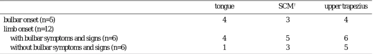
Results: Of the five patients with bulbar-onset ALS, four had abnormalities in the upper trapezius and four in the tongue by needle EMG. In contrast, of the 12 patients with limb-onset ALS, 11 had abnormalities in the upper trapezius, and only five in the tongue. When MEP was performed, it was found that three of the five patients with bulbar symp- toms and three of the six patients with isolated limb involvement had abnormal MEP findings.
Conclusions: Electrophysiological studies of the upper trapezius are more sensitive those of the tongue in patients without bulbar symptoms. Thus, needle EMG and MEP of the upper trapezius are alternative tools for assessing bulbar and rostral neuraxial involvement in the diagnosis of ALS.
Key Words: Amyotrophic lateral sclerosis, Needle EMG, MEP, Upper trapezius
Address for correspondence Kwang-Woo Lee, M.D., Ph.D.
Department of Neurology, Seoul National University Hospital,
28 Yongon-dong, Chongno-gu, Seoul, 110-744, Korea
Tel: +82-2-2072-3215 Fax: +82-2-3672-7553
E-mail : [email protected]
were to compare the relative utilities of EMG of the tongue, SCM, and upper trapezius muscles in the evaluation of LMN dysfunction, and to inves- tigate MEP by transcranial magnetic stimulation (TMS) in the upper trapezius muscles of ALS patients, finally we examined the value of elec- trophysiological studies of the upper trapezius muscle as alternative tools for assessing cranio- bulbar involvement in the diagnosis of ALS.
Material & method s
1. Patients and normal subjects
The study was performed in 17 ALS patients (12 males and 5 females; aged 35~72 years, mean±
S D = 5 4 . 7±11.0). Their mean duration of illness was 20.9±11.8 months at the time of this investi- gation. A diagnosis of ALS was made based on clinical and electrophysiological examinations based according to E1 Escorial criteria.
1To deter- mine the normal parameters of MEP, 21 healthy subjects (7 males and 14 female; aged 30~71 years, mean±S D = 5 2 . 9±12.7) were included.
These controls had no neurological abnormalities, and had electrophysiological study results within normal limits. All subjects provided informed consent, and the study was approved by our institutional ethics committee.
2. Procedures
1) Needle EMG of the upper trapezius
Needle EMG was performed as previously d e s c r i b e d .
5 , 6Though the number of muscles stu- died per patients varied, needle EMG was per- formed in bulbar, limbs, and paraspinal muscles.
In particular, attention was focused on the tongue, SCM, and upper trapezius muscles. EMG abnormalities in muscles were assessed by fol- lowing a standard protocol.
7 , 8We first looked for abnormal spontaneous activity in the form of positive sharp waves and fibrillation potentials with muscles at rest. Abnormal spontaneous activity was defined as abnormal activity in two separate sites within a muscle. Motor unit action potential (MUAP) configuration was assessed during a minimal muscle contraction, and was considered complex when polyphasic (greater than four action potentials crossing the baseline) or polyturn (greater than five turns without crossing the baseline).
7The pattern of MUAP
recruitment was excluded due to faster firing rates of the bulbar muscles.
62) MEP of the upper trapezius
Corticobulbar projections were investigated by TMS, and evoked potentials were recorded via surface electrodes placed on the upper trapezius muscle, as previously described.
9In brief, the active electrode was taped 1~2 cm below the upper margin of the muscle, approximately mid- way between the acromion and the C7 spinous process. The reference electrode was placed over the acromion. For TMS, a standard circular coil (diameter 90 mm, Cadwell MES-100) with a peak magnetic field of 2.0 tesla was used and poten- tials were registered using a conventional unit (Cardwell Excel EMG instrument). The coil was applied tangentially to the skull with its center placed over the vertex or slightly laterally toward the stimulated hemisphere. In order to evoke muscle responses with shortest latencies and largest amplitudes, the subjects were asked to contract the target muscle by lifting the shoulder of interest against resistance applied by an examiner to the wrist.
The parameter analyzed was the central motor conduction time (CMCT). CMCT was defined as the difference between the shortest onset latency of the MEP and the latency of the compound muscle action potentials (CMAP). Peripheral CMAP was obtained by the spinal accessory nerve (SAN), which was electrically stimulated with surface electrodes slightly above the midpoint between the clavicle and mastoid process along the posterior border of the SCM.
1 0Strictly speak- ing, this parameter was not a true central con- duction time, since it included the proximal seg- ment of the accessory nerve. An UMN-lesion was assumed when either the cortically evoked response was absent or the CMCT was delayed (>
mean+2SD) with the normal nerve conduction study of spinal accessory nerve. The stimulation intensity was increased to 100% of the maximal output of the stimulator before a response was regarded absent. Absence of potential was defined as no reproducible response in four consecutive t r i a l s .
11,123) Statistical analysis
Correlations between bulbar dysfunction and
MEP findings were analyzed using Fisher’ s test and Sperman’ s correlation coefficients. The clini- cal severity of bulbar dysfunction was evaluated by using Appel ALS rating scale (AALSRS).
1 3T o compare MEP findings and AALSRS score, we divided the patients into two groups according to MEP abnormalities. The difference of bulbar AAL- SRS score between two groups were statistically investigated using the Mann-Whitney U test, and the level of statistical significance was set at P
<0.05. Statistical analyses were performed using the SPSS statistical software package.
Re s u l t s
1. Normal subjects
The mean CMAP amplitude of the upper trapez- ius in normal subjects was 13.2±4.6 mV (range:
4.8~25.5 mV), and its mean latency was 2.1±0 . 3 ms (range: 1.4~3.0 ms). MEP was easily elicited from the upper trapezius muscle in all subjects although maximal MEP amplitudes often required a higher level of voluntary contraction, and a higher magnetic stimulation strength. The mean MEP latency was 8.7±0.85 ms (range: 7.0~10.8 ms), and the mean CMCT was 6.42±1.25 ms (range: 1.8~85 ms) and normal limiting values were defined as the mean value + 2SD (≤8.92 ms).
2. Patients
1) Needle EMG of the upper trapezius
EMG abnormalities in limb-onset ALS were
more frequent in the upper trapezius (11/12) than in the SCM (8/11) or in the tongue (5/12). Patients with limb-onset ALS were further divided into those who had no bulbar symptoms (n=6) and those who had bulbar symptoms (n=6). In patients with no bulbar symptoms, EMG abnormalities of the upper trapezius and SCM were detected in five and three ALS patients, respectively, but EMG tongue abnormalities were found in only one patient. In patients with bulbar symptoms, EMG abnormalities were found in the upper trapezius (n=6) and tongue (n=4). We also analyzed our data in an alternative way by dividing the 17 patients into those with no bulbar symptoms (n=6) and those with bulbar symptoms (n=11), regardless of the location of symptom-onset (Table 1, Table 2).
2) Upper trapezius MEP
The upper trapezius MEP was abnormal in 12 of 17 patients. Four patients had a prolonged CMCT with a normal CMAP latency value. MEP included no potential or only poor potential in eight patients. A clinical examination performed at the time of trapezius MEP revealed that six patients had the symptoms and signs of UMN involve- ment, such as, hyperactive jaw reflexes or reduced tongue motility with spastic dysarthria and dysphagia. Three of the five patients with bulbar symptoms such as tongue atrophy and fasciculation showed evidence of additional sub- clinical coricobulbar tract involvement. In three of six patients with isolated limb involvement,
Table 1. EMG abnormalities* in 17 ALS patients
tongue SCM
†upper trapezius
bulbar onset (n=5) 4 3 4
limb onset (n=12)
with bulbar symptoms and signs (n=6) 4 5 6
without bulbar symptoms and signs (n=6) 1 3 5
* EMG abnormalities mean denervation potentials and/or giant motor unit action potentials
†
sternocleidomastoid muscle
Table 2. Electrophysiological findings of 17 ALS patients
EMG abnormalities* upper trapezius MEP
tongue SCM
†upper trapezius abnormal normal
with bulbar symptoms and signs (n=11) 8 5 10 9 2
without bulbar symptoms and signs (n=6) 1 3 5 3 3
* EMG abnormalities mean denervation potentials and/or giant motor unit action potentials
†