• 검색 결과가 없습니다.

Electrophysiological Studies in the Diagnosis of Amyotrophic Lateral Sclerosis

N/A
N/A
Protected

Academic year: 2021

Share "Electrophysiological Studies in the Diagnosis of Amyotrophic Lateral Sclerosis"

Copied!
13
0
0

로드 중.... (전체 텍스트 보기)

전체 글

(1)

Amyotrophic lateral sclerosis (ALS) is a relent- lessly progressive neurodegenerative disorder involving primarily motor neurons in the cerebral cortex, brainstem, and spinal cord. A complete understanding of the pathogenesis of ALS remains elusive. The variability in clinical find- ings early in the course of ALS and the lack of any biological diagnostic marker make absolute diagnosis difficult and compromise the certainty of diagnosis in clinical practice, therapeutic trials and other research purposes.

1 - 4

Electrophysiological studies are the centerpiece of the diagnostic evaluation of ALS since L a m b e r t’s criteria at 1957. Traditionally, those studies has been used to document lower motor neuron (LMN) loss and has been most valuable in

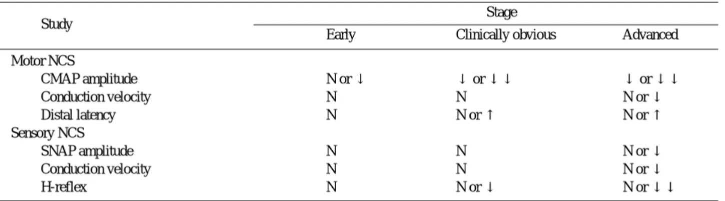
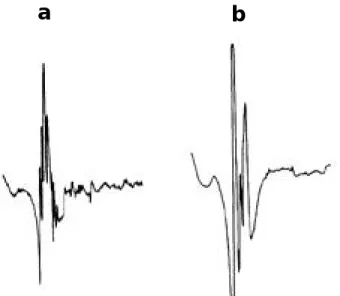
the diagnostic process. Electrophysiological test- ing provides unique information on the compen- satory or secondary changes that occur in muscle with denervation and consequently is a sensitive measure of LMN loss. Electrophysiological studies has also provided insight into the pathological changes that occur in muscle with disease pro- gression, and a number of those tests can be employed to follow disease progression and are useful in clinical trials. More recently, special electrophysiological methods have been developed to study upper motor neuron (UMN) loss.

2 , 3 , 5 - 7

1. Diagnostic criteria

1) Clinical diagnosis

The World Federation of Neurology (WFN) con- vened in El Escorial, Spain, in 1994 to formulate a set of research criteria for the diagnosis of ALS.

The criteria were reviewed in 1998 at a subse- quent WFN meeting, which led to a revised set of criteria with a limited number of changes. The criteria focus on four key features; (1) evidence

근위축성 측삭경화증의 진단에 있어서 전기진단학적 검사

대구가톨릭대학교 의과대학 신경과학교실

이 동 국

Electrophysiological Studies in the Diagnosis of Amyotrophic Lateral Sclerosis

Dong Kuck Lee, M.D.

Department of Neurology, School of Medicine, Catholic University of Daegu

Amyotrophic lateral sclerosis (ALS) is a progressive neurodegenerative disorder that primarily affects motor neurons.

The characteristic features of this devastating disorder are the simultaneous presence of upper and lower motor neuron (LMN) signs with progression from one region of the neuraxis to the next and eventual death, typically from respiratory compromise.

Electrophysiological studies are an indispensible part of the ALS evaluation, especially serving as an extension of the clinical examination, and most useful in identifying LMN dysfunction. Not only may electrodiagnostic studies reveal characteristic changes in those regions clinically manifesting signs, but it also serves to disclose asymptomatic areas of involvement.

Key Words: Amyotrophic lateral sclerosis, Electrophysiological studies

Address for correspondence Dong Kuck Lee, M.D.

Department of Neurology, School of Medicine Catholic University of Daegu

3056-6 Daemyung 4 Dong, Namgu, Daegu, 705-718, Korea

Tel: +82-53-650-4267 Fax: +82-53-654-9786

(2)

for UMN loss, (2) evidence for LMN loss, (3) evi- dence for progression within a region and to other regions, and (4) no evidence for an alterna- tive explanation for motor dysfunction (Table 1, 2 ) .

1 , 5

The neuraxis is divided into four regions:

head (bulbar), cervical, thoracic, and lumbosacral.

Electrophysiological testing is an important diagnostic tool because it is an extension of the neurological examination to verify and document the extent of LMN loss. At the end of the evalua- tion process, the distribution of UMN and LMN leads to levels of diagnostic certainty: clinically definite, clinically probable, clinically probable- laboratory supported, and clinically possible ALS (Table 3).

1 , 5

2) Electrophysiological diagnosis

ALS is primarily a clinical diagnosis. However, electromyography (EMG) and nerve conduction

studies (NCS) can be extremely important in establshing the diagnosis when insufficient clini- cal evidence is available. A number of criteria for electrophysiological diagnosis have been suggest- ed. But there are certain guidelines should be remembered. First, evidence of muscle denerva- tion should be diffuse; there is, fibrillation and large motor units should be present in multiple muscles of multiple extremities. Most criteria require denervation to be present in three of four areas of the neuraxis, with the four areas being bulbar, cervical, thoracic, and lumbosacral.

Second, there should be no evidence of motor conduction block. Because conduction block implies that there is an area of axon through which action potentials are not conducted, even though viable axon exists both proximal and dis- tal to that area. Third, motor and sensory con- duction velocity and compound sensory action potential amplitudes should be normal or nearly so from both arm and leg. If these conditions are fulfilled, the diagnosis of LMN disease can be considered confirmed. But to make a diagnosis of ALS, clinical evidence of UMN disease must also be present.

4,8

2. Electrophysiological tests

Electrophysiological studies are very helpful in the diagnostic process in suspected ALS because they can exclude other neuromuscular diagnoses, show evidence that clinically affected regions suffer from LMN loss, and demonstrate that there is LMN loss in clinically uninvolved regions.

2 , 5

Within the WFN diagnostic criteria a set of electrophysiological criteria designed to identify Table 1. Clinical signs of upper and lower motor neuron

involvement in ALS Signs of upper motor neuron loss

Labile emotional affect Forced yawning

Exaggerated snout reflex Pathological jaw jerk Pathological gag reflex

Spastic tone to passive limb movements Pathological tendon reflexes

Preserved tendon reflexes in weak and atrophic limb Clonus

Hoffmann responses Extensor plantar responses Signs of lower motor neuron loss

Muscle weakness Muscle atrophy Fasciculations

Table 2. Revised World Federation of Neurology requirements for the diagnosis of ALS

Features present Features absent

Evidence of lower motor neuron degeneration Electrophysiological or pathological evidence by clinical, electrophysiological or of other disease processes that might neuropathological examination explain signs of lower motor neuron or upper motor degeneration or both

Evidence of upper motor neuron Neuroimaging evidence of other disease degeneration by clinical examination processes that might explain the observed

clinical and electrophysiological signs Progressive spread of signs within a region,

or to other region as determined by history

or examination

(3)

active and ongoing denervation consistent with a neuropathic process (Table 4).

1 , 5

The most impor- tant electrophysiological test is needle elec- tromyography (EMG). However, there is a spec- trum of tests that can be used to exclude other diagnoses and help support the diagnosis of A L S .

5 , 9

1) Nerve conduction studies

NCS are an integral part of the evaluation of ALS patients and are useful in distinguishing ALS from disorders that mimic it. So the electrophysi- ological evaluation starts with NCS to look for evidence of focal or diffuse neuropathies. NCS can help identify these neuropathies by recording slowed conduction velocity. Motor studies com- prise conventional assessments of evoked motor responses amplitudes, distal latencies, nerve con- duction velocities, F-wave latencies, and sequen- tial evoked motor amplitudes with repetitive nerve stimulation. Sensory studies comprise con- ventional assessments of sensory nerve action potential amplitudes, distal latencies, conduction velocities, and H reflexes.

Motor nerve conduction values reflect the largest and hence fastest conducting nerve fibers.

Consequently, conduction velocity values will fall in ALS with loss of neurons with large diameter fibers. Despite very low compound muscle action potential (CMAP) amplitudes in some nerves in

ALS, conduction velocity was never less then 70%

of the lower limit of normal, and distal motor latencies and F wave latencies were rarely greater than 125% of the upper limit of normal.

But in studies of motor function, conduction velocity in ALS patients has been shown consis- tently normal or near normal until muscle atro- phy becomes extreme. However, even when motor conduction velocity is normal, distal motor laten- cy often is prolonged. Distal latencies that are prolonged out of proportion to proximal conduc- tion velocity is a finding often associated with dying-back neuropathies in which the terminal axon is more affected than the cell body or proxi- mal axon.

2 , 5 , 1 0

A pattern of motor nerve conduction abnormal- ities in intrinsic hand muscles called the “s p l i t h a n d”pattern has been described in ALS patients.

The observation is that the CMAP amplitudes recorded from the lateral side of the hand (medi- an nerve-innervated thenar muscles) are fre- quently lower than from the medial side of the hand (ulnar nerve-innervated hypothenar mus- cles), which is opposite to the normal pattern of amplitudes. Among ulnar nerve-innervated mus- cles, the first dorsal interosseous muscle may show the greatest loss of CMAP amplitude.

1 1

Although many patients with ALS report vauge sensory symptoms, clinical sensory examination usually is normal. Therefore, it is not surprising

Table 3. Levels of dignostic certainty in ALS based on clinical signs of upper and lower motor neuron loss

Clinically definite ALS

Upper and lower motor neuron clinical signs in bulbar plus two spinal regions, or in three spinal regions

Clinically probable ALS

Upper and lower motor neuron clinical signs in at least two regions with some upper motor neuron signs rostral to lower motor neuron signs

Clinically probable laboratory-supported ALS

Upper and lower motor neuron clinical signs in one region, or upper motor neuron signs in one region and lower motor neuron signs by electrophysiological criteria in at least two limbs

Clinically possible ALS

Upper and lower motor neuron clinical signs in one region, or upper motor neuron clinical signs in two or more regions, or lower motor neuron clinical signs rostral to upper motor neuron signs

Table 4. Electrodiagnostic features of lower motor neuron loss in ALS

Signs of active denervation Fibrillation potentials Positive sharp waves

Fasciculation potentials (common in active and chronic denervation, but may be absent in some muscles)

Signs of chronic denervation

Motor unit action potential morphology Increased amplitude

Increased duration

Increased proportion of polyphasia Motor unit instability

Motor unit recruitment

Rapid firing rates (usually greater than 10 Hz unless there is a significant upper motor neuron component, when rates can be less than 10 Hz)

Reduced interference pattern

(4)

that routine sensory NCS are normal or nearly normal in most ALS patients. However, sensory nerve action potential amplitudes and conduction velocities are modestly but statistically signifi- cantly lower in ALS patients, and show further deterioration over time. This is consistent with autopsy studies, which have found up to 30%

reductions in the number of dorsal root ganglion cells. The most common sensory abnormality will be focal slowing from entrapment syndromes and reduced or absent distal leg sensory responses in elderly patients

4 , 5 , 1 2

(Table 5, 6).

1 , 2

2) Repetitive nerve stimulation test

Electrophysiological tests of neuromuscular junction function may be performed as part of an electrodiagnostic protocol for weakness. Newly reinnervated muscle fibers mimic a postsynaptic defect in neuromuscular junction transmission.

Abnormalities may also be found in repetitive nerve stimulation studies in ALS patients. Tests of low-rate repetitive nerve stimulation (2 to 3

Hz) in ALS subjects show decrement in approxi- mately half of proximal (trapezius) muscles and distal (hypothenar) muscles (Fig. 1).

1 3

These find- ings suggests that functional alterations of the neuromuscular junction accompany this dis- e a s e .

2 , 5 , 1 3

3) Needle electromyography

Needle EMG is the most important electrophys- iological test for the diagnosis of ALS. EMG can provide evidence of generalized motor neuron degeneration early in the course of disease. It adds essentially to the clinical examination and is superior to it in as far as it can provide early signs of denervation in unaffected extremities.

The typical finding is a combination of denerva- tion and collateral reinnervation. The key is the distribution of the neurogenic changes, which extend beyond the area of single spinal roots or peripheral nerves and do not necessarily show a distal preponderance, as is the case in most p o l y n e u r o p a t h i e s

2 , 5

(Table 7).

2

Table 5. Nerve conduction studies in ALS

Features consistent with ALS Features inconsistent with ALS Motor conduction times should Evidence of motor conduction block

be normal, unless the CMAP is small

Sensory nerve conduction studies Motor conduction velocities lower than 70%,

can be abnormal in the presence and distal motor latencies over 30%, of the lower of entrapment syndromes and and upper limit of normal values, respectively coexisting peripheral nerve disease

Lower extremity sensory nerve Abnormal sensory nerve conduction studies*

responses can be difficult to F-wave or H-reflex latencies more than 30%

elicit in the elderly above established normal values

Decrements greater then 20% on repetitive nerve stimulation at 2 Hz

* With the exception of age, entrapments, and coexisting sensory polyneuropathy.

CMAP = compound muscle action potential

Table 6. Nerve conduction study changes at different stages of ALS

Stage Study

Early Clinically obvious Advanced

Motor NCS

CMAP amplitude N or ↓ ↓ or ↓↓ ↓ or ↓↓

Conduction velocity N N N or ↓

Distal latency N N or ↑ N or ↑

Sensory NCS

SNAP amplitude N N N or ↓

Conduction velocity N N N or ↓

H-reflex N N or ↓ N or ↓↓

NCS = nerve conduction studies; N = normal; ↓/↑ = decreased/ increased

one arrow = mild; two arrows = moderate

(5)

Figure 1. Repetitive nerve stimulation of the trapezius muscle baseline (phase I), immediately following 30 s of exercise (phase II), and at 3 min postexercise (phase III) in a patient with ALS. The response shows a typical decremental pattern of 13% (phase I), 14%

(phase II), and 15%(phase III) with no facilitation immediate postexercise (phase II). Calibration = 2 mV/5 ms.

Table 7. Needle electrode examination changes at different stages of ALS

Stage Study

Early Clinically obvious Advanced

At rest

Fasciculations N or + N to ++ N to ++

Insertional PSWs + or ++ + to +++ + to +++

Fibrillations N or + + to +++ + to +++

MUPs

Recruitment N or ↓ ↓ or ↓↓ ↓↓ or ↓↓↓

Duration N or ↑ ↑ or ↑↑ ↑↑ or ↑↑↑

Amplitude N ↑ or ↑↑ ↑↑ or ↑↑↑

Complexity N + or ++ + or ++

MUPs = motor unit potentials; N = normal/none; ↓/↑ =decreased/increased; one arrow = mild; two arrows = moderate, three

arrows = marked. + = mild; ++ = moderate; +++ = marked

(6)

(1) Abnormal spontaneous activity

The first sign of motor neuron degeneration is pathological spontaneous activity such as fibrilla- tion potentials, positive sharp waves (PSW), and complex repetitive discharges (CRD) found in fully relaxed muscles. Fibrillation potentials and PSW are common findings as they occur in early den- ervation. But CRD are less often found and are more typical of chronic denervation. Initially, pathologic spontaneous activity is found focally and asymmetrically in line with the clinical pre- sentation. But relatively soon, those activities become generalized. Denervation may occur in both neurogenic and myopathic diseases. Finding fibrillation potentials and PSW in multiple mus- cles supports a diffuse process. The full needle EMG study is usually relied on to distinguish between myopathic and neuropathic processes.

The presence of fibrillation potentials and PSW is a supportive diagnostic finding, but abnormal spontaneous activity may not be observed in cra- nial innervated muscles or early on in limb mus- c l e s .

5 , 9

(2) Fasciculation potentials

One of the most characteristic abnormalities seen in ALS patients is the presence of fascicula- tion potentials. Those have long been observed clinically in ALS, and are included in the electro- physiological criteria. But the origin of fascicula- tion is yet unclear. Fasciculations are common in other disorders and in normal individuals, in whom they are considered to be benign. Several features separate benign fasciculations from those observed in ALS; on needle EMG examina- tion benign fasciculations tend to have normal motor unit configurations for amplitude, dura- tion, and number of phases, are stable from dis- charge to discharge, and have a higher discharge rate. And also showes restricted distribution, infrequent firing, and does not present any other neurologic dysfunctions. But fasciculations found in ALS differ in that they may have more complex configurations, and the degree of complexity may be greater in weaker muscles. Furthermore, among fasciculations in ALS, less complex exam- ples are more readily activated by voluntary acti- vation. These findings suggest that complex and unstable fasciculations reflect enlarged motor

units. And also showes abundant frequency, and other abnormal neurologic or electrophysiologic s i g n s .

In ALS, fasciculations may be observed in clini- cally strong muscles with no positive waves and fibrillation potentials and whose only signs of denervation is increased fiber density. This implies that fasciculations may constitute one of the earliest signs of LMN changes in ALS. The WFN criteria emphasize caution in interpreting fasciculations; their absence raises diagnostic doubts, and they may be benign or occur in other denervating disorders.

4 , 5 , 8 , 9 , 1 4 , 1 5

(3) Motor unit recruitment

Normal recruitment refers to the orderly acti- vation of more motor units as the effort and fir- ing rate of individual units increases. Reduced motor unit recruitment reflects the primary pathological process of LMN loss. Clinical assess- ment of recruitment is subjective and is based on estimates of the discharge frequency and the number of motor units on the EMG oscilloscope screen. Frequency estimation is commonly carried out at low levels of muscle force because individ- ual units become difficult to discern in a field of m a n y .

Discrimination of discharge frequency is usually aided by the sound of the motor unit discharge over a loudspeaker. LMN loss in ALS results in higher discharge frequencies and a greater degree of discharge variability. And motor unit recruit- ment is also affected by UMN loss.

2 , 4 , 5 , 9

(4) Motor unit amplitude

With progression of the ALS, collateral sprout- ing of the surviving motor units leads to an enlargement of the motor unit action potentials, i.e. an increase of duration, amplitude, and of the rate of polyphasic configuration. The giant potentials recorded with concentric needle EMG represent the summation of single muscle fiber action potentials within an individual motor unit of increased size and capacities; this is probably evidence of fiber-type grouping. These signs of chronic denervation are often seen in muscles with no clinical apparent weakness or atrophy.

Motor unit amplitude is most commonly estimated

at low levels of force in conjunction with assess-

ment of recruitment.

4 , 5 , 9 , 1 6

(7)

(5) Motor unit duration

Motor unit duration, including the initial part, main spike, and terminal part, but excluding satellite potentials, is increased in ALS. The com- plexity of the motor unit waveform is character- ized by the number of turns and phases. In ALS, the number of polyphasic (more than four phases) motor units is increased. In addition, polyphasic and polyturn (more than five turns) motor units show instability from discharge to discharge, reflecting greater variability in neuromuscular transmission related to reinnervation and vari- abilities of conduction along muscle fibers (Fig.

2 ) .

1 7

This variability has been termed “j i g g l e”( F i g . 3 ) .

1 8

Motor units in ALS tend to have a greater number of satellite potentials.

(6) Examination of special muscles

The level of WFN diagnostic certainty is based in part on the distribution of LMN symptoms and signs. Determination of the distribution of LMN

loss in asymptomatic muscles is aided by needle EMG. Accordingly, needle EMG evidence for LMN loss in bulbar and thoracic paraspinal muscles increases the level of diagnostic certainty. Studies of the tongue in ALS show signs of active dener- vation (abnormal motor recruitment and increased proportion of polyphasia) in two thirds of patients studied. Studies of the frontalis, tem- poralis, mentalis, masseter, and sternocleidomas- toid muscles show active denervation in a third and chronic denervation in one half to two thirds.

Needle EMG study of thoracic paraspinal muscles reveals active denervation in up to 75% of ALS patients when two to four spinal segments are examined (Table 8).

1 , 2

Figure 2. Examples of motor unit potentials: (a) polyturn poten- tial and (b) polyphasic potential.

a b

Figure 3. Examples of superimposed MUPs from a control sub- ject (upper) and a patient with ALS (lower). There is a marked difference in the variability of shape of the two MUPs. Note also the different gain. CAD and CCC, the new parameters of jiggle, successfully express the degree of variability. Vertical bars indicate the ±2.5 ms analysis window.

Table 8. Topography of active and chronic denervation in ALS: Signs of LMN dysfunction should be found in at least 2/4 regions to support diagnosis of ALS

Region of CNS Considered positive when

Brainstem One muscle is involved (eg, tongue)

Thoracic spinal cord Paraspinal muscles at or below T6 or abdominal muscles are involved Cervical spinal cord At least two muscles innervated by different roots

and peripheral nerves are involved

Lumbosacral spinal cord At least two muscles innervated by different roots

and peripheral nerves are involved

(8)

3. Special electrophysiological techniques

Special electrophysiological techniques can be used to aid the diagnosis of ALS. The WFN elec- trophysiological criteria list several quantitative tests that can detect chronic partial denervation.

The single-fiber EMG (SFEMG) electrode provides a detailed view of the motor unit, and can be used to show early signs of denervation and reinner- vation by measuring muscle fiber density, neuro- muscular jitter, and a view of the whole motor unit with macro-EMG (Table 9).

1 , 5

1) Fiber density

SFEMG uses needle electrodes with a very small recording site. This allows the detection of signals from single muscle fibers of motor unit. The most important parameters analysed are fiber density and jitter. Fiber density is the count of single potential components recorded at a site and expressed as mean of 20 measurements. The recording or uptake radius of a single-fiber elec- trode is about 300 um. Normal values have been determined empirically and vary between muscles and with subject age, and average 1.15 to 1.80.

Elevated fiber density values are felt to reflect greater numbers of fibers within the motor unit due to collateral reinnervation.

In ALS, increased fiber density values are the first sign of denervation, before changes in rou- tine needle EMG are recorded and before changes in muscle strength can be appreciated. Fiber den- sity values tend to be higher in weaker muscles and increase as muscle weakness increases due to collateral axonal sprouting of the surviving motor

neurons. But at late stages, fiber density values can fall. The fall is felt to reflect marked loss of motor units

5 , 9 , 1 9 , 2 0

(Fig. 4).

2 0

2) Jitter

SFEMG can be used to assess neuromuscular junction transmission across individual junctions and is the most sensitive measure of transmission abnormalities. The jitter quantifies the instability of neuromuscular transmission. In ALS, jitter values are increased in most muscles, including those that are clinically strong. Jitter in ALS is felt to be abnormal because of a reduced safety factor in neuromuscular junction transmission due to fetal acetylcholine receptor subunits in reinnervated muscle

5 , 9 , 1 9

(Fig. 5).

1 9

3) Macroelectromyography

Macro-EMG is a two-channel technique using needles with a large recording surface. The resulting potentials are appropriate measures the size of the number of muscle fibers of a motor unit. The macro-EMG electrode uses 15 mm of the cannula of a single-fiber electode as the active electrode. The macro-EMG signal is a motor unit potential that has less distinct proper- ties than those of the motor unit potential recorded from concentric and monopolar elec- trodes, and the measures of interest are macro- motor unit potential amplitude and area. Macro- EMG is a good means to detect increased and decreased motor unit sizes and is more sensitive than concentric EMG in the estimation of motor s i z e .

The macro-EMG motor unit potential amplitude is increased in approximately 50% of muscles in ALS. Amplitude values are normal in subjects with primary UMN signs and highest in subjects with slow progression. In serial studies, the macro-EMG potential fell in some, attributed to a collapse or decompensation of the motor unit with disease progression

5 , 9 , 2 1 - 2 3

(Fig. 6).

2 3

4) Quantitative electromyography

The technique of quantitative EMG entails the assessment of motor unit parameters of ampli- tude, duration, number of phases and turns, and derived parameters, such as area and area-to- amplitude ratio, in a statistically sound manner.

This is in contrast to routine needle EMG studies, Table 9. Special electrophysiological techniques to demonstrate

chronic partial denervation that supplement routine EMG techniques

Single-fiber electrodes Fiber density Jitter Macro-EMG

Concentric or monopolar electrode Quantitative motor unit analysis Analysis of turns and amplitude Motor unit number estimation

A number of techniques available

(9)

which rely on qualitative assessment of these parameters. The usefulness of quantitative EMG is that small differences that support chronic partial neurogenic denervation can be gathered.

In neurogenic disorders, a comparison of many motor unit parameters indicates that amplitude is the most discriminating feature separating neu- rogenic from normal and myopathic disorders, and that the number of phases and turns is the feature indicating motor unit irregularity.

Interestingly, motor unit duration is a less sensi- tive discriminator, but tends to be greater in neurogenic disorders.

2 4 , 2 5

5) Interference pattern analysis

Interference pattern analysis is a useful tool in

the description of muscle activity, muscle fatigue, occupational work, chronic muscle pain, disused muscle, and dystonic muscles treated with botu- linum toxin and in the diagnosis of patients with neuromuscular disorders. As muscle force increases, more motor units are recruited, and an interference pattern of the oscilloscope screen grows and becomes full. Identification of individ- ual motor units on which to make specific mea- surements is no longer feasible. However, the interference pattern contains information about the number, amplitude, and complexity of motor units. Analysis of the interference pattern can identify chronic partial neurogenic denervation.

As might be expected in ALS, measures of amplitude are increased in many but not all

Figure 4. Anataomical basis of fiber density. 1, One fiber is due to one muscle fiber activated by axon A. 2, Two fibers are due to

two muscle fibers activated by axon B. 3, Three fiber are due to three muscle fibers activated by axon A. Axon A has axon-

al sprouts to two muscle fibers that were originally innervated by a completely denervated axon B. 4, Five fibers are due to

four activated muscle fibers by axon A. Axon A has axonal sprouts to four muscle fibers that were originally innervated by

(10)

interference patterns, which contrasts with myo- pathic disorders, in which amplitude measures are reduced or normal.

2 6

6) Motor unit number estimation (MUNE) Being the smallest functional units under neur- al control, motor units play an integral role in muscle physiology. However, at the present time, there does not exist any widely accepted tech- nique for quantifying or estimating the number of motor units in a muscle. MUNE is an electro- physiological technique that measures the appro- priate number of LMN innervating a single mus- cle or a small group of muscles. MUNE is a unique electrophysiological method that can assess the number of surviving motor units innervating a muscle or group of muscles. MUNE is based on a simple ratio:

Figure 5. Single fiber EMG terminologies. Jitter is expressed as MCD (mean consecutive difference). SF, single fiber;

TP, triggering potential; SP, slave potential; a, the shortest interpotential interval; b, the longest interpo- tential interval; c, jitter.

Figure 6. Macro electromyography. Schematic description of recording principle. A. The modified single-fiber electromyographic (SFEMG) electrode with distal 15-mm bare cannula inserted into the muscle. One motor unit territory is encircled. B . SFEMG signals (derived between SFEMG surface and the cannula) obtained during voluntary contraction, used to trigger the sweep and the averager. C. Macro electromyography derived between the cannula and a remote surface electrode. The sweep is triggered from the SFEMG channel. D. The macro motor unit potential (MUP) extracted after averaging the can- nula signal.

A

B

C

D

(11)

The maximal CMAP is obtained by routine per- cutaneous electrical nerve stimulation.

Determination of the average amplitude of a sin- gle motor unit is more problematic because single motor units have a range of sizes in both normal and denervated muscle. As a consequence, a number of different MUNE techniques have been developed to address how to obtain a suitable sample of single motor unit values from the motor unit population.

MUNE is well suited to the study of motor neu- ron disease because it is the only electrophysio- logical test unaffected by collateral reinnervation.

A number of studies show that MUNE values fall before CMAP amplitude, and performing MUNE in asymptomatic muscles with normal CMAP values can be useful to document diffuse LMN loss.

Accordingly, finding low MUNE values in an asymptomatic and strong muscle can be strongly supportive of diffuse denervation.

2 , 1 7 , 2 7 - 3 2

4. E l e c t rophysiologic features sug- gesting other disease proc e s s e s

1,7

1) Evidence of motor conduction block.

2) Motor conduction velocities lower than 70%

and distal motor latencies higher than 30% of the lower and upper limits of normal values, r e s p e c t i v e l y .

3) Sensory NCS that are abnormal. Entrapment syndromes, peripheral neuropathies, and advanced age may render sensory nerve action potentials difficult to elicit in the lower extremities.

4) F-wave or H-wave latencies more than 30%

above established normal values.

5) Decrements greater than 20% on repetitive s t i m u l a t i o n .

6) Somatosensory evoked response latency greater than 20% above established normal v a l u e s .

7) Full interference pattern in a clinically weak m u s c l e .

8) Significant abnormalities in autonomic func- tion or electronystagmography.

5. E l e c t rophysiological studies of UMN l o s s

Activation of corticospinal neurons was origi- nally accomplished by high-voltage transcranial stimulation, but this was painful and was sup- planted by painless magnetic stimulation in which cortical neurons are activated by a collapsing large magnetic field located over the motor cor- t e x .

There are differences between the two tech- niques; electrical stimulation activates cor- ticospinal neurons directly, whereas magnetic stimulation activates cortical interneurons and corticospinal neurons indirectly. Transcranial magnetic stimulation can be used in the study of central conduction time and changes in excitabil- ity of both UMN and LMNs.

Several latency measurements can be made: (1) motor evoked latency, reflecting the total con- duction time from cortex to the muscle, (2) peripheral conduction time from spinal cord to the muscle, measured by stimulating at the appropriate root or derived using F-wave laten- cies, and (3) central motor delay, calculated by subtracting peripheral time from total time. The amplitude of the cortical evoked response can be expressed as an absolute value or as a percentage of maximal peripheral evoked CMAP.

In ALS, transcranial magnetic stimulation has been used to document UMN involvement.

Abnormal mesures include an inability to evoke a response, modest but significantly prolonged motor evoked latencies, low amplitude evoked responses, and stimulation threshold.

Transcranial magnetic stimulation can be used to assess synaptic excitability of UMN and LMNs.

In normal subjects, there is an age-dependent, linear decrease in the potential amplitude thought to reflect UMN attrition or dysfunction. In most ALS subjects, excitatory potentials are lower than expected for age or are absent. However, in some ALS subjects, the potentials are larger than nor- mal. The reduced amplitude is interpreted as indicating a decrease in the number of function- ing UMNs, whereas the increased amplitude is interpreted as possibly reflecting enhanced corti- Maximal CMAP amplitude or area

Average single motor unit amplitude or area

(12)

Somatosensory evoked potential studies revealed abnormalities in the posterior columns and thalamocortical projections in familial ALS.

3

6. E l e c t rophysiological measure m e n t of ALS pro g ression and pro g n o s i s

1) Disease progression

Electrophysiological tests are used primarily to make the diagnosis of ALS. Serial testing is important to document diffuse denervation when the diagnosis is not secure because initial studies show denervation restricted to one or two regions. After the diagnosis is made, progression is usually monitored qualitatively by assessing a p a t i e n t’ s functional abilities and measuring mus- cle strength.

Needle EMG is sensitive to the presence of den- ervation, and permits a choice of muscles to test to exclude focal lesions. However, needle EMG does not lend itself to quantification of the degree of denervation as a measure of progression.

CMAP amplitude is an appropriate measure but amplitude is influenced by the compensatory effects of collateral reinnervation and the techni- cal issue of reproducible placement of the record- ing electrodes. Serial CMAP studies show a decline in amplitude over time, but there is a high degree of variability for individual patients, mak- ing CMAP measurements less sensitive.

MUNE may be the most sensitive electrophysio- logical test to measure disease progression because it is uninfluenced by collateral reinner- vation and measures the primary pathological process of LMN loss. MUNE values have been found to fall in a muscle before CMAP amplitude.

Serial MUNE studies have shown that the rate of LMN loss is exponential, with an initial rapid loss followed by a slower rate of loss.

3 2 , 3 6 , 3 7

2) Prognosis

Predicting rates of progression or survival in ALS is challenging because the rate of motor neuron loss varies widely among ALS patients, as reflected in the range of rates of loss of strength and survival times. There are also external vari- ables related to survival, such as use of gastric feeding tubes and noninvasive ventilation.

7. Summary

Electrophysiological studies are a powerful tool for assessing LMN loss. Since LMN loss is the principal clinical feature of ALS, electrophysio- logical studies have a prominent role in the diag- nosis of ALS. The electrophysiological criteria focus on the needle EMG study to demonstrate ongoing neurogenic denervation.

Other electrophysiological tests are helpful in providing evidence for denervation but are less specific for neurogenic denervation. Measuring progression by electrophysiological testing is dif- ficult because the compensatory effects of collat- eral reinnervation blunt the changes caused by LMN loss. MUNE is a unique electrophysiological test that can estimate the number of surviving LMN innervating a muscle, and it is well suited for following disease progression. But electro- physiological measurement of UMN loss is yet c h a l l e n g i n g .

REFERENCES

11. Brooks BR, Miller RG, Swash M, Munsat TL, for the World Federation of Neurology Research Group on Motor Neuron Disease. El Escorial revisited: revised criteria for the diagnosis of amytrophic lateral sclerosis. ALS and other motor neuron disorders 2000;1:293-299.

12. Chad DA. Electrodiagnostic approach to the patient with suspected motor neuron disease. In: Katirji B. Neurologic C l i n i c s. Vol. 20. Philadelphia: W.B. Saunders Company.

2002; 527-555.

13. Murray B, Mitsumoto H. Amyotrophic lateral sclerosis.

In: Katirji B, Kaminski HJ, Preston DC, Ruff RL, Shapiro BE. Neuromuscular disorders in clinical practice. Boston:

Butterworth Heinemann. 2002;417-444.

14. Shefner JM. Amyotrophic lateral sclerosis. In: Samuels MA, Feske SK. Office practice of neurology. 2nd ed. New York: Churchill Livingstone. 2003;548-553.

15. Bromberg MB. Amyotrophic lateral sclerosis. In: Brown WF, Bolton CF, Aminoff MJ. Neuromuscular function and disease. 1st ed. Vol. 2. New York. 2002;1307-1325.

16. Behnia M, Kelly JJ. Role of electromyography in amy- otrophic lateral sclerosis. Muscle Nerve 1 9 9 1 ; 1 4 : 1 2 3 6 - 1241.

17. Wilbourn AJ. Clinical neurophysiology in the diagnosis of amyotrophic lateral sclerosis: the Lambert and El Escorial criteria. J Neurol Sci 1998;160:S25-S29.

18. Bertorini TE, Narayanaswami P, Senthinkumar K.

Important neuromuscular disorder diagnostic criteria. In:

(13)

Bertorini TE. Clinical evaluation and diagnostic tests for neuromuscular disorders. 1st ed. Boston: Butterworth Heinemann. 2002;763-766.

19. Troger M, Dengier R. The role of electromyography (EMG) in the diagnosis of ALS. ALS and other motor neu - ron disorders 2000;S2:S33-S40.

10. Cornblath DR, Kuncl RW, Mellits ED. Nerve conduction studies in amyotrophic lateral sclerosis. Muscle Nerve 1992;15:1111-1115.

11. Wilbourn AJ. The “split hand syndrome”. Muscle Nerve 2000;23:138.

12. Mondelli M, Rossi A, Passero S, Guazzi GC. Involvement of peripheral sensory fiber in amyotrophic lateral sclero- sis: electrophysiological study of 64 cases. Muscle Nerve 1993;16:166-172.

13. Killian JM, Wilfong AA, Burnett L, Appel SH, Boland D.

Decremental motor responses to repetitive nerve stimula- tion in ALS. Muscle Nerve 1994;17:747-754.

14. Carvalho M, Swash M. Fasciculation potentials: a study of amyotrophic lateral sclerosis and other neuromuscular dis- orders. Muscle Nerve 1998;21:336-344.

15. Ross MA. Clinical features and diagnosis of amyotrophic lateral sclerosis. Continuum 2002;8:9-31.

16. Swash MS, Schwartz MS. Neuromuscular diseases. 3rd ed. London: Springer. 1997;89-120.

17. Zelewska E, Rowinska-Marsinska K, Hausmanowa- Petrusewicz I. Shape irregularity of motor unit potentials in some neuromuscular disorders. Muscle Nerve 1998;21:1181-1187.

18. Stalberg EV, Sonoo M. Assessment of variability in the shape of the motor unit action potential, the “j i g g l e”, at consecutive discharges. Muscle Nerve 1994;17:1135-1144.

19. Yuen EC, Olney RK. Longitudinal study of fiber density and motor unit number estimate in patients with amy- otrophic lateral sclerosis. Neurology 1997;49:573-578.

20. Oh SJ. Electromyography. 1st ed. Baltimore: Williams &

Wilkins, 1988;181-199.

21. Stalberg EV. Macro EMG. Muscle Nerve 1983;6:619-630.

22. Tackmann W, Vogel P. Fiber density, amplitudes of macro EMG motor unit potentials and conventional EMG recordings from the anterior tibial muscle in patients with amyotrophic lateral sclerosis. A study on 51 cases. J Neurol 1988;235:149-154.

23. Aminoff MJ. Clinical electromyography. In: Aminoff MJ.

Electrodiagnosis in clinical neurology. 4th ed. New York:

Churchill Livingstone. 1999;223-252.

24. Stewart C, Nandedkar S, Massey J, et al. Evaluation of an automatic method of measuring features of motor unit action potentials. Muscle Nerve 1989;12:141-148.

25. Bromberg M, Smith A, Bauerle J. A comparison of two commercial quantitative electromyographic algorithms with manual analysis. Muscle Nerve 1999;22:1244-1248.

26. Fuglsang-Frederiksen A. The utility of interference pattern analysis. Muscle Nerve 2000;23:18-36.

27. Slawnych MP, Laszlo CA, Hershler C. A review of tech- niques employed to estimate the number of motor units in a muscle. Muscle Nerve 1990;13:1050-1064.

28. Felice KJ. Thenar motor unit number estimates using the multiple point stimulation technique: reproducibility stud- ies in ALS patients and normal subjects. Muscle Nerve 1995;18:1412-1415.

29. Felice KJ. A longitudinal study comparing thenar motor unit number estimates to other quantitative tests in patients with amyotrophic lateral sclerosis. Muscle Nerve 1997;20:179-185.

30. Armon C, Brandstarter ME. Motor unit number estimate- based rates of progression of ALS predict patient survival.

Muscle Nerve 1998;22:1571-1575.

31. Lomen-Hoerth C, Olney RK. Comparison of multiple point and statistical motor unit number estimation. Muscle Nerve 2000;23:1525-1533.

32. Schulte-Mattler WJ, Georgiadis D, Tietze K, Zierz S.

Relation between maximum discharge rates on the elec- tromyography and motor unit number estimates. M u s c l e Nerve 2000;23:231-238.

33. Eisen A, Enterzari-Taher M, Stewart H. Cortical projec- tions to spinal motoneurons: changes with aging and amy- otrophic lateral sclerosis. Neurology 1996;46:1396-1404.

34. Triggs WJ, Menkes D, Onorato J, et al. Transcranial mag- netic stimulation identifies upper motor neuron involve- ment in motor neuron disease. N e u r o l o g y 1 9 9 9 ; 5 3 : 6 0 5 - 611.

35. Oh SJ. Clinical electromyography. 3rd ed. New York:

Lippincort Williams & Wilkins. 2003;504-559.

36. Bromberg MB, Spiegelberg T. The influence of active electrode placement on CMAP amplitude. E l e c t r o m y o g r Clin Neurolphysiol 1997;105:385-389.

37. Bromberg MB. Electrodiagnostic studies in clinical trials

for motor neuron disease. J Clinical Neurophysiol 1 9 9 8 ;

15(2):117-128.

수치

Table 2. Revised World Federation of Neurology requirements for the diagnosis of ALS
Table 6. Nerve conduction study changes at different stages of ALS
Table 7. Needle electrode examination changes at different stages of ALS
Figure 3. Examples of superimposed MUPs from a control sub- sub-ject (upper) and a patient with ALS (lower)
+3

참조

관련 문서