394
Alterations of 9p21-22 Region Encoding Genes in Primary Glioblastomas
Hong Jik Doh, Seong Il Suh
1, Dong Won Kim, Il Man Kim, Man Bin Yim Eun Ik Son, Kun Young Kwon
2, Sang Sook Lee
2and Sang Pyo Kim
2Departments of Neurosurgery,
1Microbiology and
2Pathology, Keimyung University School of Medicine, Daegu, Korea
Background : Glioblastomas are one of the most common and aggressive malignant glial tumors occuring in the central nervous system. This study analyzed the status of p15INK4b, p14ARF, p16INK4a, MTAP, IFNA, and IFNB genes in 36 primary glioblastomas to investi- gate whether the inactivation of these genes participate in primary glioblastoma tumorigene- sis. Methods : We used polymerase chain reaction, polymerase chain reaction/single strand conformational polymorphism (PCR/SSCP) analysis, and methylation-specific PCR. Results : Homozygous deletions at the p16INK4a gene were detected in 11 cases (30.5%) of 36 pri- mary glioblastomas, and the promoter hypermethylation was found in 3 cases (8.3%) of 36 primary glioblastomas. In mutational analysis for the p16INK4a gene by PCR/SSCP, there was no abnormal mobility-shifted band in 36 cases of primary glioblastomas. The overall fre- quency of p16INK4a alterations including homozygous deletion and promoter hypermethyla- tion in 36 primary glioblastomas was 38.8% (14 of 36). Deletions of p15INK4b were noted in 4 cases (11.1%), whereas deletions of the p14ARF and MTAP genes were detected in 1 case of 36 cases of primary glioblastomas. But deletions of the INFA and B genes were not found. Conclusions : These results suggest that alterations of the p16INK4a gene can be important mechanisms of the tumorigenesis of primary glioblastomas, and the p16INK4a gene is inactivated by mechanisms including homozygous deletion and promoter hyperme- thylation.
Key Words : Brain Neoplasms-Glioblastoma-Chromosomes, Human, Pair 9-Genes, p16
도홍직∙서성일1∙김동원∙김일만임만빈∙손은익∙권건영2∙이상숙2 김상표2
394
일차성 아교모세포종에서 염색체 9p21-22 부위 유전자의 이상
아교모세포종은 원발성 악성 뇌종양 중에서 높은 빈도를 차지 하며 예후가 가장 불량한 뇌종양이다. 진단 후 약 90%의 환자 가 모든 치료에도 불구하고 2년 이내에 사망하며 우리나라에서 는 뇌 신경아교종 중 1/3을 차지한다. 일반적으로 아교모세포종 은 Scherer
1
에 의해서 제안된 일차성과 이차성 아교모세포종으 로 세분할 수 있으며, 병리조직학적으로 많은 역형성 별아교세 포들(anaplastic astrocytes)의 증식, proliferating cell nuclear antigen과 MIB-1에 높은 증식지수, 다양한 괴사 및 혈관내피세 포의 증식 등이 관찰된다. 아교모세포종의 분자생물학적인 발생 기전은 일차성의 경우 epidermal growth factor receptor (EGFR)의 증폭, 재배열 및 과발현이 주된 요인이며 이차성은 p53 유전자의 돌연변이가 가장 중요한 것으로 알려져 있다.2
한편 9p21 부위에 존재하며 세포주기 조절에 관여하는 대표
적인 cyclin-dependent kinase (CDK) 억제인자인 p16INK4a 와p15INK4b는 CDK 4/6와 결합하여 CDK에 의한 pRb 단백 의 인산화를 저해시킴으로써 pRb 단백의 기능을 보호하는 역할 을 한다.
3,4
만약p16INK4a와 p15INK4b 유전자의 이상이 초래 되면 pRb 항암단백의 인산화가 촉진되어 pRb와 결합하고 있던 전사조절인자인 E2F가 유리되어 세포주기를 G1에서 S기로 이 행시키는 유전자들을 활성화시켜 세포증식을 유도한다.5
아교모 세포종에서p16INK4a 유전자의 결손은 일차성 및 이차성이 각 각 36%와 4%의 빈도를 보이는 것으로 알려져 있다.6
또한 p16INK4a 유전자가 결손된 경우에는 p16INK4a 유전자의 종 말체 쪽에 위치하며 purine 및 methionine 대사에 관여하는 유 전자인MTAP (methylthioadenosine phosphorylase)의 결손 을 동반하는 경우가 많다.7,8
그리고p16INK4a와 p15INK4b 유394
394
접 수 : 2002년 8월 5일게재승인 : 2002년 10월 21일
책임저자 : 김 상 표
우 700-712 대구광역시 중구 동산동 194 계명대학교 의과대학 병리학교실 전화: 053-250-7855
Fax: 053-250-7852 E-mail:[email protected] 계명대학교 의과대학 신경외과학교실
1미생물학교실, 2병리학교실
전자의 결손이 없는 경우에 촉진자의 CpG island 부위의 메틸 화가 유전자의 비활성화을 유발하는 또 다른 기전으로 알려져 있다. 이러한 epigenetic 변화는 여러 가지 암에서 보고되어 있 다.
9
9p21 부위에 존재하는 또 다른 유전자인p14ARF 유전자 는p16INK4a 유전자의 중심점 쪽에 위치하며 p16INK4a 유전 자와는 첫번째 exon인 1 를 제외한 나머지 두 개의 exon을 공 유하고 MDM2에 의한 p53 단백의 분해를 억제하여 wild-type 의 p53 단백을 보호하는 기능을 한다.10
만약 p14ARF 유전자 의 변이가 생기면 p53 단백의 분해가 촉진되어 세포자멸사, DNA 복구 및 세포주기의 억제가 이루어지지 않아 악성 종양 발생에 기여할 것으로 생각된다.본 연구는 상기의 문헌적인 지견을 바탕으로 하여 아교모세포 종의 발생 기전에 기여하는 분자생물학적 자료를 얻기 위하여 일차성 아교모세포종 36예에 대하여 9p21-22에 위치하는 p15INK4b, p14ARF, p16INK4a, MTAP와 interferon (IFN) A 및B 유전자의 결손과 p16INK4a 유전자의 돌연변이 빈도를 조 사하였다. 그리고p16INK4a 유전자에 결손 및 돌연변이가 없 는 경우에는 촉진자의 메틸화 유무를 알아보고자 하였다.
재료와 방법 연구대상
본 연구의 대상은 계명대학교 동산의료원 신경외과학교실에 내원하여 임상 및 병리조직학적 검사 결과 일차성 아교모세포종 으로 확진된 36예의 종양 조직을 사용하였다. 이 때에 이용된 일차성 아교모세포종의 진단 기준은 3개월 이내의 임상적인 병 력을 가지며 방사선영상촬영이나 병리조직 검사상 저등급 별아 교세포종 혹은 역형성 별아교세포종으로부터 이행된 증거 없이 처음부터 아교모세포종의 소견을 보이는 경우로 하였다. 환자의 나이 분포는 13-72세 사이로 평균 52.3세였으며 남녀 비율은 21:15였다.
일차성 아교모세포종 조직으로부터
DNA
분리일차성 아교모세포종 종양절제술 후 만들어진 대표적인 파라 핀 블록을 이용하여 Blin 등
11
의 방법으로 DNA를 분리하고 13 mM Tris-0.2 mM EDTA액에 DNA를 녹여 ultra-violet spectrophotometer로 농도 및 순도를 측정한 후 4℃
에 보관하 면서 사용하였다. 추출된 DNA를 검증하기 위하여 3q23 부위에 있는 pseudo-MTAP 유전자에 대한 polymerase chain reac- tion (PCR)을 실시하였다.PCR
을 이용한9p21-22
부위 유전자들의 결손 검색 염색체 9p21-22 부위에 있는 유전자들의 동형접합성 결손을파악하기 위해서 D9S171, p15INK4b exon 2, p14ARF exon 1 , p16INK4a exon 2 및 3, MTAP exon 8 및 1, 그리고 IFNA 및 B에 대한 primer를 이용하여 PCR을 실시하였으며, 이 때에 양성 대조군으로는 W138 세포에서 분리한 DNA를, 음성 대조군으로는 Jurkat 세포로부터 분리한 DNA를 이용하 였다. 이 실험에 사용된 primer 염기서열은 Table 1과 같다.
PCR/SSCP (Single Strand Conformational Polymorphism)
분석PCR/SSCP 분석은 Orita 등
12
의 방법에 준하여 p16INK4a exon 2 및 exon 3의 돌연변이 유무를 검색하였다, 먼저 PCR을 시행하고 합성된 PCR 산물 1 L를 9 L의 98% formamide, 20 mmol EDTA, 0.05% bromphenol blue, 0.05% xylene cyanol 용액과 잘 섞고 90℃
에서 2분간 두었다. 그 다음 6%non-denaturing polyacrylamide gel에서 cooling system을 이 용하여 10
℃
를 유지하면서 TBE buffer로 10 watt에서 16시간 전기영동을 하였다. 전기영동된 gel은 Whatmann filter paperMTAP: methylthioadenosine phosphorylase, IFNA: interferon alpha, IFNB: interferon beta.
Primer Sequence
PseudoMTAP
sense 5
′
AGGGACCTCGTTTTATCTCTTGA3′
antisense 5
′
CTAGCATTTTCTTTCGGGGTCTG3′
D9S171sense 5
′
AGCTAAGTGAACCTCATCTCTGTCT3′
antisense 5
′
ACCCTAGCACTGATGGTATAGTCT3′ p15INK4b exon 2
sense 5
′
GCTCTGACCATTCTGTTCTC3′
antisense 5
′
CAGATCATCAGTCCTCACCT3′ p14ARF exon 1
sense 5
′
CTGCTCACCTCTGGTGCCAA3′
antisense 5
′
TTCTACGAAGCGGCTGCTGC3′ p16INK4a exon 2
sense 5
′
GCTCTGACCATTCTGTTCTC3′
antisense 5
′
CAGATCATCAGTCCTCACCT3′ p16INK4a exon 3
sense 5
′
GGATGTTCCCACACATCTTTTG3′
antisense 5
′
ATGAAAACTACGAAAGCGGG3′ MTAP exon 8
sense 5
′
AGTTTTCTGTTTTATTACCAAG3′
antisense 5
′
GTCATTTGCTTTTCTTCTGTATT3′ MTAP exon 1
sense 5
′
GGGGAGGAAGAGGAGGGAGTCAAG3′
antisense 5
′
AAGAAGAATCGGGCAGGGCGAACC3′ IFNA
sense 5
′
ACCCTTCTAGATGAATTCTA3′
antisense 5
′
GGTCTCATTCCTTACTCTTC3′ IFNB
sense 5
′
GGCACAACAGGTAGTAGGCG3′
antisense 5
′
GTAACCTGTAAGTCTGTTAAT3′
Table 1. Primer sequences used for this experiment
상에서 gel dryer를 사용하여 건조시킨 후 autoradiography를 시행한 다음 돌연변이 유무를 확인하였다. p16INK4a 유전자의 exon 2에 돌연변이가 있다고 알려진 HL60 세포주를 양성 대조 군으로 사용하였다.
p16INK4a
유전자 촉진자의CpG Island
에 메틸화 유무 검색Herman 등
13
의 methylation-specific PCR 방법에 준하여 메틸화 유무를 검색하였다. p16INK4a 유전자의 결손이 없는 15 예에 대하여 추출한 DNA를 sodium bisulfite, hydroquinone 및 sodium hydroxide와 반응시킨 후 탈아미노작용을 유도하여 메틸화되어 있지 않은 cytosine을 uracil로 변화시켰다. 그 다음 에 Wizard DNA purification kit (Promega, Madison, WI, U.S.A.)를 이용하여 DNA를 순수 분리한 후 sodium hydrox- ide로 탈술폰화반응을 시킨 다음 에탄올로 침전시켜 PCR을 실 시하였다. p16INK4a 유전자의 촉진자의 CpG island에 메틸화 가 되어 있는 DNA를 증폭하기 위해서 p16MM-S (5、
TTAT TAGAGGGTGGGGCGGATCGC3、
) 및 p16MM-AS (5、
GACCCCGAACCGCGACCGTAA3、
)를 시동체로 이용하였 으며, 메틸화되어 있지 않는 DNA를 증폭하기 위해서는 p16UM- S (5、
TTATTAGAGGGTGGGGTGGATTGT3、
) 및 p16UM-AS (5、
CAACCCCAAACCACAACCATAA3、
)를 시동체로 사용하였다.결 과
9p21-22
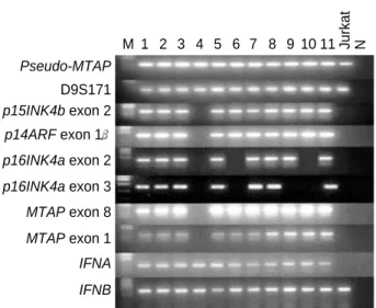
에 위치하는 유전자들의 결손Pseudo-MTAP 유전자에 대한 PCR에서 분명하게 증폭된 DNA band가 출현하는 일차성 아교모세포종 36예를 가지고 9p21-22에 위치하는 D9S171, p15INK4b exon 2, p14ARF exon 1 , p16INK4a exon 2 및 3, MTAP exon 8 및 1, 그리고 INFA 및 B 유전자 결손을 조사한 결과, p15INK4b exon 2에서 MTAP exon 1까지 결손된 경우가 1예 관찰되었다. p16INK4a 유전자의 exon 2 결손은 7예(19.4%)에서 관찰되었고p16INK4a 유전자 exon 3의 결손은 exon 2의 결손이 있는 6예를 포함하여 10예(27.7%)에서 관찰되었다. 따라서p16INK4a 유전자의 결손 은 36예 중 11예(30.5%)에서 관찰되었으며INFA 및 B 유전자 의 결손은 관찰할 수 없었다. 그리고p15INK4b 유전자의 결손은 4예(11.1%)에서 관찰되었다(Fig. 1).
p16INK4a
유전자의 돌연변이p16INK4a 유전자의 돌연변이 유무를 알아보기 위하여 PCR/SSCP를 실시한 결과, p16INK4a 유전자의 exon 2에 돌
연변이가 있다고 알려진 HL60 세포를 제외하고는 mobility shift가 있는 검체를 확인할 수 없었다(Fig. 2).
p16INK4a
유전자 촉진자의CpG Island
에 메틸화 유무 p16INK4a 유전자의 결손이 없는 15예에 대한 메틸화 유무를Fig. 1. Detection of homozygous deletions of p15INK4b, p14ARF, p16INK4a, MTAP, and INF genes in primary glioblastomas. DNA from primary glioblastoma tissues amplified by polymerase chain reaction (PCR) using the primers indicated. The PCR products were separated in 2% agarose gel and visualized by staining with ethidi- um bromide. Lane numbers (1-11) indicate representative cases of primary glioblastoma. M: molecular size marker, N: no template, MTAP: methylthioadenosine phophorylase, IFNA: interferon alpha, IFNB: interferon beta.
Pseudo-MTAP
M 1 2 3 4 5 6 7 8 9 10 11 Jurkat N
D9S171 p15INK4b exon 2 p14ARF exon 1 p16INK4a exon 2 p16INK4a exon 3 MTAP exon 8 MTAP exon 1 IFNA IFNB
Fig. 2. Polymerase chain reaction/single strand conformational poly- morphism (PCR/SSCP) analysis of p16IN4a exon 2 in the represen- tative cases of primary glioblastomas (1-9). There is no evidence of mobility shifted band.
1 2 3 4 5 6 7 8 9
HL60 Fibroblast
검색하기 위하여 methylation-specific PCR을 실시한 결과, 15 예 중 3예에서 촉진자에 메틸화가 관찰되었다. 3예 중에 2예는 부분적인 메틸화가 관찰되었고 1예는 완전한 메틸화가 관찰되었 다(Fig. 3).
고 찰
아교모세포종에서 가장 흔히 변이가 관찰되는 염색체는 7, 9p, 10 및 17p로 알려져 있고 9p의 결실은IFNA와 B 표지자 를 사용하여 처음으로 규명되었다.
14,15
별아교세포종의 세포주와 고등급 별아교세포종에서IFN 유전자의 동형접합성 및 반접합 체(hemizygous) 결손이 보고되어 있으나,16
IFN 유전자가 9p 결손의 표적인지 단순히 표적유전자의 부근에 위치하여 결손이 같이 동반되는 지는 분명치 않았다. 그러나 1994년에p16INK4a 및p15INK4b 유전자가 9p21에 위치하면서 여러 가지 종양에서 동형접합성 결손을 동반하여 종양억제유전자로서 알려지게 되었 다.17
본 연구 결과도IFN 유전자의 결손이 36예의 일차성 아교 모세포종에서 관찰되지 않아 표적유전자의 가능성은 배제할 수 있었다.p16INK4a는 CDK4/6의 기능을 억제하는 16 kDa 단백을 부 호화하는 유전자로서 세포주기의 진행을 막는 세포주기 조절억 제유전자로 알려져 있다.
18
그리고p15INK4b는 CDK 억제인자 로서p16INK4a와 많은 상동성을 보이고 p16INK4a의 upstream 30 Kb에 위치하며 9p21의 세포주기 조절억제유전자로 간주된 다.19
여러 연구자로부터 보고된p16INK4a 유전자의 이상은 아 교모세포종에서 약 30-60%의 결손을 나타내며 p15INK4b 유 전자는 10% 내외의 결손빈도를 보인다.20-23
이와 더불어 반접 합체 결손이나 9p 이형접합성 소실은 아교모세포종에서 20%의 빈도가 보고되어 있다.24
p16INK4a 및 p15INK4b의 동종접합 성 결손 빈도는 저등급성 별아교세포종에서는 동형접합성 결손 이 보고되지 않아 악성 종양 발생과 밀접한 관계가 있을 것으로 생각된다.21
또한p16INK4a 유전자의 동형접합성 결손이 동반 된 역형성 별아교세포종은 동형접합성 결실이 없는 경우보다MIB-1 증식지수가 높다는 것
25
이 밝혀져, p16INK4a 유전자의 결손이 일차성 아교모세포종에서 더욱 더 활발한 종양 세포 증 식과 밀접한 관계가 있을 것으로 생각되며 급속하게 성장하는 종양의 생물학적 양상과 임상적으로 나쁜 예후를 보이는 경우와 상관관계가 있을 것으로 추정할 수 있다. 특히p16INK4a 유전 자의 동형접합성 결손이 있는 경우는 현저하게 나쁜 예후를 보 인다는 최근의 보고23
도 있다. 본 연구에서 시행한 p16INK4a 유전자의 동형접합성 결손은 총 36예 중 11예(30.5%)에서 관찰 되어, p16INK4a 유전자의 결손에 의한 세포증식 조절의 이상 이 일차성 아교모세포종의 발생에 부분적인 기전임을 다시 알 수 있었다. 그리고p15INK4b 유전자의 결손도 4예(11.1%)에 서 관찰되어 Izumoto 등21
의 보고와 유사한 소견을 얻을 수 있 었으며, 이는p16INK4a 유전자의 결손 때에 동반 결손되는 것 으로 생각할 수 있다. 결국 다른 악성 종양과 비슷하게 p16INK4a 유전자의 결손이 p15INK4b 유전자의 결손보다 많 은 빈도를 보여p16INK4a 유전자의 이상이 일차성 아교모세포 종의 발생에 더욱 더 관여하리라 생각된다. 한편p16INK4a 유 전자의 돌연변이가 일차성 아교모세포종에서 관찰되는지를 알아 보고자 PCR/SSCP를 시행한 결과, p16INK4a 유전자의 exon 2에 돌연변이가 있다고 알려진 HL60 세포에서만 돌연변이를 관찰할 수 있었고 일차성 아교모세포종에서는 돌연변이를 관찰 할 수 없었다.p16INK4b 유전자의 불활성화는 점 돌연변이에 의한 p53의 불활성화와는 그 기전이 달라 본 연구에서처럼 동형접합성 결손 이 주된 기전이지만 그 외에 촉진자 부위의 CpG islands에 메 틸화로 인하여 전사가 억제되는 것이 다른 하나의 기전이다. 촉 진자 부위의 CpG islands의 메틸화는 일반적으로 그 유전자의 결손과 돌연변이와 같은 효과를 낼 수 있는 것으로 알려져 있 다.
9
Methylation-specific PCR 방법으로p16INK4a 유전자의 촉진자 부위의 메틸화 유무를 조사한 결과 p16INK4a 유전자 결손이 없는 15예 중 3예에서 메틸화가 관찰되었다. 이 3예 중 2예는 부분적인 메틸화가, 1예는 완전한 메틸화가 관찰되었다.따라서 36예의 일차성 아교모세포종을 분석한 결과를 종합하면 p16INK4a 유전자의 이상은 촉진자의 메틸화(8.3%)보다 동형 접합성 결손(30.5%)이 일차성 아교모세포종의 발생에 기여하는 빈도가 더 높을 것으로 생각된다. 이 결과는 Schmidt 등
26
이 제 시한 성적과 유사한 소견이다.p14ARF 유전자의 발현은 Myc, E1A 및 E2F의 과발현에 의해서 상향조절되며, MDM2 단백과 직접적으로 결합하여 MDM2에 의한 p53 분해를 억제하는 역할을 한다.
27
p14ARF 유전자의 exon 2와 3은 p16INK4a 유전자와 공유하기 때문에 alternative splicing 방법으로 p14ARF 유전자가 발현된다.28
본 연구에서는p14ARF 유전자의 exon 1 에 대한 결손은 비 록 1예에서 관찰되었지만p14ARF와 공유하고 있는 p16INK4a 유전자의 exon 2와 3의 결손이 30.5%로 관찰되어, p14ARF 유전자의 기능 이상이 일차성 아교모세포종의 발생에 어느 정도Fig. 3. Methylation-specific polymerase chain reaction (PCR) anal-
ysis of the p16IN4a promoter was performed in primary glioblas- toma cases (1-11). Reverse images of the ethidium bromide stained gels are displayed. Bisulfite treated DNA from W138 cell lines was used as a normal control. M: molecular size marker.
M 1 2 3 4 5 6 7 8 9 10 11 W138 Methylated
Unmethylated
기여할 것으로 추측된다. 최근 Nakamura 등
29
에 의하면 p14ARF 유전자의 동형접합성 결손과 메틸화가 50예의 아교모 세포종 중 29예(58%)에서 관찰되어 상당한 인과관계가 있음을 보고하였다. p16INK4a 유전자의 종말체 쪽에 위치한 MTAP 유전자의 결손이p16INK4a 유전자의 결손과 동반되어 있는 경 우를 암종에서 흔히 볼 수 있다.8
MTAP은 adenine 및 methio- nine의 salvage 합성에 관여하는 중요한 효소로 알려져 있다.7
본 연구에서MTAP 유전자의 결손이 1예만 관찰되어 일차성 아교모세포종의 발생과 관계가 적은 것으로 생각된다. 이상의 결과로 볼 때 일차성 아교모세포종에서 9p21-22부위의 유전자 들 중p16INK4a 유전자의 동형접합성 결손과 메틸화가 이 종 양 발생에 가장 중요한 기전임을 알 수 있었다.참고문헌
1. Scherer HJ. Cerebral astrocytomas and their derivatives. Am J Can- cer 1940; 40: 159-98.
2. Watanabe K, Tachibana O, Sata K, Yonekawa Y, Kleihues P, Ohgaki H. Overexpression of the EGF receptor and p53 mutations are mutu- ally exclusive in the evolution of primary and secondary glioblas- tomas. Brain Pathol 1996; 6: 217-23.
3. Peter M, Herskowitz I. Joining the complex: cyclin-dependent kinase inhibitory proteins and the cell cycle. Cell 1994; 79: 181-4.
4. Sherr CJ, Roberts JM. Inhibitors of mammalian G1 cyclin-dependent kinases. Genes Dev 1995; 9: 1149-63.
5. von Deimling A, Louis DN, Wiestler OD. Molecular pathways in the formation of gliomas. Glia 1995; 15: 328-38.
6. Biernat W, Tohma Y, Yonekawa Y, Kleihues P, Ohgaki H. Alterations of cell cycle regulatory genes in primary (de novo) and secondary glioblastomas. Acta Neuropathol 1997; 94: 303-9.
7. Batova A, Diccianni MB, Nobori T, et al. Frequent deletion in the methylthioadenosine phosphorylase gene in T-cell acute lymphoblas- tic leukemia: strategies for enzyme-targeted therapy. Blood 1996; 88:
3083-90.
8. Hori Y, Hori H, Yamada Y, et al. The methylthioadenosine phospho- rylase gene is frequently co-deleted with the p16INK4a gene in acute type adult T-cell leukemia. Int J Cancer 1998; 75: 51-6.
9. Herman JG, Merlo A, Mao L, et al. Inactivation of the CDKN2/p16/
MTS1 gene is frequently associated with aberrant DNA methylation
in all common human cancers. Cancer Res 1995; 55: 4525-30.10. Sharpless NE, DePinho RA. The INK4A/ARF locus and its two gene products. Curr Opin Genet Dev 1999; 9: 22-30.
11. Blin N, Stafford DW. A general method for isolation of high molecu- lar weight DNA from eukaryotes. Nucleic Acids Res 1976; 3: 2303-8.
12. Orita M, Suzuki Y, Sekiya T, Hayashi K. Rapid and sensitive detection
of point mutations and DNA polymorphisms using the polymerase chain reaction. Genomics 1989; 5: 874-9.
13, Herman JG, Graff JR, Myohanen S, Nelkin BD, Baylin SB. Methyla- tion-specific PCR: a novel PCR assay for methylation status of CpG islands. Proc Natl Acad Sci USA 1996; 93: 9821-6.
14. James CD, He J, Carlbom E, Nordenskjold M, Cavenee WK, Collins VP. Chromosome 9 deletion mapping reveals interferon and inter- feron -1 gene deletions in human glial tumors. Cancer Res 1991; 51:
1684-8.
15. Olopade OI, Jenkins RB, Ransom DT, et al. Molecular analysis of dele- tions of the short arm of chromosome 9 in human gliomas. Cancer Res 1992; 52: 2523-9.
16. Miyakoshi J, Dobler KD, Allalunis-Turner J, et al. Absence of IFNA and IFNB genes from human malignant glioma cell lines and lack of correlation with cellular sensitivity to interferons. Cancer Res 1990;
50: 278-83.
17. Kamb A, Grui NA, Weaver-Feldhaus J, et al. A cell cycle regulator potentially involved in genes of many tumor types. Science 1994; 264:
436-40.
18. Ruas M, Peters G. The p16INK4a/CDKN2A tumor suppressor and its relatives. Biochim Biophys Acta 1998; 1378: F115-77.
19. Heyman M, Einhorn S. Inactivation of the p15INK4B and p16INK4 genes in hematologic malignancies. Leuk Lymphoma 1996; 23: 235- 45.
20. Jen J, Harper JW, Bigner SH, et al. Deletion of p16 and p15 genes in brain tumors. Cancer Res 1994; 54: 6353-8.
21. Izumoto S, Arita N, Ohnishi T, Hiraga S, Taki T, Hayakawa T.
Homozygous deletions of p16INK4A/MTS1 and p15INK4B/MTS2 genes in glioma cells and primary glioma tissues. Cancer Lett 1995;
97: 241-7.
22. Ueki K, Ono Y, Henson JW, Efird JT, von Deimling A, Louis DN.
CDKN2/p16 or RB alterations occur in the majority of glioblastomas
and are inversely correlated. Cancer Res 1996; 56: 150-3.23. Kamiryo T, Tada K, Shiraishi S, et al. Analysis of homozygous dele- tion of the p16 gene and correlation with survival in patients with glioblastoma multiforme. J Neurosurg 2002; 96: 815-22.
24. Hegi ME, zur Hausen A, Ruedi D, Malin G, Kleihues P. Hemizygous or homozygous deletion of the chromosomal region containing the
p16INK4a gene is associated with amplification of the EGF receptor
gene in glioblastomas. Int J Cancer 1997; 73: 57-63.25. Ono Y, Tamiya T, Ichikawa T, et al. Malignant astrocytomas with homozygous CDKN2/p16 gene deletions have higher Ki-67 prolifer- ation indices. J Neuropathol Exp Neurol 1996; 55: 1026-30.
26. Schmidt EE, Ichimura K, Messerle KR, Goike HM, Collins VP. Infre- quent methylation of CDKN2A (MTS1/p16) and rare mutation of both
CDKN2A and CDKN2B (MTS2/p15) in primary astrocytic tumours.
Br J Cancer 1997; 75: 2-8.
27. Lohrum MA, Ashcroft M, Kubbutat MH, Vousden KH. Contribution of two independent MDM2-binding domains in p14 (ARF) to p53 stabilization. Curr Biol 2000; 10: 539-42.
28. Simon M, Koster G, Menon AG, Schramm J. Functional evidence for
a role of combined CDKN2A (p16-p14 (ARF))/CDKN2B (p15) gene inac- tivation in malignant gliomas. Acta Neuropathol 1999; 98: 444-52.
29. Nakamura M, Watanabe T, Klangby U, et al. p14ARF deletion and methylation in genetic pathways to glioblastomas. Brain Pathol 2001;
11: 159-68.