600
In this study, we investigated the mechanism of inhibition of pathogenic bacteria by Pediococcus pentosaceus strain SH-10 isolated from hard Clam Meretrix meretrix sikhae. When P. pentosaceus SH-10 was co-cultured in MRS broth with pathogenic bacteria, including Bacillus cereus, Listeria monocytogenes, Salmonella choleraesuis and Staphyloccus aureus, no viable pathogenic cells were detected after 18 h of incubation. However, pediocin or a pediocin-like bacteriocin was not detected in cultures of P. pentosaceus SH-10 by the agar diffusion method.
Organic acids were produced in MRS broth in proportion to the incubation time of P. pentosaceus SH-10. These results indicate that P. pentosaceus SH-10 inhibited the growth of pathogenic bacteria by lowering the pH of the growth medium through the production of organic acids, including sodium lactate, sodium acetate, and sodium ci- trate.
Key words: Probiotics, Pediococcus pentosaceus SH-10, Pathogenic bacteria, Organic acids
Article history;
Received 8 October 2012; Revised 23 October 2012; Accepted 21 November 2012
*Corresponding author: Tel: +82. 63. 469. 1822 Fax: +82. 63. 469. 1821 E-mail address: [email protected]
Kor J Fish Aquat Sci 45(6) 600-605, December 2012 http://dx.doi.org/10.5657/KFAS.2012.0600 pISSN:0374-8111, eISSN:2287-8815
ⓒ The Korean Society of Fishereis and Aquatic Science. All rights reserved
Dong-Min Shin, Hee-Dai Kim¹, Jae-Geun Koo and Kwon-Sam Park *
Inhibition of Pathogenic Bacteria by Pediococcus pentosaceus Strain SH-10 Isolated from Hard Clam Meretrix meretrix Sikhae
Department of Food Science and Biotechnology, Kunsan National University, Kunsan 573-701, Korea
¹Department of Biotechnology and Biomedicine, Chungbuk Provincial College, Okcheon 373-807, Korea
백합( Meretrix meretrix) 식해에서 분리한 Pediococcus pentosaceus SH-10에 의한 병원성 세균의 억제 기작
군산대학교 식품생명공학과, 1충북도립대학 바이오생명의약과
신동민·김희대
1·구재근·박권삼*
서 론
생균제
(probiotics)
란사람과가축의장내균총의능력을개 선하여숙주건강에유익한효과를 주는살아있는미생물이 라고R. Fuller
에의해정의가제안되었다(Fuller, 1989).
생균 제로개발되어사용되고있는미생물에는Lactobacillus spp., Bifidobacterium spp., Lactococcus spp., Streptococcus spp., Bacillus spp., Enterococcus spp.
및효모등이며(Guarner and Schaafsma, 1998),
이들미생물은위산및담즙산에비교적잘 견딜뿐만아니라병원성세균에대한길항작용을가지는경우 가많기때문이다(Fuller, 1989).
생균제는장내균총의균형유 지라는일차적인기능과함께다음과같은몇가지특성도요 구된다.
위와십이지장등의소화기관을통과하는과정에서위 산및담즙산등에의해죽지않고살아서장까지도달한다음장에서정착하고증식하여야한다
.
또한장관내에서유용한 효과를나타내어야하며다양한항생제에대한내성이있으며 독성이없는비병원성미생물이어야한다.
지금까지알려진생균제의주요기능에는숙주에대한장내 세균수의안정화
(Lidbeck and Nord, 1987),
위장관내병원균 의증식억제(Fernandes et al., 1992),
혈중콜레스테롤의저하(Suzuki and Kaizu, 1991),
특이및비특이면역반응의유도및 영양소이용성의향상(Fernandes et al., 1992),
암퇴화및장 내효소활성감소에의한결장암의예방효과(Goldin and Gor- bach, 1984)
및비타민과같은인체유용물질의합성등이보 고되어있다.
국내의경우
,
생균제는인체의약품인정장제나유산균제제,
사료첨가제로서의생균제그리고식품공전에서정의한건강 식품의일종인유산균식품으로사용하고있는데어느경우든유산균에 의한 병원성 세균의 억제기작
601
생균제미생물은유산균이주종을이루고있는실정이다
.
국내 의생균제에대한최근연구는한국인의장내용물또는전통 발효식품으로부터우수한유산균을분리하여이를식품이나 의약품으로이용하고자하는노력이활발하게진행되고있는 실정이다(Kang et al., 2001; Ha et al., 2004; Kim et al., 2005;
Shin et al., 2008; Lim and Im, 2009).
최근농림수산식품부에서는축산물에대한소비자의신뢰제고를위해
2011
년하반기부터축산용사료에항생제첨가를전면금지한다고발표하였 다
(MIFAFF, 2010).
따라서축산업계에서는항생제대체물질 로서동물에게유익한미생물을계획적으로투여할수있는생 균제의개발및이용분야의관심과연구가활발하게진행되고 있는실정이다(Lee et al., 2008; Kim et al., 2010; Park et al.,
2010).
생균제가항생제대체물질로서각광을받는이유에는항생제는장내정상적인균총형성을억제하는데비해생균제 는장내정상적인균총형성에도움이될뿐만아니라병원성 미생물의항생제내성획득에도관계가없기때문이다
(Isolauri et al., 2001).
최근
Song et al.(2011)
은백합식해에서분리한유산균을대 상으로생균제로서의가능성을검토한결과Pediococcus pen- tosaceus SH-10
균주는내산성,
담즙내성,
다양한항생제에대 한내성, biofilm
형성능및병원성세균의발육억제능등이뛰 어나생균제로서개발가치의가능성을보고하였다. P. pento-
saceus
는김치또는젓갈발효에관여하는그람양성의호염성
,
사련쇄상구균으로주로항균성peptide
인pediocin
을생 산하며, pediocin
은병원성세균중에서도그람양성의Listeria monocytogenes
및Staphyloccus aureus
에효과적으로작용한 다고보고되어있다(Fleming et al., 1975; Cintas et al., 1998;
Osmanagaoglu et al., 2001; Wu et al., 2004). Pediocin
은저분 자의내열성을지니는Class IIa bacteriocin
으로표적세균의 세포벽합성을억제하거나세포막에구멍을형성하여살균효 과를나타내며(Sablon et al., 2000: Hechard and Sahl, 2002:
Bauer and Dicks, 2005), pediocin
을암호화하는유전자는일 반적으로plasmid DNA
에operon
형태로존재한다고보고되 어있다(Miller et al., 2005).
본연구는백합식해에서분리한Pediococcus pentosaceus SH-10
균주의병원성세균에대한 발육억제기작에관하여검토하였다.
재료 및 방법
균주 및 배지
사용 균주는백합식해에서분리한
Pediococcus pentosa- ceus SH-10
균주를사용하였으며(Song et al., 2011),
한국 미생물보존센타에서구입한Bacillus cereus KCCM40138, Enterobacter aerogenes KCCM12177, Listeria monocyto- genes KCCM40307, Salmonella choleraesuis KCCM11806,
Staphylococcus aureus KCCM12214
및Vibrio cholerae KCCM41626
등의병원성균주는P. pentosaceus SH-10
에의 한발육억제능을검토하기위한대조균으로사용하였다.
유산 균의배양배지로는Lactobacilli MRS (Difco, USA)
평판배 지 또는액체배지를사용하였으며,
병원성균주의 배양에는Brain heart infusion (Difco, USA), Brilliance Bacillus cereus agar (Oxoid, UK), LPM agar (Fluka, Switzerland), XLD agar (Difco, USA), Staphylococcus 110 agar (Difco, USA)
및TCBS agar (Difco, USA)
를사용하였다.
각균주는35℃
에서 정치또는진탕배양하였다.
병원성균 억제능 측정
유산균에의한병원성균의억제능실험은
MRS broth
에병원성균과유산균을혼합배양후병원성균의변화를측정하여 검토하였다
.
유산균과4
종의병원성균은각각의적절한배지 에서하룻밤배양한후유산균과병원성균의농도를대략10
6- 10
7CFU/mL
가되도록조정하여MRS broth
에혼합접종하 면서0, 6, 12, 18
및24
시간배양후의병원성세균의변화는 생균수로측정하였다.
대조구는병원성세균만을MRS broth
에접종하여35℃
에서24
시간배양한후각각의선택배지에서 균수를측정하여비교하였다.
Pediocin 유전자의 확인
Pediocin
유전자확인을위한primers
는기존의보고(Shin et al., 2008)
를참고하여Bioneer (Daejon, Korea)
에의뢰합성 하였다(Ped-F, 5'-TTGTGATGAAAAAAATTGAAAAAT- TA-3'
및Ped-R, 5'-GCATTTATGATTACCTTGAT- GTCC-3'). PCR
반응에는Takara kit (Japan)
를사용하였으 며, PCR
반응액은0.5 µL (2.5 U) Taq polymerase, 5.0 µL Taq polymerase buffer (10×), 4.0 µL 2.5 mM dNTP, 34.5 µL dH
20
에20 pmol
의각primer 2.0 µL
와2.0 µL
의주형DNA
를 첨가하였다. DNA
증폭은95℃ 5
분간1
회변성하고, 95℃ 30
초, 55℃ 30
초및72℃
에서1
분을30
회반복하였다. PCR
증 폭산물은1.5% agarose gel (Sigma, USA)
에서전기영동후ethidium bromide
로염색하여DNA
의증폭여부를확인하였 다.
효소활성 조사
P. pentosaceus SH-10
균주가생산하는효소및효소활성을 조사하기위하여API ZYM kit (BioMerieux, France)
를사용 하였다.
균주는Lactobacilli MRS
고체배지에서하룻밤배양 한후균체를회수하여PBS (phosphate buffered saline, 137
mM NaCl, 2.7 mM KCl, 10 mM Na
2HPO
4·2H
2O, 2.0 mM
KH
2PO
4, pH 7.4)
로현탁하여Mcfarland
탁도5-6
으로조정하 였다.
현탁한균은ZYM kit
각cupule
에65 µL
씩분주하고37
℃
에서4
시간배양한후색깔변화로효소활성을조사하였다. 유기산 분석
Lactobacilli MRS broth
에P. pentosaceus SH-10
을접종후35℃
에서정치배양하면서배양개시0, 6, 12
및24
시간후의 배양액을8,000 rpm
에서10
분간원심분리하여상층액을회 수하여0.45 μM membrane filter
로여과하고그여액을high performance liquid chromatography (515 series, Waters, USA)
로유기산을분리및정량하였다.
사용기기,
컬럼및분 석조건은Table 1
에제시하였다.
유기산분석을위한표준물질 로사용한sodium acetate, sodium citrate
및sodium lactate
은Sigma (USA)
제품을사용하였다.
Pediocin 생산능 검토
Pediocin
생산은기존의방법(Shin et al., 2008)
을약간변형 하여검토하였다. Lactobacilli MRS broth
에P. pentosaceus SH-10
균주를접종하여35℃
에서정치배양하면서배양12, 24
및48
시간후의배양액을8,000 rpm
에서2
분간원심분리 하여상층액을회수하였다.
상층액은1 N NaOH
를사용하여pH
를6.5
로조정하고0.45 μM membrane filter
로여과한다음 적절한단계까지희석하였다.
하룻밤배양한병원성시험균주 는10
6-10
7CFU/mL
의농도가되도록배지에섞은후고체화 하였다.
제조된고체배지는화염멸균된punch
를사용하여직 경3 mm
크기의구멍을만들고여기에희석한상층액20 μL
를접종하여35℃
에서24
시간배양후병원성세균의억제대 로pediocin
생산및활성을검토하였다.
결과 및 고찰
P. pentosaceus SH-10 균주의 병원성 세균 억제능 검토
유산균에의한병원성세균의억제기작에는대사산물
(
각종 유기산, H
2O
2 등)
의생성,
박테리오신과같은항균물질의생산,
장내상피세포의부착장소경쟁등이알려져있다
(Reid and Burton, 2002). P. pentosaceus SH-10
에의한병원성세균( B.
cereus KCCM40138, L. monocytogenes KCCM40307, S.
choleraesuis KCCM11806
및S. aureus KCCM12214)
의성 장억제능력은P. pentosaceus SH-10
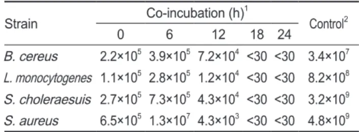
와각각의병원성세균과 의혼합배양후병원성세균의균수변화로측정하였다. Table 2
에나타낸바와같이각각의병원성세균을MRS broth
에서24
시간단독배양하였을경우,
균수는3.4×10
7CFU/mL
에서4.8×10
9CFU/mL
의범위로측정되었다.
그러나
P. pentosaceus SH-10
와혼합배양할경우4
종의병 원성세균은배양시간경과에따라감소하다가배양18
시간후 에는검출되지않았다(Table 2).
이는P. pentosaceus SH-10
이 생산하는살균활성을나타내는물질(
박테리오신,
각종유기산,
과산화수소수등)
에의해병원성세균은사멸한것으로판단된 다.
P. pentosaceus SH-10의 효소활성
병원성세균의발육억제기작을검토하기위한예비실험으 로
P. pentosaceus SH-10
균주가어떤효소활성을나타내는지 는API ZYM kit
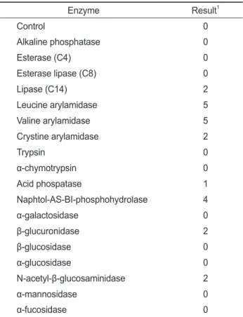
를사용하여검토하였다. Table 3
에나타낸바 와같이P. pentosaceus SH-10
균주는19
종류의효소중8
종류 의효소활성이관찰되었다. Leucine arylamidase
와valine ar- ylamidase
는다른효소들에비해생산량이높아약40 nmol
정 도를, naphtol-AS-BI-phosphohydrolase
는30 nmol, crystine arylamidase, β-glucuronidase, lipase
및N-acetyl-β-glucosa- minidase
는10 nmol
를생산하는것으로파악되었다.
그리고acid phospatase
는약5 nmol
을생산하는것으로분석되었다. API ZYM kit
를사용하여P. pentosaceus
의효소활성을측정 한기존의연구결과가없기때문에P. pentosaceus SH-10
와 비교할수는없으나서로유사할것으로예상된다. Lim et al.
(2007)
이보고한Lactobacillus salivarius CPM-7
와비교해보 면, P. pentosaceus SH-10
균주가생산하는효소의종류는L.
salivarius CPM-7
에비해적고효소활성에도차이가많았다.
Table 1. HPLC instruments and analysis conditions for organicacids
Item Analysis condition
HPLC system Waters 515 HPLC
Detector Waters 2489 UV/visible detector Column temperature 40℃
Flow rate 0.6 mL/min
Column Aminex HPX-87H 300×7.8 mm (Bio-Rad) Injection volume 20 µL
Mobile phase 4 mM sulfuric acid
Table 2. Inhibitory of pathogenic bacteria by co-incubation with Pediococcus pentosaceus strain SH-10
Strain Co-incubation (h)1
Control2
0 6 12 18 24
B. cereus 2.2×10⁵ 3.9×10⁵ 7.2×10⁴ <30 <30 3.4×10⁷ L. monocytogenes 1.1×10⁵ 2.8×10⁵ 1.2×10⁴ <30 <30 8.2×10⁸ S. choleraesuis 2.7×10⁵ 7.3×10⁵ 4.3×10⁴ <30 <30 3.2×10⁹ S. aureus 6.5×10⁵ 1.3×10⁷ 4.3×10³ <30 <30 4.8×10⁹
1Each strain was grown with pathogenic bacteria in MRS broth.
2Only pathogenic bacteria were grown in MRS broth after 24 h.
유산균에 의한 병원성 세균의 억제기작
603
P. pentosaceus SH-10균주의 pediocin 생산능의 검토
P. pentosaceus
는박테리오신의일종인pediocin
을생산한 다고보고되어있으며(Fleming et al., 1975; Wu et al., 2004;
Shin et al., 2008), pediocin
은L. monocytogenes
및S. aureus
와같은일부병원성세균에대하여항균활성을나타낸다고보 고되어있다(Clintas et al., 1998). P. pentosaceus SH-10
균주 의pediocin
생산여부및활성을검토하기위하여MRS broth
에P. pentosaceus SH-10
균주를접종하여35℃
에서정치배양하면서배양개시
12, 24
시간및48
시간후의배양액을회수하 여6
종의병원성세균에대한생육저지대생성여부로pediocin
의활성을확인하였다.
배양12, 24
시간및48
시간배양액의 원액 및희석액공히6
종의모든병원성균주( B. cereus, E.
aerogenes, L. monocytogenes, S. choleraesuis, S. aureus
및V. cholerae)
에대한억제능력이전혀관찰되지않았다(
결과 미제시).
따라서PCR assay
로pediocin
유전자의확인을검토 하였으나예상크기의DNA
산물은증폭되지않았다.
이는P.
pentosaceus SH-10
균주는pediocin
유전자미보유균주이기 때문에pediocin
이생산되지않는것으로확인되었다(
결과미 제시). P. pentosaceus
는균주에따라pediocin
의생산량이매 우적거나없는경우가있는데그원인은관련유전자의소실 에의한다는기존의연구결과(Diep et al., 2006)
와일치한다. P. pentosaceus SH-10의 배양시간에 따른 pH의 변화
P. pentosaceus SH-10
균주는pediocin
을생산하지않음에 도불구하고병원성세균에대한발육억제능력이우수한이 유는각종유기산생산에따른배지의pH
저하일가능성이높 다고판단되어배양시간에따른pH
변화를검토하였다. MRS broth
에P. pentosaceus SH-10
균주를접종하여35℃
에서정치 배양하면서2
시간간격으로24
시간까지균증식과pH
변화를 검토하였다(Fig. 1).
균증식은배양4
시간부터대수적으로증 식하기시작하여10
시간후에는정점에도달한다음거의일정 한농도를유지하였다.
균증식에따른pH
변화는초기배지의pH
는6.5
에서시작하여균증식과비례하여pH
는감소하여배 양6
시간후pH
는5.30
를, 12
시간후에는4.13
의pH
를나타내 었으며,
배양24
시간후배지의pH
는3.90
였다.
배양시간경과 에따른pH
저하는P. pentosaceus SH-10
가생산하는각종유 기산에의한결과로판단된다.
Table 3. Enzyme activities of Pediococcus pentosaceus strain SH- 10 by API ZYM analysis
Enzyme Result1
Control 0
Alkaline phosphatase 0
Esterase (C4) 0
Esterase lipase (C8) 0
Lipase (C14) 2
Leucine arylamidase 5
Valine arylamidase 5
Crystine arylamidase 2
Trypsin 0
α-chymotrypsin 0
Acid phospatase 1
Naphtol-AS-BI-phosphohydrolase 4
α-galactosidase 0
β-glucuronidase 2
β-glucosidase 0
α-glucosidase 0
N-acetyl-β-glucosaminidase 2
α-mannosidase 0
α-fucosidase 0
10, 0 nmol; 1, 5 nmol; 2, 10 nmol, 3, 20 nmol; 4, 30 nmol; 5, 40 nmol.
3.0
OD600
OD600Absorbance at 210 nm
Time (h)
Time (min)
pH pH
2.5 2.0 1.5 1.0
0 2 4 6 8 10 12 14 16 18 20 22 24 26 0.5
0.0
0.20 0.15 0.10 0.05
0.00
0.00 2.00 4.00 6.00 8.00 1
0.014 0.097 12.717 15.487
3
2
10.00 12.00 14.00 16.00 18.00 7.0 6.5 6.0 5.5 5.0 4.5 4.0 3.5
Fig. 1. Relation of cell growth and pH of Pediococcus pentosa- ceus strain SH-10.
Table 4. Production of organic acids in MRS broth by incubation time
Time (h) Concentration of organic acids (mg/100 mL) Sodium citrate Sodium lactate Sodium acetate
0 252.9 224.2 843.8
6 271.6 780.7 889.1
12 311.4 1518.4 909.3
24 265.5 1822.1 1424.2
신동민·김희대·구재근·박권삼
604
P. pentosaceus SH-10이 생산하는 유기산의 정량 P. pentosaceus SH-10
균주가생산하는유기산의종류및양 은MRS broth
에P. pentosaceus SH-10
을배양하면서배양0, 6, 12
및24
시간후의배양액중에존재하는sodium citrate, sodium lactate
및sodium acetate
함량을HPLC
로측정하 였다.
재료및방법에제시한조건으로분석한결과sodium citrate, sodium lactate
및sodium acetate
표준물질의검출시 간은8.097, 12.717
및15.487
분에검출되었다(Fig. 2).
배양 액중의sodium citrate
는배양시간이경과하여도초기농도인252.9 mg/100 mL
와비교하면6, 12
및24
시간배양액에서의sodium citrate
증가량은매우미미하였다. Sodium acetate
의 배지중의초기농도는843.8 mg/100 mL
이며,
배양6
및12
시 간후배양액에서의농도는초기농도와크게차이가없었으나24
시간배양액에서의농도는1424.2 mg/100 mL
로초기농도 에비해1.7
배증가하였다.
따라서sodium acetate
는P. pento- saceus SH-10
의배양초기에는생성량이적으나배양후기(
정 지기)
에증가하는유기산으로판명되었다. Sodium lactate
의 배지중의초기농도는224.2 mg/100 mL
이였으나배양시간에 비례하여지속적으로증가하는경향을보이며배양24
시간후 에는1822.1 mg/100 mL
로초기농도의약8
배이상의증가를 보였다(Table 4).
결과적으로P. pentosaceus SH-10
가생산하 는유기산중sodium lactate
는배양초기부터후기에이르기까 지가장많이생성되며배지의pH
저하에가장중요하게작용 한다고판단된다.
백합식해에서분리한
P. pentosaceus SH-10
균주는pediocin
을생산하지않음에도불구하고 병원성세균과혼합배양하 였을때병원성세균에대한발육억제능력이우수한이유는sodium lactate
등의유기산생산에의한배지중pH
의산성화 에의해병원성세균이사멸하는것으로확인되었다.
사 사
본연구는
2012
년군산대학교수산과학연구소학술연구비지원에의하여수행되었으며
,
이에감사드립니다.
참고문헌
Bauer R and Dicks LM. 2005. Mode of action of lipid II-target- ing lantibiotics. Int J Food Microbiol 101, 201-216.
Clintas LM, Casaus P, Fernandez MF and Hernandez PE. 1998.
Comparative antimicrobial activity of enterocin L50, pedio- cin PA-1, nisin A and lactosin S against spoilage and food borne pathogenic bacteria. Food Microbiol 62, 1764-1769.
Diep DB, Godager L, Brede D and Nes IF. 2006. Data mining and characterization of a novel pediocin-like bacteriocin system from the genome of Pediococcus pentosaceus ATCC 25745. Microbiol 152, 1649-1659.
Fernandes CF, Chandan RC and Shahani KM. 1992. Ferment- ed dairy products and health In: the Lactic acid Bacteria, Volume1, Wood, BJB(ed), London, NewYork: Elsevier Ap- plied Science, 297-342.
Fleming HP, Etchells JL and Costilow RN. 1975. Microbial inhibition by an isolate of Pediococcus from cucumber brines. Appl Microbiol 30, 1040-1042.
Fuller R. 1989. Probiotics in man and animal. J Appl Bacteriol 66, 365-378.
Goldin BR and Gorbach SL. 1984. The effect of milk and Lac- tobacillus feeding on human intestinal bacterial enzyme activity. Am J Clin Nutr 39, 756-761.
Guarner F and Schaafsma GJ. 1998. Probiotics. Int J Food Mi- crobiol 39, 237-238.
Ha CG, Cho JK, Chai YG and Heo KC. 2004. Isolation and identif ication of lactic bacteria containing superior activity of the bile salts deconjugation. Korean J Food Sci Ani Re- sour 24, 164-170.
Hechard Y and Sahl H G. 2002. Mode of action of modif ied and unmodif ied bacteriocins from Gram positive bacteria.
Biochimie 84, 545-557.
Isolauri E, Sutas Y, Kankaanpaa P, Arvilommi H and Salminen S. 2001. Probiotics: effects on immunity. Am J Clin Nutr 73. 444S-450S.
Kang DG, Kang SP, Chang DH, Kim SH and Yoon SS. 2001.
Isolation and characterization of Lactobacillus strains iso- lated from Korean feces. Korean J Food Sci Technol 33, 567-573.
Kim JD, Chung HW, Shim KS, Park SY, Ju JC, Song JJ, Lee KH, Park JK, Park DY and Kim CH. 2010. Effects of probiotics as an alternative for antibiotics on growth per- formance, nutrient digestibility, noxious gas emission and fecal microbial population in growing piglets. Korean J Org Agri 18, 527-539.
Kim SJ, Ma SJ and Kim HL. 2005. Probiotic properties of lac- tic acid bacteria and yeasts isolated from Korean traditional food, Jeot-gal. Kor J Food Preserv 12, 184-189.
Lee NK, Park YL, Kim HW, Rhim SL, Kim JM, Kim JM, Nam HM, Jung SC and Paik HD. 2008. Purification and characterization of lacticin NK34 produced by Lactococcus
OD600
OD600Absorbance at 210 nm
Time (h)
Time (min)
pH pH
2.0 1.5 1.0
0 2 4 6 8 10 12 14 16 18 20 22 24 26 0.5
0.0
0.20 0.15 0.10 0.05
0.00
0.00 2.00 4.00 6.00 8.00 1
0.014 0.097 12.717 15.487
3
2
10.00 12.00 14.00 16.00 18.00 6.0 5.5 5.0 4.5 4.0 3.5
Fig. 2. HPLC chromatogram of organic acids.
유산균에 의한 병원성 세균의 억제기작
605
lactis NK34 against bovine mastitis. Korean J Food Sci Ani Resour 28, 457-462.
Lidbeck GJ and Nord CE. 1987. Impact of Lactobacillus aci- dophilus on the normal intestinal f lora after administration of two antibiotic agents. Infection 16, 329-335.
Lim SJ, Jang SS and Kang DK. 2007. Probiotic properties of Lactobacillus salivarius CPM-7 isolated from chicken fe- ces. Kor J Microbiol Biotechnol 35, 98-103.
Lim SM and Im DS. 2009. Screening and characterization of probiotic lactic acid bacteria isolated form Korean ferment- ed foods. J Microbiol Biotechnol 19, 178-186.
Miller KW, Ray P, Steinmetz T, Hanekamp T and Ray B. 2005.
Gene organization and sequences of pediocin AcH/PA-1 production operons in Pediococcus and Lactobacillus plas- mids. Lett Appl Microbiol 40:56-62.
Ministry for food, agriculture, forestry and f isheries. 2010. Bul- letin 2010-4.
Osmanagaoglu O, Beyatli Y and Gudu U. 2001. Isolation and characterization of pediocin producing Pediococcus pento- saceus Pep1 from vacuum-packed sausages. Tr J Biol 25, 133-143.
Park SH, Choi JS, Jung DS, Auh JH and Choi YI. 2010. Effects of complex probiotics and antibiotics on growth perfor- mance and meat quality in broilers. Korean J Food Sci Ani Resour 30, 504-511.
Reid G and Burton J. 2002. Use of Lactobacillus to prevent infection by pathogenic bacteria. Microbes Infect 4, 3119- 3124.
Sablon E, Contreras and Vandamme E. 2000. Antimicrobial peptides of lactic acid bacteria: mode of action, genetics and biosynthesis. Adv Biochem Eng Biotechnol 68, 21-60.
Shin MS, Han SK, Ryu JS, Kim KS and Lee WK. 2008. Isola- tion and partial characterization of a bacteriocin produced by Pediococcus pentosaceus K23-2 isolated from kimchi. J Appl Microbiol 105, 331-339.
Song HJ, Kim KJ, Kim HD, Yoo JH, Koo JG and Park KS.
2011. Probiotic properties of Pediococcus pentosaceus SH- 10 isolated from the Hard Clam (Meretrix meretrix) shi- khae. Kor J Fish Aquat Sci 44, 605-611.
Suzuki Y and Kaizu H. 1991. Effect of cultured milk on serum cholesterol concentration in rats which were fed high cho- lesterol diets. Anim Sci Technol 62, 565-576.
Wu CW, Yin LJ and Jiang ST. 2004. Purif ication and character- ization of bacteriocin from Pediococcus pentosaceus AC- CEL. J Agric Food Chem 52, 1146-1151.