Inhibitory Effects of Illicium verum Hooker fil. Dichloromethane Fractions on Adipocyte Differentiation
Hyun Young Jeong1, In Kyo Jeong2, Nam Ju Kim2, Hee Jung Yun2,3, Jung Ha Park2,3, Byung Woo Kim1,2,3 and Hyun Ju Kwon1,2,3*
1Blue-Bio Industry regional Innovation Center, Dong-eui University, Busan 47340, Korea
2Department of Life Science and Biotechnology, Dong-eui University Graduate School, Busan 47340, Korea
3Biopharmaceutical Engineering Major. Division of Applied Bioengineering, College of Engineering, Dong-eui University, Busan 47340, Korea Received December 12, 2018 /Revised April 10, 2019 /Accepted April 11, 2019
Fat accumulation in adipocytes occurs through the process of adipogenesis in which preadipocytes differentiate into adipocytes. Obesity is a metabolic disorder caused by excessive accumulation of fat in the body, which increases the incidence of cardiovascular diseases, hypertension, type 2 diabetes, hyperlipidemia, and various cancers. Recently, inhibition of adipocyte differentiation was shown to be a potential antiobesity strategy. In this study, the inhibitory effect of dichloromethane fractions from Illicium verum Hooker fil. water extract on the differentiation of 3T3-L1 preadipocytes to adipocytes was investigated. Dichloromethane fractions from I. verum Hooker fil. significantly inhibited adipocyte differentiation when applied during the adipocyte differentiation process, as assessed by measuring fat accumulation using Oil-red O staining. In addition, dichloromethane fractions from I. verum Hooker fil. reduced important adipogenic transcription factors, such as CCAAT/enhancer binding protein α (C/EBPα), C/EBPβ, and peroxisome proliferator activated receptor γ (PPARγ). The expression of FAS and LPL, which are terminal differentiation markers of mature adipocytes, was also reduced in the 3T3-L1 adipocytes treated with dichloromethane fractions from I. verum Hooker fil. In addition, the treatment significantly inhibited mitotic clonal expansion, which is essential for adipocyte differ- entiation, by arresting the G1 phase of the cell cycle. Taken together, these results suggest that di- chloromethane fractions from I. verum Hooker fil. may be a natural material with antiobesity effects.
Key words : Adipogenic transcription factor, Illicium verum, inhibition of adipocyte differentiation, inhibition of adipogenesis, inhibition of mitotic clonal expansion
*Corresponding author
*Tel : +82-51-890-1519, Fax : +82-505-182-6871
*E-mail : [email protected]
This is an Open-Access article distributed under the terms of the Creative Commons Attribution Non-Commercial License (http://creativecommons.org/licenses/by-nc/3.0) which permits unrestricted non-commercial use, distribution, and reproduction in any medium, provided the original work is properly cited.
Journal of Life Science 2019 Vol. 29. No. 4. 447~454 DOI : https://doi.org/10.5352/JLS.2019.29.4.447
서 론
비만은 흡수된 에너지와 소비된 에너지의 불균형으로 인하 여 체내에 지방조직이 축적되는 현상을 말한다. 또한 대사성 질환으로 심혈관계 질환, 고혈압, 2형 당뇨, 고지혈증 및 각종 암의 발생 빈도를 증가시키는 것으로도 알려져 있다[18, 25, 26]. 비만은 일반적으로 지방전구세포가 성숙한 지방세포로 분화하는 과정인 adipogenesis를 통해 중성지방이 세포 내 축 적되어 유발되는 것으로 알려져 있다. Adipogenesis는 세포의 형태학적 변화, 호르몬 등에 의한 반응 및 유전자 발현의 다양 한 변화에 의해 일어나며[24], adipogenesis의 조절은 비만 예 방 또는 치료에 좋은 방법으로 알려져 있다[1 ,7, 22]. 지방세포
분화에 가장 많이 사용되는 모델은 3T3-L1 지방전구세포로써 3T3-L1 지방전구세포가 confluence상태 일 때, IBMX, dex- amethasone 및 insulin (MDI)을 처리하면 세포 내 지방전구세 포 내에 중성지방이 축적되고, 지방세포 특이 유전자들의 발 현이 일어나면서 성숙한 지방세포로 분화된다[3, 8]. 지방세포 특이 유전자들에는 대표적으로 CCAAT/enhancer binding proteins (C/EBPs), peroxisome proliferator-activated re- ceptor γ (PPARγ) 등의 전사인자들이 밝혀져 있다[9]. 지방세 포 분화 초기에 발현되는 전사 인자인 C/EBPβ는 지방세포 분화 후기에 C/EBPα와 PPARγ의 발현을 유도하여, FAS (fat- ty acid synthase), LPL (lipoprotein lipase), aP2 (adipocyte protein 2) 그리고 leptin 등의 유전자를 발현 시킨다. 이 전사 인자들의 상호 작용을 통하여 지방전구세포에서 지방세포로 분화가 이루어 진다[2, 11, 29]. Adipogenesis 과정 중 3T3-L1 지방전구세포가 confluence 상태가 되면, contact inhibition에 의해 세포의 주기가 멈추게 된다. 이 때 MDI를 처리하게 되면, 정지 되어 있는 세포 주기가 다시 움직이게 되어 세포 수가 2~3배 증가하는데, 이를 mitotic clonal expansion 단계라 한 다. 이 단계는 adipogenesis에서 필수적인 단계라고 알려져
Fig. 1. Extraction and fractionation of Illicium verum Hooker fil.
by various organic solvents.
있으며, 이 단계에서 세포 주기가 억제되면 지방전구세포에서 지방세포로의 분화는 진행되지 않는다[10, 17, 28].
팔각회향(Illicium verum Hooker fil.)은 대회향이라고 불리 며, 붓순나무과(Illiciaceae)의 열매로 8개의 골돌과가 방사상 으로 퍼져 팔각형을 이루며 적갈색을 띈다. 중국 및 베트남에 서 주로 자라고 향신료나 차로 사용되며[30], 페놀성 방향 성분 이 많아 방부성, 구취성이 있고 마취용으로 사용된다[14].
본 연구에서는 3T3-L1 지방전구세포가 지방세포로 분화될 때, 팔각회향 분획물이 어떠한 영향을 미치는지 알아보고자 팔각회향을 열수로 추출한 다음 분획하여 가장 효과가 좋은 dichloromethane 층으로 실험을 수행하였으며, 분화 관련 전 사인자의 발현 양상에 미치는 영향과 지방세포 분화 초기 단계 인 mitotic clonal expansion 단계를 억제하는지 확인하여, 팔 각회향의 항비만 천연소재로서의 가능성에 대해 고찰하였다.
재료 및 방법
실험 재료
Dulbecco’s modified Eagle’s medium (DMEM), phosphate buffered saline (PBS), fetal bovine serum (FBS), bovine calf serum (BCS)은 Welgene Inc. (Daegu, Korea)에서 구입하였다.
Oil Red O, Insulin, 3-isobutyl-1-methylxanthine (IBMX), dex- amethasone, dimethyl sulfoxide (DMSO)는 Sigma 사(St. Louis, MO, USA)에서 구입하여 사용하였다. 본 실험에서 사용한 세 포주는 3T3-L1 (mouse embryonic fibroblast cell line)을 미국 세포주은행(American Type Culture Collection: ATCC, USA) 에서 분양 받아 사용하였다. Western blotting에 사용된 C/
EBPα, C/EBPβ의 1차 항체는 Cell signaling (Cell Signaling, Beverly, MA, USA)에서 구매 하였으며, PPARγ, SREBP1c, LPL, p21, Cyclin D, Cdk2, Cdk4, Actin등의 1차 항체 및 an- ti-mouse IgG와 anti-rabbit IgG은 Santa Cruz (Santa Cruz, CA, USA)에서 구입하였다.
팔각회향 추출 및 분리
본 실험에서 사용한 팔각회향(Illicium verum Hooker fil.)은 대한 생약 (Busan, Korea)에서 구입하였고, 잘게 세절하여 분 말화한 후, 팔각회향 1 kg과 물 2 l를 넣고 100℃에서 4시간 동안 3회 추출하였다. 추출액을 Whatman No.2 paper (Whatman International Ltd., England) 로 여과하여 불순물 을 제거하였다. 3회 추출된 액을 모아 rotary evaporator (LABOROTA 4000 efficient, Heidoph Instruments GmbH &
Co. KG, Germany)를 이용하여 감압 농축한 후 건조된 물질을 증류수로 녹여서 분획을 수행하였다. 용매의 극성 증가 순으 로 분획을 실시하여 dichloromethane fraction (0.88 g), ethyl acetate fraction (26.06 g), butanol fraction (47.2 g) 및 water fraction (118.48 g)을 얻었다(Fig. 1).
3T3-L1 세포의 배양과 분화
3T3-L1 지방전구세포의 배양은 DMEM배지에 10% BCS와 1% penicillin/streptomycin을 첨가하여 37℃, 5% CO2 in- cubator 에서 배양하였다. 3T3-L1 지방전구세포를 지방세포로 분화 시키기 위해 35 mm cell culture dish에 5×10⁴ cells/dish 의 농도로 분주한 후, confluence 상태가 될 때까지 10% FBS, 1% penicillin/streptomycin이 첨가된 DMEM배지를 교환하 였다. Confluence 상태에서 2일 더 배양한 후, 500 μM IBMX, 0.25 μM dexamethasone, 10 μg/ml insulin이 첨가된 배지 (MDI배지)를 처리하여 2일간 분화를 유도하였다. 그 후 2일 마다 10 μg/ml insulin이 첨가된 DMEM (10% FBS, 1% pen- icillin/streptomycin)배지를 교체하였다. 지방세포 분화를 유 도하는 동안 시료를 처리하여 실험하였다.
Oil Red O 염색
3T3-L1 지방전구세포가 지방세포로 분화 과정에서 세포 내 축적된 중성지방을 염색하기 위해 분화가 완료된 세포의 배지 를 제거하고 PBS로 2회 세척한 다음, 10% formalin 용액으로 실온에서 1시간 고정시킨 후 formalin을 제거하고 증류수를 이용하여 세포를 세척하였다. 60% isopropanol를 처리하여 실 온에서 5분간 반응시킨 후, 60% isopropanol을 제거 한 다음 완전히 건조시켰다. Oil Red O 염색액을 처리한 다음 실온에 서 10분간 염색을 하였다. 염색액을 완전히 제거한 후 증류수 로 세척하여 건조시킨 후 현미경을 이용하여 관찰하고, 100%
isopropanol로 염색된 중성지방을 용출 시킨 다음, spectro- photometer로 490 nm에서 흡광도를 측정하였다.
WST assay를 이용한 세포 독성 평가
3T3-L1 세포에 대한 독성을 평가하기 위해 WST-1 assay (TaKaRa Bio Inc., Tokyo, Japan)를 수행하였다. 3T3-L1 세포 를 96 well plate에 5×10³ cells의 농도로 각 well에 첨가하여
A
B
Fig. 2. Effects of several fractions derived from lllicium verum Hooker fil. on lipid accumulation in 3T3-L1 adipocytes.
(A) 3T3-L1 adipocytes were stained with Oil-Red O sol- ution for 1 hr. (B) Stained oil droplets were dissolved in isopropanol and quantified by spectrophotometric analysis at 490 nm. Results are expressed as Mean ± SD of three independent experiments and each sample was measured in triplicate. : *p<0.05, **p<0.01 as compared to the control.
24시간 배양한 다음, 시료를 처리하였다. 세포와 시료를 24시 간 반응 시킨 후, WST-1 reagent를 각 well 당 20 μl씩 첨가한 후, 4시간 반응시켰다. 반응 종료 후, Microplate reader (Bio- rad, Benchmark, Hercules, CA, USA)를 이용하여 450 nm에 서 흡광도를 측정하였다.
Mitotic clonal expansion 저해 효능 평가
팔각회향 dichloromethane층이 mitotic clonal expansion 단계에서 어떠한 영향을 미치는지 알아보기 위해 trypan blue exclusion assay를 시행하였다[13]. 3T3-L1 지방전구세포를 60 mm culture dish에 1×10⁵ cells의 농도로 각 dish에 첨가하여 배양 한 다음 confluence 상태가 되면 2일간 더 배양하였다.
그 후 DMEM (MDI)으로 배지를 교환할 때 팔각회향 di- chloromethane층을 농도별(0, 50, 100, 150, 200 μg/ml)로 처리 하였다. 팔각회향 dichloromethane층 처리 후 0, 24, 48 시간에 세포를 회수한 후 trypan blue (Trypan blue solution 0.4%, SIGMA)로 1 : 1로 염색하였고 hemocytometer를 이용하여 생 세포수를 측정하였다.
세포 주기 변화 측정
Mitotic clonal expansion 단계에서 시료가 세포 주기에 어 떠한 영향을 미치는지 알아보기 위하여 세포 주기 변화를 측 정하였다. 3T3-L1 지방전구세포를 60 mm cell culture dish에 1×10⁵ cells의 농도로 각 dish에 첨가하여 배양 한 다음 con- fluence 상태가 되면 2일간 더 배양하였다. 그 후 DMEM (MDI)로 교환할 때 시료를 처리하고 24시간 후에 세포를 회수 하여 PBS로 세척하고 70% ethanol을 이용하여 고정시켰다.
고정된 세포는 MuseTM Cell Cycle Reagent (Millipore Co.
Milford, MA, USA)를 처리하여 30분간 반응시킨 후, MuseTM Cell Analyzer (Millipore Co. Milford, MA, USA)로 분석하였다.
Western blot analysis
전사인자들의 발현 양상을 알아보기 위해 western blot analysis를 수행하였다. 세포를 회수 한 뒤, lysis buffer (Cell signaling technology, Danvers, MA, USA)를 세포에 처리한 뒤, 초음파로 세포를 파쇄하였다. 14,000 rpm, 4℃에서 20분간 원심 분리하여 상등액을 얻었다. 단백질 정량을 하기 위해 상 등액을 Bio-Rad DC protein assay kit (Bio-Rad, Hercules, CA, USA)를 이용하였다. 동일한 양의 단백질을 SDS-PAGE (poly- acrylamide gel electrophoresis)하여 단백질을 크기별로 분리 하였으며, polyvinylidene fluoride (PVDF) membrane (Bio- Rad, Hercules, CA, USA)에 전사시키고, 분석하고자 하는 표 적 단백질의 1차 항체를 반응 시켰다. 2차 항체와 연속적으로 반응시킨 후, 화학발광시스템(Chemi-luminescence system;
Super Signal West Femto Maximum sensitivity Substrate, Pierce, USA)로 검출하였다.
통계분석
실험결과는 평균(mean) ± 표준편차로(SD)로 나타내었으 며, Statistical Package for the Social Sciences (SPSS 18.0; SPSS Inc., Chicago, IL, USA) 통계 프로그램을 사용하여 분석하였 다. 각 군간의 유의성 검정은 t-test를 수행하여 검증하였다.
결과 및 고찰
3T3-L1 지방전구세포에서 지방세포로의 분화 시 세포 내 중성지방 축적에 미치는 팔각회향 분획물의 영향
3T3-L1 지방전구세포에서 지방세포로 분화 될 때 가장 큰 특징은 세포 내 축적된 중성지방(triglyceride, lipid droplet)이 관찰되는 점이다[16]. 따라서 분화 과정에서 나타나는 세포 내 중성지방의 축적에 팔각회향 분획물이 어떠한 영향을 미치는 지 알아보기 위해 지방전구세포에서 지방세포로 분화 유도시, 각각의 분획물을 0, 50, 100, 150, 200 μg/ml의 농도로 동시에 처리한 다음, Oil Red O 염색을 수행하여 중성지방의 축적된 정도를 관찰하였다. 그 결과, Fig. 2에서 보듯이 MDI와 Insulin
Fig. 3. Effects of CH2Cl2 fraction derived from lllicium verum Hooker fil. on cell viability in 3T3-L1 pre-adipocytes.
3T3-L1 pre-adipocytes were treated with lllicium verum Hooker fil. CH2Cl2 fraction at various concentration for 24 hr. Cell viability was measured by the WST-1 assay.
Results are expressed as Mean ± SD of three independent experiments and each sample was measured in triplicate.
: *p<0.05, **p<0.01 as compared to the control.
만을 처리한 경우(0 μg/ml), 중성지방이 전체적으로 고루 분 포됨을 확인하였다. Control군에서 Oil Red O를 용출시켜 흡 광도를 측정한 값을 100%로 하였을 때, 팔각회향 dichloro- methane층은 농도별로 각각 13.05, 42.43, 60. 51, 79.13%의 중 성지방 축적 억제 효과를 확인하였으나, ethyl acetate, buta- nol, 수층에서는 중성지방의 축적 저해 효과를 확인하지 못하 였다. 따라서 지방세포 분화 억제 효능을 가지는 후보 분획물 로 팔각회향 dichloromethane 층을 선정하였고, 이 후 연구를 수행하였다.
3T3-L1 지방전구세포에 팔각회향 dichloromethane층의 세포 독성 유발 여부 확인
팔각회향 dichloromethane층에 의한 지방세포 내 지방축적 억제 효능이 3T3-L1 지방전구세포의 독성 유발에 의한 결과인 지 확인하기 위해, 지방전구세포에 팔각회향 dichloromethane 층을 0, 50, 100, 150, 200 μg/ml의 농도로 처리한 다음 WST-1 assay를 수행 하였다. WST-1 assay는 세포 내 소기관인 미토 콘드리아의 탈수소효소와 WST-1 reagent가 반응하여 생성되 는 formazan의 농도를 흡광도로 측정하여, 생세포의 농도를 확인하는 원리이다[15]. 그 결과 Fig. 3에서 보듯이, 팔각회향 dichloromethane층을 처리하지 않은 군의 생존율을 100%라 고 했을 때 50, 100, 150, 200 μg/ml의 농도에서 각각 101.27, 101.53, 104.74, 102.72%로 생존율의 변화가 거의 나타나지 않 았다. 이 결과로 보아 팔각회향 dichloromethane층에 의한 지 방세포내 지방축적 억제 효과가 3T3-L1 지방전구세포에 대한 독성에 의한 결과는 아닌 것으로 사료된다.
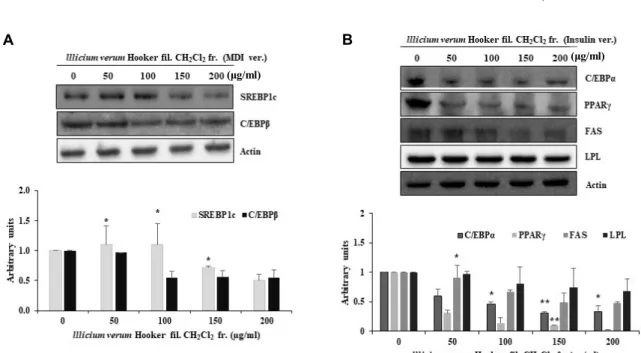
지방세포 분화 관련 전사인자들의 발현에 팔각회향 dichloro- methane층이 미치는 영향
지방전구세포에서 지방세포로의 분화하는 과정은 많은 종 류의 분화 관련 전사인자들이 단계적으로 발현되고 조절되어 유발된다. 지방세포 분화에 관련된 전사인자들의 종류에는 sterol regulator element-binding protein-1c (SREBP1c), PPAR γ와 C/EBPs family 등이 밝혀져 있다. 지방세포 분화 시 분화 촉진 인자로 인해 분화가 촉진되면 초기 단계에서 C/EBPβ가 발현된다. 발현된 C/EBPβ는 C/EBPα와 PPARγ의 발현을 활 성화시키는 인자이며 세포 내 중성지방의 생성과 축적을 촉진 시키는 역할을 한다[6, 11]. C/EBPβ에 의해 활성화된 C/EBPα 와 PPARγ는 서로 상호 작용을 하면서 중성지방의 생성을 유 도하며, 이들이 adipogenesis와 관련된 지방세포 특이적 인자 들인 FAS, LPL 등의 유전자의 발현에 깊이 관여 한다[19, 23].
활성화된 PPARγ과 SREBP1c에 의해 발현이 촉진되는 FAS는 지방산 합성 효소로 지방세포 분화의 최종 마커로 알려져 있 다[21]. LPL은 triglyceride를 fatty acid와 2-monoacylglycerol 로 가수분해하여 fatty acid를 세포 내로 유입시키는 효소이다.
에너지 대사 불균형으로 인해 완전히 소진되지 않은 fatty acid 는 triglyceride로 다시 전환되어 세포 내에 축적된다[12]. 이러 한 전사인자들의 발현에 팔각회향 dichloromethane층이 어떠 한 영향을 미치는지 알아보기 위하여 western blot analysis를 수행하였다. SREBP1c와 C/EBPβ는 지방세포 분화 촉진 인자 인 MDI를 처리할 때, 즉 지방세포 분화 초기단계에서 발현되 는 전사인자이다. 3T3-L1 지방전구세포가 confluence상태에 서 분화 촉진 인자인 MDI를 처리할 때 팔각회향 dichloro- methane층을 동시에 처리하고 48시간 뒤에 세포를 회수하여 전사 인자의 발현 양상을 확인한 결과, 팔각회향 dichloro- methane층을 처리한 군들의 발현이 감소함을 확인하였다(Fig.
4A). 또한 SREBP1c와 C/EBPβ의 발현에 의해 발현이 촉진되 는 지방 세포 분화 후기 전사인자인 C/EBPα, PPARγ의 발현 양상을 확인하기 위하여 MDI를 2일간 처리하고 insulin을 4일 간 처리한 다음 세포를 회수하여 확인한 결과, 팔각회향 di- chloromethane층을 처리한 군들에서 이들 전사인자들의 발현 이 저해되었음을 확인하였다(Fig. 4B). 또한 지방세포 분화의 최종 마커인 FAS, LPL의 발현도 팔각회향 dichloromethane층 처리에 의해 감소하였다(Fig. 4B). 지방세포 초기에 발현되는 C/EBPβ가 팔각회향 dichloromethane층에 의해 발현이 저해 됨에 따라 C/EBPβ가 발현을 촉진 시키는 C/EBPα와 PPARγ 의 발현이 저해 되었고, C/EBPα와 PPARγ에 의해 조절되는 FAS, LPL역시 발현이 저해되어 지방전구세포가 지방세포로 분화하는 것을 억제한다고 생각할 수 있다. 즉, 팔각회향 di- chloromethane층은 지방세포 분화에 관련되는 전사인자들의 발현을 저해하여 분화를 억제함으로써 세포 내 중성지방의 축적을 저해하는 것으로 생각된다.
팔각회향 dichloromethane층이 지방세포 분화 단계 중 mitotic clonal expansion 단계에서 세포 증식에 미치는 영향
A B
(μg/ml)
(μg/ml)
Fig. 4. Expression of adipogenesis-related proteins in 3T3-L1 cells treated with lllicium verum Hooker fil. CH2Cl2 fraction. (A) 3T3-L1 cells were incubated in DMEM (MDI) for 2 days with lllicium verum Hooker fil. CH2Cl2 fraction (B) 3T3-L1 cells were incubated in DMEM (MDI) for 2 days, and then in DMEM containing insulin with lllicium verum Hooker fil. CH2Cl2 fraction for 4 days. Cells were lysed and proteins were subjected to Western blot analysis. Results are expressed as Mean ± SD of three independent experiments and each sample was measured in triplicate. : *p<0.05, **p<0.01 as compared to the control.
지방세포 분화 단계 중 3T3-L1 지방전구세포가 confluence 상태가 되면 contact inhibition의 영향으로 세포 주기가 정지 가 되는데, 이때 지방세포 분화 촉진 인자인 MDI를 처리하면 정지되었던 세포 주기는 다시 움직여 세포의 수가 2~3배 늘어 나는 mitotic clonal expansion 단계가 된다. 이 단계에서 가장 중요한 역할을 하는 전사 인자가 C/EBPβ라고 알려져 있다 [17, 20, 27]. 위의 western blot analysis 결과에서 지방세포 분화 촉진 인자인 MDI에 의해 발현되는 C/EBPβ가 팔각회향 dichloromethane층에 의해 발현이 저해됨을 확인함에 따라 팔각회향 dichloromethane층이 mitotic clonal expansion단계 도 저해할 것이라 생각하고 실험을 수행하였다. 60 mm cell culture dish에 3T3-L1 지방전구세포를 confluence상태가 될 때까지 배양한 후, MDI와 팔각회향 dichloromethane층을 처리 하여 0, 24, 48시간에 세포를 회수하여 trypan blue exclusion assay로 생세포의 수를 확인하였다. 그 결과, 3T3-L1 지방전구 세포가 confluence상태일 때 MDI만 처리한 세포군들의 증식 은 0 hr을 100%로 하였을 때, 24, 48 hr의 세포의 증식이 약 1.6, 2배 증가함을 확인하였으나, 팔각회향 dichloromethane 층을 처리한 세포군들의 증식은 150 μg/ml의 농도에서는 1.14, 1.4배 증가하였으며, 200 μg/ml의 농도는 1배, 1.13배로 증가하여 세포의 증식이 억제됨을 확인하였다(Fig. 5A). 따라 서 팔각회향 dichloromethane층이 MDI처리에 의해 정지된 세포 주기를 다시 움직여 세포 증식을 유도하는 시기인 mi- totic clonal expansion을 저해한다고 사료된다. 팔각회향 di- chloromethane층에 의한 세포증식억제가 독성에 의한 결과인
지 확인하기 위해 MDI와 팔각회향 dichloromethane층을 동시 에 처리하고 48시간 후에 WST-1 assay를 수행한 결과, Fig.
5B에서 보듯이 세포 생존율에 전혀 변화가 없는 것으로 보아 mitotic clonal expansion의 억제가 세포 독성에 의한 결과는 아닌 것으로 사료된다.
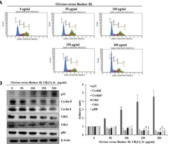
팔각회향 dichloromethane층이 mitotic clonal expansion 단계의 세포주기 변화와 cell cycle 관련 단백질의 발현에 미 치는 영향
팔각회향 dichloromethane층에 의해 mitotic clonal expan- sion단계가 저해됨을 확인함에 따라 세포 주기에 어떠한 변화 가 일어났을지 알아보고자 하였다. 3T3-L1 지방전구세포가 confluence상태일 때, 지방세포 분화 촉진인자인 MDI와 팔각 회향을 동시에 처리하여 세포를 회수하고 MuseTM Cell Analyzer를 이용하여 세포주기 변화를 관찰하였다. 팔각회향 dichloromethane층을 처리하지 않고 분화촉진인자(MDI)만 처리한 대조군 3T3-L1 지방전구세포는, G1기 개체군이 48.4%
로 확인 되었고, 팔각회향 dichloromethane층을 150, 200 μ g/ml의 농도로 처리한 군들의 G1기 개체군의 비율이 각각 55.1, 58.8%로 G1기에 머무르는 개체의 비율이 높아짐을 확인 하였다(Fig. 6A). 따라서 팔각회향 dichloromethane층이 G1기 에서 세포주기 정지를 유도하는 것으로 판단하고, 이에 관련 한 단백질의 변화를 확인하였다. 3T3-L1 지방전구세포가 con- fluence상태일 때, 지방세포 분화 촉진 인자인 MDI와 팔각회 향 dichloromethane 층을 동시에 처리하여 세포를 회수하고
A B
Fig. 5. Anti-proliferative effects of lllicium verum Hooker fil. CH2Cl2 fraction in the mitotic clonal expansion process of adipocyte differentiation. 3T3-L1 cells were incubated in DMEM (MDI) for 2 days with lllicium verum Hooker fil. CH2Cl2 fraction.
(A) Trypan blue exclusion assay was performed at 0, 24 and 48 hr. (B) Cell viability was determined by WST assay performed at 48 hr. Results are expressed as Mean ± SD of three independent experiments and each sample was measured in triplicate.
: *p<0.05, **p<0.01 as compared to the control.
A
B
Fig. 6. Cell cycle analysis of 3T3-L1 cells treated with of lllicium verum Hooker fil. CH2Cl2 fraction during the mitotic clonal expansion process of adipocyte differentiation. 3T3-l1 cells were incubated in DMEM (MDI) for 2 days with lllicium verum Hooker fil. CH2Cl2 fraction. (A) Cells were harvested and then fixed in 70% EtOH. Cells were stained with Muse™ Cell Cycle Reagent and analyzed by flow cytometer. (B) Cell were harvested and lysed and then equal volumes of proteins were de- termined by western blot analysis. Results are expressed as Mean ± SD of three independent experiments and each sample was measured in triplicate. : *p<0.05, **p<0.01 as compared to the control.
western blot analysis를 통하여 G1기에서 S기로 전이될 때 중요한 역할을 하는 단백질들의 발현 양상을 확인하였다. 세 포 주기는 S기를 준비하는 G1기, DNA를 합성하는 S기, M기
를 준비하는 G2기 및 체세포분열을 하는 M기로 나누어져 있 으며, 각 주기에 특이적인 단백질의 활성화 및 비활성화를 통 해 세포 주기가 진행된다. 특히, cyclin과 cyclin dependent
kinase (cdk)가 세포 주기의 진행을 담당하는 중요한 인자로 알려져 있다[20]. 세포주기 중 G1기에서는 cyclin D가 발현되 어 cdk4나 cdk6와 복합체를 형성하여 활성화 되면 망막아종 단백질인 retinoblastoma protein (Rb)을 인산화하여 S기 특이 적인 인자들의 발현을 촉진시킨다. 또한 cyclin E와 cdk2의 복합체 역시 G1기에서 S기로 전이되는 단계에서 Rb를 인산화 시키는 역할을 수행한다[4]. 즉, cyclin과 cdk의 발현이 저해되 면 복합체의 형성에도 영향이 미쳐서 복합체 형성이 저해되고 Rb인산화에도 영향이 미치게 되어 G1기에서 S기로의 전이가 정지가 된다. G1기에서 S기로 전이에 관여하는 단백질인 cy- clin E와 cyclin D, cdk2와 cdk4, phospho-Rb 및 cdk inhibitor 인 p21의 발현에 팔각회향 dichloromethane층이 어떠한 영향 을 미치는지 확인하였다. Fig. 6B에서 보는 바와 같이 팔각회 향 dichloromethane층에 의해 cdk inhibitor인 p21의 발현은 증가하였으며, cyclin E와 cyclin D, cdk2와 cdk4, phospho-Rb 의 발현은 감소됨을 확인하였다. 즉, cdk inhibitor인 p21의 발현이 증가되어 cyclin과 cdk의 복합체에 결합, 활성을 저해 하고, 이에 따라 Rb의 인산화가 억제되어 G1기에서 세포주기 가 정지되었을 것으로 생각된다. 이와 동일하게, 팔각회향 di- chloromethane층의 농도 의존적으로 G1기에서의 cell cycle arrest가 일어나는 것을 확인할 수 있었다. 이상의 결과를 종합 해보면 MDI만 처리한 3T3-L1 지방전구세포는 정상적으로 cell cycle이 진행되어 mitotic clonal expansion이 시작되지만, 팔각회향 dichloromethane 층을 처리하면 G1기에서 세포주 기 정지가 유도되어 mitotic clonal expansion이 억제 되고, 동시에 지방세포 분화 관련 전사 인자의 발현이 저해되어 지 방세포의 분화가 억제된다고 사료된다.
감사의 글
이 논문은 한국연구재단 연구과제(NRF-2017R1D1A1B04034994) 와 부산광역시 지원 항노화 바이오산업 육성산업(2018-항노 화-01), 동의대학교 블루바이오소재개발 및 실용화 지원센 터의 지원으로 이루어졌으며 이에 감사드립니다.
References
1. Alessi, M. C., Lijnen, H. R., Bastelica, D. and Juhan-Vague, I. 2003. Adipose tissue and atherothrombosis. Pathophysiol.
Haemost. Thromb. 33, 290-297.
2. Christy, R. J., Kaestner, K. H., Geiman, D. E. and Lane, M.
D. 1991. CCAAT/enhancer binding protein gene promoter:
binding of nuclear factors during differentiation of 3T3-L1 preadipocytes. Proc. Natl. Acad. Sci. USA. 88, 2593-2597.
3. Cornelius, P., MacDougald, O. A. and Lane, M. D. 1994.
Regulation of adipocyte development. Annu. Rev. Nutr. 14, 99-129.
4. Dorai, T. and Aggarwal, B. B. 2004. Role of chemopreventive
agents in cancer therapy. Cancer Lett. 215, 129-140.
5. Farmer, S. R. 2006. Transcriptional control of adipocyte formation. Cell Metab. 4, 263-273.
6. Fox, K. E., Fankell, D. M., Erickson, P. F., Majka, S. M., Crossno, J. T. and Klemm, D. J. 2006. Depletion of cAMP response element-binding protein/ATF1 inhibits adipogenic conversion of 3T3-L1 cells ectopically expressing CCAAT/
enhancer-binding protein (C/EBP) alpha, C/EBP beta, or PPAR gamma 2. J. Biol. Chem. 281, 40341-40351.
7. Gesta, S., Tseng, Y. H. and Kahn, C. R. 2007. Developmental origin of fat: tracking obesity to its source. Cell 131, 242-256.
8. Green, H. and Meuth, M. 1974. An established pre-adipose cell line and its differentiation in culture. Cell 3, 127-133.
9. Gregoire, F. M. 2001. Adipocyte differentiation: From fibro- blast to endocrine cell. Exp. Biol. Med. 226, 997-1002.
10. Gregoire, F. M., Smas, C. M. and Sul, H. S. 1998. Under- standing adipocyte differentiation. Physiol. Rev. 78, 783-809.
11. Hamm, J. K., Park, B. H. and Farmer, S. R. 2001. A role for C/EBPbeta in regulating peroxisome proliferator activated receptor gamma activity during adipogenesis in 3T3-L1 preadipocytes. J. Biol. Chem. 276, 18464-18471.
12. Hong, W. and Robert, H. E. 2009. Lipoprotein lipase: from gene to obesity. Am. J. Physiol. Endocrinol. Metab. 297, E271- E288.
13. Ji, H. H., Jeong, H. Y., Jin, S. J., Kwon, H. J. and Kim, B.
W. 2012. Inhibition of adipocyte differentiation by metha- nol extracts of oenanthe javanica seed in 3T3-L1 preadipo- cytes. J. Life Sci. 22, 1688-1696.
14. Kohda, H., Kohjyouma, M. and Namera, A. 1997. Production of anethole in callus of Illicium verum Hooker filius. J. Nat.
Med. 51, 519-522.
15. Lee, S. J. and An, S. K. 2016. Antioxidant and antiwrinkle effects of amentoflavone for cosmetic materials develop- ment. Asian J. Beauty Cosmetol. 14, 66-76.
16. Manickam, E., Sinclair, A. J. and Cameron-Smith, D. 2010.
Suppressive actions of eicosapentaenoic acid on lipid drop- let formation in 3T3-L1 adipocytes. Lipids Health Dis. 9, 57.
17. MacDougald, O. A. and Lane, M. D. 1995. Transcriptional regulation of gene expression during adipocyte differ- entiation. Annu. Rev. Biochem. 64, 345-373
18. Mokdad, A. H., Ford, E. S., Bowman, B. A., Dietz, W. H., Vinicor, F., Bales, V. S. and Marks, J. S. 2003. Prevalence of obesity, diabetes, and obesity-related health risk factors.
2001. JAMA. 289, 76-79.
19. Morrison, R. F. and Farmer, S. R. 2000. Hormonal signaling and transcriptional control of adipocyte differentiation. J.
Nutr. 130, 3116S-3121S
20. Nurse, P. 1994. Ordering S phase and M phase in the cell cycle. Cell 79, 547-550.
21. Palmer, D. G., Rutter, G. A. and Tavaré, J. M. 2002. Insulin- stimulated fatty acid synthase gene expression does not re- quire increased sterol response element binding protein 1 transcription in primary adipocytes. Biochem. Biophys. Res.
Commun. 291, 439-443.
22. Park, J. A., Jin, K. S., Oh, Y. N., Hyun, S. K., Choi, Y. H., Kwon, H. J. and Kim, B. W. 2013. Antiadipogenic effect of
초록:팔각회향 dichloromethane 분획물에 의한 지방세포 분화 억제 효과
정현영1․정인교2․김남주2․윤희정2,3․박정하2,3․김병우1,2,3․권현주1,2,3*
(1동의대학교 블루바이오 소재개발센터, 2동의대학교 대학원 생명응용학과, 3동의대학교 공과대학 바이오응용공학
부 바이오의약공학전공)
비만은 에너지의 불균형으로 인하여 체내 지방조직에 지방이 축적되는 대사성질환으로 심혈관계 질환, 고혈압, 2형 당뇨, 고지혈증 및 각종 암의 발생 빈도를 증가시키는 요인이다. 지방의 축적은 지방전구세포가 지방세포로 분화하는 과정을 의미하는 adipogenesis라는 과정을 거쳐서 일어난다. 지방세포로의 분화는 다양한 호르몬과 전 사인자들의 상호작용에 의해서 일어난다. 본 연구에서는 팔각회향이 항비만 소재로 활용 가능한지 확인하기 위해, 팔각회향 물 추출물을 분획하여 지방축적 억제 활성이 좋은 dichloromethane층을 선정하였다. 3T3-L1 지방전구세 포가 성숙한 지방세포로 분화할 때 팔각회향 dichloromethane 층이 어떠한 기전으로 분화를 조절하는지 확인한 결과, 지방세포 분화에서 중요한 전사인자인 C/EBP family, PPARγ의 발현이 억제되었고, 지방세포 최종 분화 마커로 알려져 있는 FAS 및 LPL의 발현 또한 감소되었다. 또한 G1기에서의 세포주기 정지를 통해 지방세포 분화 단계에서 필수적인 mitotic clonal expansion 단계를 억제한다는 결과를 얻었다. 이러한 연구 결과는 팔각회향이 항비만 효과를 가지는 천연물 소재로의 활용가능성을 보여주는 기초 자료가 될 것으로 사료된다.
Vitis amurensis root methanol extract and its solvent frac- tions in 3T3-L1 preadipocytes. J. Life Sci. 23, 69-78.
23. Park, Y. K., Lee, T. Y., Choi, J. S., Hong, V. S., Lee, J. H., Park, J. W. and Jang, B. C. 2014. Inhibition of adipogenesis and leptin production in 3T3-L1 adipocytes by a derivative of meridianin C. Biochem. Biophys. Res. Commun. 452, 1078- 1083.
24. Rosen, E. D. and Spiegelman, B. M. 2000. Molecular regu- lation of adipogenesis. Annu. Rev. Cell Dev. Biol. 16, 145-171.
25. Smith, P. D., O’Halloran, P., Hahn, D. L., Grasmick, M. and Radant, L. 2010. Screening for obesity: Clinical tools in evo- lution, a WREN study. Wisc. Med. J. 109, 274-278.
26. Spiegelman, B. M. and Flier, J. S. 2001. Obesity and the regu- lation of energy balance. Cell 104, 531-543.
27. Tang, Q. Q. and Lane, M. D. 1999. Activation and centro- meric localization of CCAAT/enhancer-binding proteins during the mitotic clonal expansion of adipocyte differ- entiation. Genes Dev. 13, 2231-2241.
28. Tang, Q. Q., Otto, T. C. and Lane, M. D. 2003. Mitotic clonal expansion: a synchronous process required for adipogenesis.
Proc. Natl. Acad. Sci. USA. 100, 44-49
29. Tzeng, T. F. and Liu, I. M. 2013. 6-gingerol prevents adipo- genesis and the accumulation of cytoplasmic lipid droplets in 3T3-L1 cells. Phytomedicine 20, 481-487.
30. Wang, G. W., Hu, W. T., Huang, B. K. and Qin, L. P. 2011.
Illicium verum: A review on its botany, tradition use, chem- istry and pharmacology. J. Ethnopharmacol. 136, 10-20.