Changes in Glucosinolate Content of Dolsan Leaf Mustard Kimchi during Fermentation and Correlation with Antioxidant, Antihypertensive, and Antidiabetic Activities
Sun-Kyung Oh1, Ki-Woong Kim2 and Myeong-Rak Choi1*
1Department of Biotechnology, Chonnam National University, Yeosu 59626, Korea
2Department of Marine Bio Food Science, Chonnam National University, Yeosu 59626, Korea Received September 28, 2018 /Revised November 9, 2018 /Accepted November 12, 2018
The glucosinolate content, antioxidant activity, and antihypertensive and antidiabetic activities were measured in a crude extract of Dolsan leaf mustard kimchi (DLMK). The glucosinolate content was low at 6.41 and 7.92 mg/g in leaves and stems of DLMK after 21 days of fermentation. The total poly- phenol and total flavonoid contents were more than 2 times higher in the leaves (211.7 mg GAE/g, 158.8 mg QE/g) than in the stem (53.7 mg GAE/g, 85.2 mg QE/g) during the fermentation period.
The 2,2'-azino-bis(3-ethylbenzothiazoline-6-sulphonic acid) (ABTS) radical scavenging activity and elec- tron donating ability (EDA) were similar to those of the control group after 14 days of fermentation, while the ferric reducing antioxidant power (FRAP) was higher in the leaves after 14 days of fermen- tation when compared to the control group. The angiotensin converting enzyme (ACE) inhibitory ac- tivity showed similar or higher inhibitory activity in the leaves when compared to the control group (0.01% captopril), and the α-glucosidase inhibitory activity was higher in the leaves and stems when compared to the control group (0.05% acarbose). The glucosinolate content and the ABTS, ACE, and α-glucosidase inhibitory activity were correlated, as determined by the observed straight line plot with a positive grade. During the fermentation period, the detected glucosinolates were sinigrin, gluco- brasicin, glucotropeolin, and progoitrin. The DLMK extract is therefore expected to be valuable as a functional food because of its effective antioxidant, antihypertensive, and antidiabetic activities.
Key words : ACE inhibition, antioxidant, Dolsan leaf mustard kimchi, α-glucosidase inhibition, glucosinolates
*Corresponding author
*Tel : +82-61-659-7303, Fax : +82-61-659-7309
*E-mail : [email protected]
This is an Open-Access article distributed under the terms of the Creative Commons Attribution Non-Commercial License (http://creativecommons.org/licenses/by-nc/3.0) which permits unrestricted non-commercial use, distribution, and reproduction in any medium, provided the original work is properly cited.
Journal of Life Science 2018 Vol. 28. No. 11. 1290~1300 DOI : https://doi.org/10.5352/JLS.2018.28.11.1290
Introduction
The recent scientific developments, increase in income level, and change in dietary habit haves generated an in- creasing interest in heath maintenance and well-being, as well as a changing awareness of health-related issues. In par- ticular, reactive oxygen species (ROS) cause various diseases including cancer, are relates to aging of the human body, and damage DNA [44]. Especially, oxidative stress is in- volved in the development of chronic diseases such as re- duced liver function, and there are various studies on bio- active substances derived from foods that have been used for its control or treatment. Such researches studies are con-
ducted by methods that involve the removal of reactive oxy- gen species, blocking ultraviolet rays, and application of an- tioxidants [19]. In addition, the increase in related diseases in advanced countries such as European countries and Japan, has led to a growing interest in developing new func- tional foods that are effective in adult [19]. In addition, the antihypertensive mechanism involving the inhibition of the angiotensin-converting enzyme (ACE) essentially prevents the formation of angiotensin Ⅱ, which contracts blood ves- sels and control the decomposition of bradykinin, which re- laxes blood vessels. Studies on natural ACE inhibitors [16, 33] have been mainly conducted using fish, tofu, tea (including green tea), protein-rich foods such as marine fer- mented food and miso. Phenol compounds isolated from plants separated from plants have been reported to mediate the inhibition of growth of various germs and yeasts as well as inhibition of enzyme activity [7, 27]. Furthermore, crucif- erous vegetables are particularly rich in glucosinolate, phe- nol, flavonoida wells, and sulfur compound [26]. Currently, efforts are continuously being made to find natural anti-
Table 1. Content of glucosinolates in DLMK extracts during the fermentation period Glucosinolates in leaves
of DLMK (mg/g)
Storage period (days)
0 7 14 21 28 35
Glucoiberin Sinigrin Gluconapin Glucoerucin Glucobrassicin Gluconasturtiin Glucoraphanin Total glucosinolates
- 21.43±0.06aA 0.21±0.05bC
- 0.03±0.02eD 1.26±0.21aB 1.04±0.04aB 23.97±0.07aA
0.66±0.48bB*
7.70±0.04cA 0.11±0.02cC
- 0.22±0.03bC 0.70±0.55bB 0.07±0.02bD 9.46±0.24cA
0.04±0.02aE 7.44±0.05cB 0.06±0.02dE 0.33±0.02aC 0.18±0.02aD
- - 8.05±0.02cA
- 6.29±0.01dA
- 0.02±0.02bB 0.08±0.02dB 0.02±0.01cB
- 6.41±0.01dA
0.04±0.03aC 12.89±0.01bA
- - 0.11±0.02cB 0.03±0.02cC
- 13.07±0.01bA
- 12.51±0.02bA 0.51±0.05aB
- - 0.02±0.02cC 0.07±0.04bC 13.11±0.02bA Glucosinolates in stems
of DLMK (mg/g)
Storage period (days)
0 7 14 21 28 35
Glucoiberin Sinigrin Gluconapin Glucoerucin Glucobrassicin Gluconasturtiin Glucoraphanin Total glucosinolates
0.07±0.02bC*
22.47±0.02aA 1.07±0.02aB
- 0.03±0.02bC
- 0.05±0.03cC 23.69±0.01aA
0.11±0.02aB 11.57±0.01cA 0.07±0.02bC
- 0.05±0.03bC
- - 11.80±0.01bA
0.13±0.02aC 12.25±0.04bA 0.03±0.02bD 0.13±0.04bC 1.09±0.04aB
- 0.08±0.03cD 13.71±0.01bA
0.02±0.01cB 7.78±0.02dA
- - 0.08±0.03bB
- 0.04±0.03cB 7.92±0.01cA
0.09±0.02bC 14.00±0.02bA
- 0.06±0.04cC
- - 0.12±0.03bB 14.27±0.02bA
0.06±0.01bC 10.04±0.02cA
- 1.04±0.02aB
- - 1.05±0.04aB 12.19±0.01bA
*Data are the mean ± SD of triplicate experiment. Different superscript lower-case letters in the same row (a-e) and in the same column (A-D) indicate significant differences by Duncan’s multiple range test (p<0.05).
carcinogenic, antioxidant, antibiotic, and anti-mutagenic ma- terials from foods including some vegetables, fruits, grains, and seaweed, as well as their fermented foods products, and novel bioactive substance are constantly being discovered [15, 43]. In particular, leaf mustard which is specially pro- duced in Dolsan-eup, Jeollanam-do province, South Korea, has a tangy flavor and texture that does not soften easily and, therefore, has emerged as a well-known main or minor ingredients of Kimchi [6, 28]. Glucosinolate, which gives leafy mustard its tangy flavor, and its by-product, iso- thiocyanate have long been studied globally mainly by the evaluation of crude plant extracts [22, 36]. Leaf mustard kim- chi is expected to have physiological functionality owing to the bioactive substances formed during fermentation. How- ever, this study was designed to investigate the relationship between the antioxidant and biological activity and the glu- cosinolates content of crude Dolsan leaf mustard kimchi (DLMK) extract. The DLMK extract was prepared by sepa- rating the crude extract isolated during the fermentation pe- riod using analytical high-performance liquid chromatog- raphy (HPLC) and LC-PDA/MS/MS.
Materials and Methods Materials and reagents
The Dolsan leaf mustard used in this study was harvested in March by the Dolsan Leaf Mustard Farming Association located in Dolsan-eup, City of Yeosu, Southern Jeolla Pro- vince. The additional ingredients including garlic, green on- ion, pepper, and ginger were purchased from a local market, and the spices were mixed in the proportions shown in Table 1. The acetonitrile (CH3CN) and ethanol were from J. T.
Baker (Center Valley, PA, USA). The sinigrin, 1,1-diphenyl-2- picrylhydrazyl (DPPH), 2,2-azino-bis (3-ethyl-benzothizo- line-6-sulfonic acid) (ABTS), ACE, Hippuryl-His-Leu (HHL), ethyl acetate, sodium chloride (NaCl), α-glucosidase, potas- sium phosphate buffer (pH 7.0), p-nitrophenyl--D-glucopyr- anoside (p-NPG), sodium carbonate, captopril, and acarbose were purchased from Sigma-Aldrich Co. (St. Louis, MO, USA).
Preparation of Dolsan leaf mustard kimchi (DLMK) After washing under flowing water and then completely removing the water by air drying for 1 hr, Dolsan leaf mus- tard (DLM) was brined in a 10% salt solution for 3 hr, wash- ed twice, and the water was removed by air drying for 30 min. The Dolsan leaf mustard kimchi (DLMK) recipe in- cluded brined leaf mustard (75%), pepper powder (12%), fer- mented sand lance juice (4%), and 1% each of sugar, garlic, salt, ginger, seasoning powder, onion, and glutinous rice
paste. The pre-mixed spices were mixed with the brined leaf mustard, and a 500 g sample was packed in food-grade poly- ethylene bags. The prepared DLMK was fermented at 0℃
for 35 days after preparation, with experimental measure- ments carried out at 7 days intervals.
Glucosinolates extracted from the DLMK
To measure the glucosinolates content of the different parts of the crude plant material used to prepare the DLMK, we placed 20 g of the leaves and stems in thimble filters, added 200 ml of 50% CH3CN to the round-bottom flasks connected to a Soxhlet extractor, and extracted the material in a water bath at 90℃ for 24 hr [40]. Then, the extract were cooled to room temperature and filtered through Whatman No. 4 filters (Whatman International Ltd., England). The fil- trates were concentrated using a vacuum rotary evaporator (N-1000, Eyela, Tokyo, Japan) to a final volume of 100 ml.
The final extracts were stored at -18℃ for use in subsequent experiments.
Glucosinolates standard curve
The standard sinigrin and 6 species reagent was prepared at concentrations of 125, 250, 500, and 1,000 ppm. All HPLC measurements were made in triplicate, and the mean values were used to create a standard curve. The standard curve was used to measure the changes in glucosinolates content in DLMK during storage.
HPLC operating conditions
DLMK was extracted, and the extracts were filtered using 0.45 μm syringe filters (Toyo Roshi Kaisha, Ltd., Tokyo, Japan). The filtrates were placed in vials, and triplicate HPLC measurements were obtained. Each peak was fractio- nated based on the retention time (RT) of DLMK leaves and stems. The fractionation was done to use the functionality of the respective peaks in the experiment. The analytical- HPLC measurements were conducted using a Shimadzu JP/
LC-10A HPLC equipped with a CDD-6A detector and a Shim-pack VP octadecylsilane (ODS) column (250×4.6 mm i.d.; Shimadzu, Tokyo, Japan). The mobile phase consisted of water (A) and acetonitrile (B) (60:40, v/v). The total flow rate was 1.0 ml/min, the injection volume was 10 μl, the oven tem- perature was 40℃, and the detection wavelength was 228 nm.
Measurement of total polyphenol content
The total polyphenol content in different parts of DLM
was measured using colorimetry by using Folin-Ciocalteu’s phenol reagent [34]. Distilled water of 2.6 ml and 200 μl of Folin-Ciocalteu’s phenol reagent to 200 μl of the sample and mixed together; the mixture was allowed to react for 6 min at room temperature, and then, 2 ml of 7%(w/v) Na2CO3
solution was added. The mixture was then allowed to react for 90 min, and the absorbance was measured using a spec- trophotometer (Ultraspec 2000; Pharmacia Biotech, Cam- bridge, England) at 750 nm. A standard curve was con- structed using gallic acid as the standard, and the poly- phenol content was reported in mg gallic acid equivalents (GAE)/g extract.
Measurement of total flavonoid content
The total flavonoid content in different parts of DLM was measured as by the method of Jia et al. [12]. One mililiter of the sample, 3.2 ml of distilled water, and 150 μl of 5%
(w/v) NaNO2 were mixed together and were allowed to re- act for 5 min. Then, 10% AlCl3 solution was added and the mixture was reacted for 1 min, and 1M NaOH was added;
subsequently, the absorbance was measured at 510 nm. A standard curve was constructed using quercetin as a stand- ard substance, and the flavonoid content was reported in mg quercetin equivalents (QE)/g extract.
2,2'-Azino-bis(3-ethylbenzothiazoline-6-sulphonic acid) (ABTS) radical scavenging activity measurement
The 2,2'-Azino-bis(3-ethylbenzothiazoline-6-sulphonic acid) (ABTS) radical scavenging activity was measured using a modification of the method reported by Kriengsak et al. [17].
The stock solutions containing 1.8 and 0.63 mM ABTS and potassium persulfate solutions, respectively were kept at 37
℃ for 24 hr in dark, then the ABTS free radical concen- tration, generated by the ABTS solution, was adjusted to an absorbance of 1.4±0.1 at 735 nm. The ABTS radical solution and the sample (5 and 0.1 ml, respectively) were reacted at room temperature for 7 min and then the absorbance was measured at 735 nm. Ascorbic acid was used as a control and the percentage (%) scavenging activity was calculated using the following equation:
ABTS radical scavenging activity (%) = (1 - absorbance of the solution with the sample added/absorbance of the sol- ution without sample) ×100
Electron donating ability (EDA)
Electron donating ability (EDA), which is the radical scav-
enging effect of DPPH, was shown as the reducing power of the sample. 0.5 ml of 0.5 mM DPPH solution was added to a test tube containing 1 ml of the sample, 1 ml of meth- anol, and 0.99 ml of 100 mM sodium acetate buffer (pH 5.5), and the mixture was agitated. Afterward, the mixture was reacted in dark for 5 min; the concentration of the remaining radicals was measured at room temperature and at 517 nm and was reported according to the following equation [37].
Ascorbic acid was used for showing the positive control group:
EDA (%) = (1 - absorbance of the solution with the sample added/absorbance of the solution without sample) ×100
Measurement of ferric reducing antioxidant power (FRAP)
We modified the method reported by Benzie et al. [1] to measure the FRAP activity. 30 mM acetate buffer (pH 3.6), 10mM TPTZ (2,4,6-tripyridyls-triazine) dissolved in 40 mM HCl, and 20 mM Iron (III) chloride hexahydrate were mixed in a 10:1:1 (v:v:v) ratio. 150 μl of the sample and 2,850 μl of the reaction mixture were mixed together and were left for 30 min; afterward, absorbance was measured at room temperature and at 593 nm. A standard curve was con- structed using Iron (II) sulfate heptahydrate (FeSO4․H2O), and FRAP content was reported in mg FeSO4 equivalents/g extract.
Angiotensin I-converting enzyme (ACE) inhibitory activity measurement
The ACE inhibitory activity was measured according to the methods of Cushman and Cheung [5] with slight a modification. The ACE (peptidyldipeptide hydrolase, EC 3.4.15.1) 0.38 mg was dissolved in 0.1 M sodium borate buf- fer (pH 8.3) 10 ml Hippuryl-His-Leu (HHL) substrate, which was by mixing the substrate solution to obtain a (25 mM HHL solution). Then, 100 μl of the 25 mM HHL solution was mixed with a 50 μl sample, preincubated at 37℃ for 10 min, and the reaction was initiated by adding 150 μl of ACE dissolved in sodium borate buffer (pH 8.3), followed by further incubation of the mixture at 37℃ for 60 min. The reaction was finally stopped by adding 250 μl of 0.5 N HCl, and the hippuric acid was extracted with 1.5 ml of ethyl acetate. The extracts were centrifuged at 2,500 rpm for 10 min. 0.5 ml of the supernatant was dried in an oven at 140℃
for 10 min, dissolved in 3 ml of 1 M NaCl, and then the
absorbance was measured at 228 nm to evaluate the degree of inhibition of the ACE activity. In addition, 0.01% captopril was used as the control and the inhibitory effect was calcu- lated using the following equation:
Inhibitory activity (%) = ([Abscontrol - Abssample]/[Abscontrol
- Abssampleʹ]) ×100
Where, Abssampleʹ is the absorbance of the control solution with 0.5N HCl added to stop the reaction initiated by the ACE enzyme addition.
Measurement of α-glucosidase inhibitory activity sThe α-Glucosidase inhibitory activity was determined by using a modification of the procedure previously reported by Watanabe et al. [42]. Briefly, 50 μl each of the test sample, 0.7 unit/ml α-glucosidase enzyme solution, and 100 mM po- tassium phosphate buffer (pH 7.0) were mixed, and then preincubated at 37℃ for 5 min. Then, the reaction solution with 100 μl of 3 mM pNPG (p-nitrophenyl-α-D-glucopyrano- side) was incubated at 37℃ for 15 min, and the reaction was stopped by 750 μl of 100 mM sodium carbonate. The absorb- ance was measured at 405 nm and 0.05% acarbose was used as a control while the percentage inhibitory activity was cal- culated using following equation:
Inhibitory activity (%) = (1- Abssample/Abscontrol) ×100
LC-PDA/MS/MS analysis
Analysis of different parts of DMS extracts was performed through LC-PDA/MS/MS conducted using a Thermo LTQ Orbitrap XL system connected to a Shimadzu UFLC; sinigrin and its derivatives were investigated in positive ion mode.
Phenomenex KINETEX C-18 columns (150×2.1 mm, 2.6 μm) were used as analysis columns; the column temperature was maintained at 40℃, the detection wavelength was 228 nm (Shimadzu, SPD-M20A), and the flow rate was 0.15 ml/min.
10 μl of the sample was injected using an automatic injector.
Solvent A (water) and Solvent B (acetonitrile) were used as mobile phase solvents. Solvent B was increased to 2% for 5 min, from 2% to 32% for 30 min, and was maintained at 100% for 5 min; the total running time was 40 min. The oper- ating conditions of LC-PDA/MS/MS were as follows: scan range m/z 250-800, source voltage 5.40 kV, capillary voltage 48.00 V, capillary temperature 275℃, sheath gas flow 50.00 L/min, aux gas flow 10.00 L/min, source current 1,000 μA, and tube lens 100 V.
A
B
Fig. 1. Changes in total polyphenol (A) and total flavonoid (B) content in DLMK during the fermentation period.
Vertical bars represents standard deviation (n=3). Same small letters (a-b) are not significantly different (p<0.05) by Duncan’s multiple test.
Statistical analysis
The results are expressed as mean ± standard deviation (SD). One way analysis of variance (ANOVA) and Duncan’s test were used for multiple comparisons using the SPSS ver- sion 21.0 (SPSS Institute, Chicago, IL, USA). All tests and analyses were repeated at least three times. Difference were considered statistically significant in all experiment means at p<0.05. The correlation between the glucosinoletes content and the correlations of ABTS, EDA, ACE and α-glucosidase is schematiced using the Microsoft Office Excel 2013.
Results and Discussion
Glucosinolates contents
Sinigrin and 6 species standards was used as the standard reagent for quantitative analysis, and a standard curve was obtained from the 7 species reagent manufactured at differ- ent concentrations using HPLC (r2=1.000). As shown in Table 1, leaves and stems of DLMK was similar on day 0 (21.43±0.06 and 22.47±0.02 mg/g, respectively). The contents decreased during the fermentation period and reached mini- mum values (7.44±0.06 and 7.78±0.02 mg/g, respectively) on day 21. Cieślik et al. [4] reported fresh white cauliflower and green cauliflower have similar sinigrin contents of 34.1 mg/100 g and 38.1 mg/100 g, respectively. However, our results differed from the findings of Lim [21], who reported that the sinigrin content of Dolsan leaf mustard kimchi (DLMK) during the initial fermentation stages and the post- ripe stages were similar, and that the maximum content was observed during the optimally ripe stage (pH 4.2, acidity 0.5-0.75%). In this study, DLMK fermentation was carried out at a low temperature, unlike the typical kimchi fermenta- tion temperature (5-14℃). Different results were obtained ac- cording to the added ingredients and fermented temper- ature, as the total glucosinolate content, which gives the spi- cy taste, gradually decreased during DLMK ripening [38].
Glucosinolates has changed into other forms such as gluco- napin, glucoraphanin, and gluconasturtiin.
Total polyphenol and total flavonoid content Phenolic compounds, which are secondary metabolites widely distributed in plants, are known to have antioxidant and anti-cancer activities [8], most antioxidant compounds in plants are polyphenols and compounds acts as natural antioxidants [27]. In this study, during DLMK fermentation and the total polyphenol contents in the samples were
investigated. The total polyphenol contents in DLMK leaves and stems during the fermentation period were 100.4-211.7 mg GAE/g extract and 53.7-102.3 mg GAE/g extract, re- spectively (Fig. 1A). In particular, leaves and stems showed the lowest total polyphenol contents on day 21(p<0.05).
According Choi et al. [2] and Lee and Cheigh [20], the total polyphenol content is affected by the components of kimchi and the solvent of the kimchi extract. In this study, sig- nificant polyphenol contents were obtained as the kimchi gradually fermented throughout the 35 days storage period.
Natural flavonoids, which include various pigments in plants, are reported to suppress pathogenic bacteria, block ultraviolet rays, and have antimicrobial, antioxidant, and an- ti-inflammatory effects; moreover, multiple reports have suggested that flavonoids suppress the growth and differ- entiation of various tumor cells [3]. Fig. 1B shows the total flavonoid contents measured in the DLMK extracts; the con- tents measured on 21 days, which were 89.6 mg QE/g ex- tract and 85.9 mg QE/g extract in leaves and stems, re- spectively, were lower than the contents measured immedi- ately after manufacture (p<0.05). The total flavonoid content
A
B
C
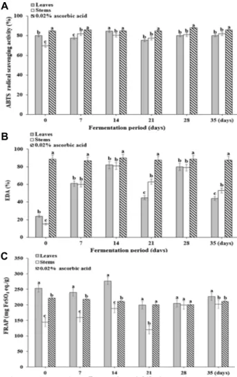
Fig. 2. Changes in 2,2'-azino-bis(3-ethylbenzothiazoline-6-sulphonic acid) (ABTS) radical scavenging activity (A), electron do- nating ability (EDA) (B) and ferric reducing antioxidant power (FRAP) (C) in DLMK during the fermentation period. Vertical bars represents standard deviation (n=3).
Same small letters (a-c) are not significantly different (p<0.05) by Duncan’s multiple test.
of DLM has been reported [24], with a total flavonoid con- tent of DLM of 387.33-391.00 mg QE/100 g. The total poly- phenol content and total flavonoid content of the Chinese cabbage kimchi fermented at 5℃ for 24 days gradually in- creased from 57.9-102.5 mg GAE/100 g extract and 4.1-12.6 mg QE/g extract during the fermentation period. Jung et al. [14] this study such as showed a tendency to increase slightly during 24 days of fermentation, but this seems to be the result of the amount of bioactive substance depending on fermentation temperature or fermentation period.
2,2-azino-bis(3-ethyl-benzothiazoline-6-sulfonic acid) (ABTS) scavenging activity and electron donating ability (EDA)
As the ABTS free radical scavenging activity can measure the scavenging activity of polar and non-polar samples, it can be applied more broadly than the DPPH free radical scavenging activity [30]. The activities measured immedi- ately after manufacture of DLMK were 79.74% and 70.43%
in leaves and stems, respectively, with the highest activities observed on day 14 (Fig. 2A). Overall, there was no statisti- cally significant difference between the ABTS radical scav- enging activities of the leaves and stems (p<0.05). The anti- oxidant capacities measured for the fermentation of cabbage leaves consumed commonly in Korea were higher for fer- mented cabbage [10, 35], it shows that the antioxidant activ- ity varies with the fermentation temperature and storage period.
In this study, the EDA is used as a measure of the sup- pression of oxidation by electrons donated to the oxidizing free radicals involved in lipid peroxidation chain reactions (Fig. 2B). The EDA values for DLMK extract to DPPH were found to be 24.38-81.51% and 14.62-81.14% in leaves and stems, respectively, with no significant difference observed between leaves and stems (p<0.05). Jang [13] found that DLM and DLMK extracts have concentration-dependent DPPH radical-scavenging activities. The concentration at which 50% DPPH radical scavenging was observed for DLM and DLMK were obtained, and DLM (5.79 mg) showed greater activity than DLMK (9.17 mg). In this study, the electron donating effect from DLMK extract to DPPH was low be- cause the antioxidant components contained in leaf mustard were degraded during the kimchi manufacturing process.
DLMK exhibited high scavenging activity in ABTS during fermentation and showed low scavenging activity in DPPH, and the free radical scavenging ability of these samples was
compared between the two methods. These results suggest that the opposite result can be obtained because the ABTS radical is removed according to the type of polyphenol but the DPPH radical is not eliminated as reported by Wang et al. [41].
Ferric reducing antioxidant power (FRAP)
In contrast to the ABTS free radical scavenging activity, the FRAP activity measures the antioxidant activities through oxidizing and reducing reactions via a mechanism in which a ferric complex is reduced to a ferrous complex by an antioxidant. As the results can differ according to the type, distribution, and biochemical properties of the antioxidants
A
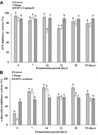
B
Fig. 3. Changes in angiotensin-converting enzyme (ACE) (A) and α-glucosidase (B) inhibitory activities of DLMK dur- ing the fermentation period. Vertical bars represents standard deviation (n=3). Same small letters (a-c) are not significantly different (p<0.05) by Duncan’s multiple test.
present in a sample, the antioxidant activity of the sample should be measured using both methods [32]. The FRAP ac- tivity showed similar trends to the ABTS free radical scav- enging activity (Fig. 2C). The FRAP activity measured in DLMK leaves and stems was 199.03-276.87 mg FeSO4 equiv/
g and 144.17-201.77 mg FeSO4 equiv/g, respectively; the ac- tivity was higher than the activity of ascorbic acid (221.30 mg FeSO4 equiv/g), which was used as the control (p<0.05).
The FRAP activities measured for various types of cabbage, an essential element of the kimchi commonly consumed by Koreans, were 87.0-714.5 mg FeSO4 equiv/100 g, indicating a stronger antioxidant effect than that obtained this study for DLMK [11]. Thus, the overall antioxidant activity is re- duced on day 21 of fermentation because antioxidant compo- nents are transformed into different substances through the fermentation of leaf mustard.
Angiotensin Converting Enzyme (ACE) inhibitory activity
ACE is an important enzyme in the rennin-angiotensin- aldosterone system, which generates dipeptide (His-Leu) by hydrolyzing the inactive angiotensin-I at the C-terminal, thereby creating angiotensin-II that causes powerful vaso- constriction. Angiotensin II eventually raises the blood pres- sure and, therefore inhibition ACE activation is a useful strategy for controlling disorders such as those involving the destruction of blood vessels and stroke [23]. The ACE in- hibitory activity of the single extract of the leaves and stems obtained by through separating the crude DLMK extract during the fermentation period is shown in Fig. 3A (p<0.05).
In addition, 0.01% captopril was used as the standard anti- hypertensive, and it showed an ACE inhibitory activity of 90%, while that of the leaves and stems extracts was also more than 80%. Previous studies have demonstrated a high inhibitory effect [9] of 70% or more with broccoli, celery, Chinese cabbage, cabbage, and chives, which suggest they a similar tendency as that of the DLMK leaves and stems extracts during the fermentation period. The higher ACE in- hibitory activity in leaves seems to contain various physio- logically active substances and it is considered that in depth study is needed.
α-Glucosidase inhibitory activity
α-glucosidase is an enzyme that exists in the small intes- tine and breaks down oligosaccharide, into monosaccharides after carbohydrate have been broken down by amylase or
other enzymes. α-glucosidase facilitates the absorption of sugar and inhibitors of this enzyme obstruct the generation of monosaccharides. Furthermore, it controls the absorption of sugar and reduces postprandial increase in blood glucose and, therefore, α-glucosidase inhibitors are used for the pre- vention and treatment of diabetes [18]. The DLMK of leaves and stems extract during the fermentation period showed the inhibition rate of 81% and 78% on day 14 (Fig. 3B). The positive control, 0.05% acarbose, showed a 57% of inhibition than the positive control did [31]. This result indicates that the extracts could effectively inhibit increases in blood glu- cose concentrations.
Correlation of EDA, ABTS, ACE and α-glucosidase inhibitory activities and glucosinolates content
The correlation of the EDA and ABTS radical scavenging and ACE and α-glucosidase inhibitory activities of the GSL extract obtained and with the GSL content of the DLMK
A
B
C
D
Fig. 4. Glucosinolates content of leaves and stems extracts of DLMK and correlations between measured electron donating ability (EDA) and 2,2'-azino-bis(3-ethylbenzothiazoline-6-sulphonic acid) (ABTS) radical scavenging (A, C) and angiotensin-converting enzyme (ACE) and α-glucosidase inhibitory activities (B, D).
leaves and stems extracts is shown in Fig. 4. The correlation coefficient (R2) values of the GSL content of the DLMK leaves extracts with the EDA and ABTS radical scavenging and ACE, and α-glucosidase inhibitory activities were 0.9705, 0.9461, 0.9694, and 0.8758, respectively, while those of the stems extracts were 0.9601, 0.9425, 0.9381, and 0.9734, re- spectively. Therefore except for the correlation coefficient be- tween GSL and ABTS radical scavenging activity (R2 = 0.8249) and the correlation coefficient of ACE inhibitory ac- tivity (R2 = 0.8353), it is not high and does not have a direct influence [39]. In addition, GSL content showed a high corre- lation with ACE inhibitory activity and α-glucosidase in- hibitory activity in stem extracts of DLMK showed a gentle slope.
LC-PDA/MS/MS analysis
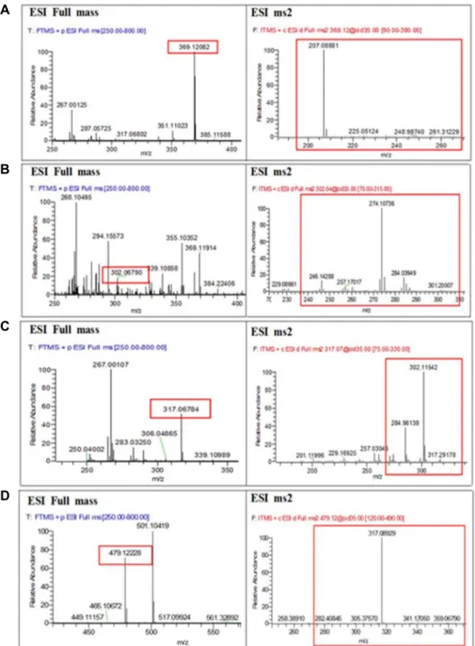
Leaves and stems extract of Dolsan leaf mustard kimchi during the fermentation period, analyzed by LC-PDA/MS/
MS, is shown in Fig. 5. Glucosinolates identified in the ex- tract of the leaves and stems of DLMK during the fermenta- tion period, include sinigrin, glucobrassicin, glucotropaeolin, progotrin. Glucobrassicin was detected in the form of [SO3- M+H]+ on day 14 of fermentation. On days 21 and 28 of
fermentation, glucobrassicin was detected in the form of [M-SO3-CH2OH+2H]+. Sinigrin, on day 14 of fermentation was detected in the form of [SO3-M+H]+ and on days 21 and 28 of fermentation period, it was found in the form of [M-SO3-H2O+Na]+ [25]. Glucosinolates are hydrolyzed by myrosinase, but some have epithiospiere protein and myr- osinase binding proteins, although the role of these proteins has not yet been clarified.
Acknowledgement
This work was supported by the BK21 program.
References
1. Benzie, M. S. 1958. Antioxidants determination by the use of a stable free radical. Nature 181, 1199-1200.
2. Choi, S. W., Kang, W. W., Chung, S. K. and Cheon, S. H.
1996. Antioxidative activity of flavonoids in persimmon leaves. Food Sci. Biotechnol. 2, 119-123.
3. Choi, Y. M., Gu, J. B., Kim, M. H. and Lee, J. S. 2008.
Antioxidant and antiproliferative activities of methanolic ex- tracts from thirty Korean medicinal plants. Food Sci. Bio- technol. 17, 1235-1239.
4. Cieslik, E., Leszczynska, T., Filipiak-Florliewicz, A., Sikora,
A
B
C
D
Fig. 5. Mass spectra showing retention times and MS/MS fragments at m/z 369.12, 302.07, 317.07, and 479.11, representing gluco- brassicin (A), sinigrin (B), glucotropaeolin (C), and progoitrin (D), respectively, in DLMK extract. It represents ESI full scan mode and ESI ms2.
E. and Pisulewski, P. 2007. Effects of some technological processes on glucosinolate contents in cruciferous vegetables.
Food Chem. 105, 976-981.
5. Cushman, D. W. and Cheung, H. S. 1971. Spectrophotometric assay and properties of the angiotensin conververting en- zyme of rabbit lung. Biochem. Pharmacol. 20, 1637-1648.
6. David, S. S. 1995. Plant Secondary Metabolism. KLUWER ACADEMIC PUBLISHER. pp. 300-310.
7. Fernandes, C. F. and Shahani, K. M. 1990. Anticarcinogenic and immunological properties of dietary. J. Food Prot. 53,
704-707.
8. Ferreres, F., Gomes, D., Valentao, P., Goncalved, R., Pio, R., Chagas, E. A., Seabra, R. M. and Andrade, P. B. 2009.
Improved loquat (Eriobotrya japonica Lindl.) cultivars:
Variation of phenolics and antioxidative potential. Food Chem. 114, 1019-1027.
9. Fisher, P. B., Karlsson, G. B., Dwek, R. A. and Platt, F. M.
1996. N-butyldeoxynojirimycin-mediated inhibition of hu- man immunodeficiency virus entry correlates with impaired qp120 shedding and qp41 exposure. J. Virol. 70, 7153-7160.
10. Harbaum, B., Hubbermann, E. M., Zhu, Z. and Schwarz, K. 2008. Impact of fermentation on phenolic compounds in leaves of pak choi (Brassica campestris L. ssp. chinensis var.
communis) and Chinese leaf mustard (Brassica juncea Coss).
J. Agric. Food Chem. 56, 148-157.
11. Huang, D., Ou, B. and Prior, R. L. 2005. The chemistry be- hind antioxidant capacity assays. J. Agric. Food Chem. 53, 1841-1856.
12. Jia, Z., Tang, M. and Wu, J. 1999. The determination of fla- vonoid contents in mulberry and they scavenging effects on super-oxide radicals. Food Chem. 64, 555-559.
13. Jang, M. Y. 2013. A study on the nutrient composition and antioxidants of leaf mustard (Brassica juncea). MS thesis, Chonnam University, Korea
14. Jung, S. J., Kim, M. J. and Chae, S. W. 2016. Quality and functional characteristics of kimchi made with organically cultivated young Chinese cabbage (olgari-baechu). J. Ethnic Foods. 3, 150-158.
15. Kim, J. O., Kim, M. N., Park, K. Y., Moon, S. H., Ha, Y.
L. and Rhee, S. H. 1993. Antimutagenic effects of 4-decanol identified from mustard leaf. J. Kor. Agric. Chem. Soc. 36, 424-427.
16. Kim, S. B., Lee, T. G., Park, Y. B., Yeum, D. M., Kim, O.
K., Byun, H. S. and Park, Y. H. 1993. Characeristic of angio- tensinⅠconverting enzyme inhibitors derived from fer- mented fish product. Bull. Kor. Fish. Soc. 26, 416-417.
17. Kriengsak, T., Unaroj, B., Kevin, C., Luis, C. Z. and David, H. B. 2006. Comparison of ABTS, DPPH, FRAP, and ORAC assays for estimating antioxidant activity from guava fruit extracts. J. Food Comp. Anal. 19, 669-675.
18. Lee, B. B., Park, S. R., Han, C. S., Han, D. Y., Park, E. J., Park, H. R. and Lee, S. C. 2008. Antioxidant activity and inhibition activity against α-amylase and α-glucosidase of viola mandshurica extracts. J. Kor. Soc. Food Sci. Nutr. 37, 405-409.
19. Lee, J. N., Kim, S. W., Yoo, Y. K., Lee, G. T. and Lee, K.
K. 2006. Antiwrinkle effect of Morinda citrifolia (Noni) extracts. J. Kor. Soc. Cosmet. Scientists 32, 227-231.
20. Lee, Y. O. and Cheigh, H. S. 1996. Antioxidant activity of various solvent extracts from freeze dried kimchi. J. Life Sci.
6, 66-71.
21. Lim, H. S. 2002. The study for contents of sinigrin in Dolsan Leaf Mustard kimchi during fermentation periods. J. Life Sci.
12, 523-527.
22. Lim, H. S., Yoo, E. J. and Choi, M. R. 2000. Changes physio- logical activity of Mustard Leaf during its fermentation period. J. Microbiol. Biotechnol. 10, 43-47.
23. Noh, H. and Song, K. B. 2001. Isolation of an angiotensin converting enzyme inhibitor from Oenanthe javanica. Agric.
Chem. Biotechnol. 44, 98-99.
24. Oh, S. K., Kim, K. W. and Choi, M. R. 2016. Antioxidant activity of different parts of Dolsan leaf mustard. Food Sci.
Biotechnol. 25, 1463-1467.
25. Oh, S. K., Tsukamoto, C., Kim, K. W. and Choi, M. R. 2017.
Investigation of glucosinolates, and the antioxidant activity of Dolsan leaf mustard kimchi extract using HPLC and
LC-PDA-MS/MS. J. Food Biochem. e 12366.
26. Park, K. Y. 1995. The nutritional evaluation, and anti- mutagenic and anticancer effects of kimchi. J. Kor. Soc. Food Nutr. 24, 169-182.
27. Park, K. Y., Baek, K. A., Rhee, S. H. and Cheigh, H. S. 1995.
Antimutagenic effect of kimchi. Foods Biotech. 4, 141-143.
28. Park, S. K., Cho, Y. S., Park, J. R., Chun, S. S. and Moon, J. S. 1993. Non-volatile organic acid, mineral, fatty acid and fiber compositions in Dolsan Leaf Mustard (Brassica juncea).
J. Kor. Soc. Food Nutr. 22, 53-57.
29. Pratt, D. E., Huang, M. T., Ho, S. T. and Lee, C. Y. 1992.
In phenolic compound in food and their effects on health (Ⅱ), antioxidants and cancer prevention. PP 54-71, Washing- ton DC.
30. Roberty, R., Anna, P., Catherine, R. E. and Min, P. Icolettap.
1999. Antioxidant activity applying an improved ABTS radi- cal cation decolorization assay. Free Radic. Biol. Med. 26, 1231-1237.
31. Sa, Y. J., Kim, J. S., Kim, M. O., Jeong, H. J., Yu, C. Y., Park, D. S. and Kim, M. J. 2010. Comparative study of electron donating ability, reducing power, antimicrobial activity and inhibition of α-glucosidase by Sorghum bicolor extracts. J. Kor.
Food Sci. Biotechnol. 42, 598-604.
32. Seong, G. U., Hwang, I. W. and Chung, S. K. 2016. Antiox- idant capacities and polyphenolics of Chinese cabbage (Brassica rapa L. ssp. Pekinensis) leaves. Food Chem. 199, 612-618.
33. Shin, J. I., Ahn, C. W., Nam, H. S., Lee, H. J., Lee, H. J.
and Moon, T. H. 1995. Fractionati-on of angiotensin convert- ing enzyme (ACE) inhibitory peptides from soybean paste.
J. Kor. Food Sci. Technol. 27, 230-234.
34. Singleton, V. L. and Rossi, J. A. Jr. 1965. Colorimetry of total phenolics with phosphomoly bdicphosphotungstic acid reagent. Am. J. Enol. Viticult. 16, 144-158.
35. Son, H. R., Oh, S. K., Bae, S. O. and Choi, M. R. 2016. Analysis of physicochemical property and antioxidative activity of Napa cabbage pickle. J. Life Sci. 26, 1275-1281.
36. Song, E. S., Jeon, Y. S. and Cheigh, H. S. 1997. Changes in chlorophylls and carotenoids of mustard leaf Kimchi dur- ing fermentation and their antioxidative activities on the lip- id oxidation. J. Kor. Soc. Food Sci. Nutr. 26, 563-568.
37. Song, H. N. 2013. Quality analysis for recycle of the drained soybean boiling water discarded in the mass production of fermented soy foods. Kor. J. Food Cookery Sci. 29, 525-531.
38. Song, L. and Thornalley, P. J. 2007. Effect of storage, process- ing and cooking on glucosinolate content of Brassica vegeta- bles. Food Chem. Toxicol. 45, 216-224.
39. Tanielian, C. and Wolff, C. 1988. Mechanism of physical quenching of singlet molecular oxygen by chlorophylls and related compounds of biological interest. J. Photochem. Photo- biol. 3, 277-280.
40. Tsao, R., Yu, Q., Potter, J. and Chiba, M. 2002. Direct and simultaneous analysis of sinigrin and allyl Isothiocyanate in mustad samples by High-Performance Liquid Chroma- tography. J. Agric Food Chem. 50, 4749-4753.
41. Wang, M. F., Shao, Y., Li, J. G., Zhu, N. Q. and Ho, C. T.
초록:발효기간에 따른 돌산갓김치의 glucosinolates 함량변화와 항산화, 항고혈압 및 항당뇨활성과의 상관관계
오선경1․김기웅2․최명락1*
(1전남대학교 생명산업공학과, 2전남대학교 해양바이오식품학과)
돌산갓김치의 조추출물을 이용하여 glucosinolates함량, 항산화활성, 항고혈압 및 항당뇨활성을 측정하였다.
Glucosinolates 함량은 DLMK의 잎과 줄기에서 6.41, 7.92 mg/g으로 발효기간 중 낮은 함량을 나타냈다. Total polyphenol과 total flavonoid함량은 줄기보다 잎에서 발효기간 동안 2배 이상의 함량을 나타냈다. ABTS라디컬 소거활성과 EDA은 발효 14일째 대조군과 유사한 활성을 나타냈고, FRAP는 발효 14일째 잎에서 대조군보다 높은 함량을 나타냈다. 그리고 항고혈압 활성(ACE 저해활성)은 잎에서 대조군인 0.01% captopril과 비슷하거나 높은 저해활성을 나타냈으며, 항당뇨활성(α-glucosidase 저해활성)은 대조군인 0.05% acarbose보다 잎, 줄기에서 높은 저해활성을 나타냈다. 또한 glucosinolates 함량과 ABTS 및 EDA, ACE 저해활성 및 α-glucosidase 저해활성의 상 관관계는 줄기 추출물 보다 잎 추출물이 높은 양의 상관관계를 나타냈다. 발효기간 중 glucosinolates는 sinigrin, glucobrassicin, glucotropaeolin, progoitrin 검출되었다. 이 결과를 통해 돌산 갓김치 추출물은 항산화, 항고혈압 및 항당뇨활성에 효과가 높은 것으로 나타냈기에 기능성 식품으로서 가치가 높을 것으로 기대된다.
1998. Antioxidative phenolic compounds from sage (Salvia officinalis). J. Agric. Food Chem. 46, 4869-4873.
42. Watanabe, J., Kawabata, J., Kurihara, H. and Niki, R. 1997.
Isolation and identification of alpha-glucosidase inhibitors from tochu-cha (Eucommia ulmoides). Biosci. Biotechnol. Bio- chem. 61, 177-178.
43. Wattenberg, W. and Loud, W. D. 1987. Inhibition of poly- cyclic aromatic hydrocarbon induced neopasa by naturally occurring indoles. J. Cancer Res. 38, 1410-1413.
44. Wolff, S. P., Jiang, Z. Y. and Hunt, J. V. 1991. Protein glyca- tion and oxidative stress in diaetes mellitus and ageing. Free Radic. Biol. Med. 10, 339-352.