• 검색 결과가 없습니다.

Effect of n-3 Polyunsaturated Fatty Acids on Regression of Coronary Atherosclerosis in Statin Treated Patients Undergoing Percutaneous Coronary Intervention

N/A
N/A
Protected

Academic year: 2022

Share "Effect of n-3 Polyunsaturated Fatty Acids on Regression of Coronary Atherosclerosis in Statin Treated Patients Undergoing Percutaneous Coronary Intervention"

Copied!
9
0
0

로드 중.... (전체 텍스트 보기)

전체 글

(1)

Korean Circulation Journal

Introduction

Atherosclerosis is a major mechanism of coronary artery disease (CAD). Although statins are currently considered the mainstay of CAD prevention, marine-derived n-3 polyunsaturated fatty acids (ω-3 PUFA), mainly composed of eicosapentaenoic acid (EPA) and docosahexaenoic acid (DHA), have received attention over the last two decades for their antiatherosclerotic effects. These effects appear to be mediated by antithrombogenic or triglyceride- lowering activities, alteration or interference in adhesion molecule metabolism, and the suppression of inflammatory mediators.

1–3)

Another study has shown a relationship between a lower ω-3 PUFA serum content and the presence of lipid-rich atherosclerotic

Print ISSN 1738-5520 • On-line ISSN 1738-5555

Effect of n-3 Polyunsaturated Fatty Acids on Regression of Coronary Atherosclerosis in Statin Treated Patients Undergoing Percutaneous Coronary Intervention

Jinhee Ahn, MD 1 , Seo Kwang Park, RT 1 , Tae Sik Park, RT 1 , Jin Hee Kim, MD 2 , Eunyoung Yun, PhD 3 , Sang-Pil Kim, MD 4 , Hye Won Lee, MD 1 , Jun-Hyok Oh, MD 1 , Jung Hyun Choi, MD 1 , Kwang Soo Cha, MD 1 , Taek Jong Hong, MD 1 , Sang Yeoup Lee, MD, 5,6 and Han Cheol Lee, MD 1

1

Division of Cardiology, Department of Internal Medicine, Pusan National University Hospital,

2

Department of Internal Medicine, Busan Medical Center,

3

Department of Biostatistics, Pusan National University Hospital,

4

Department of Cardiovascular Surgery, Pusan National University Hospital, Busan,

5

Family Medicine Clinic and Research Institute of Convergence of Biomedical Science and Technology, Pusan National University Yangsan Hospital,

6

Medical Education Unit and Medical Research Institute, Pusan National University School of Medicine, Yangsan, Korea

Background and Objectives: Statins remain the mainstay of secondary coronary artery disease (CAD) prevention, but n-3 polyunsaturated fatty acids (ω-3 PUFA) display biological effects that may also reduce the risk of atherosclerosis and CAD. However, data on the possible antiatherosclerotic benefits of adding ω-3 PUFA to statin therapy are limited. This study aimed to investigate the potential additive effects of ω-3 PUFA on regression of atherosclerosis in CAD patients receiving statin therapy and stent implantation.

Subjects and Methods: Seventy-four CAD patients undergoing percutaneous coronary intervention (PCI) with stent implantation were enrolled, prescribed statins, and randomly assigned to two groups: n-3 group (ω-3 PUFA 3 g/day, n=38) or placebo group (placebo, n=36).

All patients completed the study follow-up consisting of an intravascular ultrasound at baseline and at 12 months.

Results: There was no difference in the baseline characteristics and distribution of other medications. No significant differences were observed in primary endpoints, including changes in atheroma volume index (-12.65% vs. -8.51%, p=0.768) and percent atheroma volume (-4.36% vs. -9.98%, p=0.526), and in secondary endpoints including a change in neointimal volume index (7.84 vs. 4.94 mm

3

/mm, p=0.087).

Conclusion: ω-3 PUFA had no definite additional effect on the regression of coronary atherosclerosis when added to statin in CAD patients undergoing PCI. (Korean Circ J 2016;46(4):481-489)

KEY WORDS: n-3 polyunsaturated fatty acids ; Atherosclerosis; Coronary artery disease; Statin.

Received: July 15, 2015

Revision Received: September 3, 2015 Accepted: October 13, 2015

Correspondence: Han Cheol Lee, MD, Division of Cardiology, Department of Internal Medicine, Pusan National University Hospital, 179 Gudeok-ro, Seo-gu, Busan 49241, Korea

Tel: 82–51–240–7794, Fax: 82–51–240–7795 E-mail: [email protected]

• The authors have no financial conflicts of interest.

This is an Open Access article distributed under the terms of the Creative Commons Attribution Non-Commercial License (http://creativecommons.

org/licenses/ by-nc/3.0) which permits unrestricted non-commercial use,

distribution, and reproduction in any medium, provided the original work

is properly cited.

(2)

plaques using integrated backscatter intravascular ultrasound (IVUS).

4)

Moreover, Nozue et al. reported that decreased ω-3 PUFA levels are associated with atheroma progression, despite low levels of low-density lipoprotein (LDL) cholesterol.

5)

However, recent clinical trials failed to show that ω-3 PUFA had significant protective cardiovascular benefits.

6)7)

These conflicting results may be explained by the fact that ω-3 PUFA shows little effect in the presence of an aggressive medical treatment background, including statins (the first-line option for primary and, especially, secondary prevention in patients with CAD).

8)

In this study, we examined whether ω-3 PUFA combined with statins results in additive effects that may potentially lead to a reduction of the atherosclerotic burden in CAD patients requiring coronary stent implantation.

Subjects and Methods

Study design and population

This study was a prospective randomized placebo-controlled trial conducted between September 2010 and May 2012. A total of 74 CAD patients requiring percutaneous coronary intervention (PCI) with stent implantation were enrolled (38 patients in the n-3 group and 36 patients in the placebo group). Patients underwent IVUS examinations before and after stent implantation. The exclusion criteria were as follows: no atherosclerotic plaque detected by IVUS, target lesion containing significantly big branches or calcification with a significant echo-drop out behind, age <18 years or >80 years, acute myocardial infarction (MI) within the last 72 hours, left main coronary artery disease, severe liver or renal dysfunction, left ventricular ejection fraction (LVEF)<40%, uncontrolled hypertension or diabetes, bleeding tendency, or unwilling participants. All eligible patients were prescribed statins according to clinician preference, and then randomly assigned to two groups: n-3 group (3 g of ω-3 PUFA containing 1395 mg of EPA and 1125 mg of DHA per day, n=38) and placebo group (placebo, n=36). All patients showed good compliance to their medications and completed a 12-month clinical follow-up. Blood samples were collected on the day of PCI and at the 12-month follow-up visit, and evaluated for total cholesterol, high-density lipoprotein (HDL) cholesterol, LDL cholesterol, and triglyceride levels. This study was approved by the Institutional Review Board of Pusan National University Hospital, and informed consent was obtained from all patients prior to their enrolment.

Randomization

Simple randomization was carried out using random number

tables to assign each participant to the intervention or control group. Participants were assigned randomization numbers sequentially on recruitment to the study, and the randomization codes were retained by the clinical research coordinator of the present study. The personnel responsible for randomization as well as those performing laboratory measurements were blinded to the randomization assignments.

Coronary angiography and quantitative coronary angio graphic analysis

Coronary angiography (CAG) was performed using the standard radial or femoral approaches. Before the procedure, all patients received a loading dose of 300 mg of aspirin, 600 mg of clopidogrel, and 100 U/kg of heparin. Additionally, intravenous heparin was administered to maintain the activated clotting time within 250-300 s. Quantitative measurements of coronary arteries were obtained using a computer-based image-analysis system with best-shown segments. The reference diameter and minimal lesion diameter (MLD) were measured offline, and percentage diameter stenosis was calculated from these values. Late loss was defined as the difference between the MLD immediately post-PCI and the MLD at the 12-month follow-up.

Intravascular ultrasound imaging and analysis

Patients underwent an IVUS examination before and after the stenting procedure. Patients received intracoronary nitroglycerin (200 μg) and the IVUS examination was performed using a commercially available system (CVIS; Boston Scientific Corporation, San Jose, CA, USA). The system consisted of a single-element 40-MHz transducer mounted on the tip of a flexible shaft rotating at 1800 rpm within a 2.6-F rapid exchange/common distal lumen imaging sheath. The examination was carried out with an automated pullback device at a rate of 0.5 mm/s. The images were digitized to perform morphometric analysis with commercially available planimetry software (echoPlaque, IndecMedical Systems, Santa Clara, CA, USA).

We estimated the change in atheroma volume in a target lesion

located more than 10 mm away from the stent and in the neointimal

proliferation of the stent area. In one subset of patients, the lumen

and vessel cross-section areas (CSAs) were measured at 1.0-mm

increments from a point at least 10 mm away from both stent

edges. Similarly, lumen and stent CSAs were measured throughout

the stented segment at 1.0-mm increments. The atheroma CSA

was calculated as the vessel CSA minus the lumen CSA (Fig. 1). The

neointimal CSA was then calculated as the difference between the

stent CSA and lumen CSA. The atheroma volume and neointimal

volume were calculated by the Simpson’s method. The atheroma

volume index and neointimal volume index were calculated as the

(3)

volume divided by the length of the measured atheroma, whereas percent atheroma volume was calculated as the ratio of atheroma volume to vessel volume×100.

Study endpoints

The primary endpoints included changes in atheroma volume index and percent atheroma volume from baseline in the target lesion, while secondary endpoints included changes in neointimal volume index and serum lipid profiles at the 12-month follow-up.

Statistical analysis

A sample size of 36 patients per group was calculated for an 80% power to detect a difference in the mean percent atheroma volume index of 4, assuming a standard deviation of 8 in the primary outcome variables and an alpha error of 5%, and a dropout rate of 10%. The D’Agostino Pearson test was used to evaluate for variable normality. Baseline descriptive data were expressed as mean±standard deviation for normally distributed continuous variables, as medians (interquartile ranges) for other continuous variables, and as percentages for categorical variables.

The distributions of continuous variables were compared using the Student t-test or Mann-Whitney U test, as appropriate. Chi- square tests were used to compare the distributions of categorical variables. A p<0.05 was considered statistically significant. All statistical analyses were performed using the SPSS version 18.0 software package (SPSS Inc., Chicago, IL, USA).

Results

Baseline clinical and angiographic characteristics

No differences were observed in the baseline clinical characteristics

between the two groups. The mean ages were 59.6 and 60.7 years in the n-3 and placebo groups respectively, and more than 60% of patients were men. While the rate of stable angina was high in both groups, the incidence of non-ST-segment elevation MI was higher in the n-3 group; however, this difference was not statistically significant. No difference in the distribution of medications, including lipid-lowering, antiplatelet, or antihypertensive agents, between the groups (Table 1) was observed. Similarly, between the groups, there was no significant difference in the baseline angiographic characteristics (Table 2) and the type of implanted stents. The left anterior descending artery appeared to be the most commonly obstructed vessel.

Quantitative coronary angiographic analyses at baseline and at the 12-month follow-up

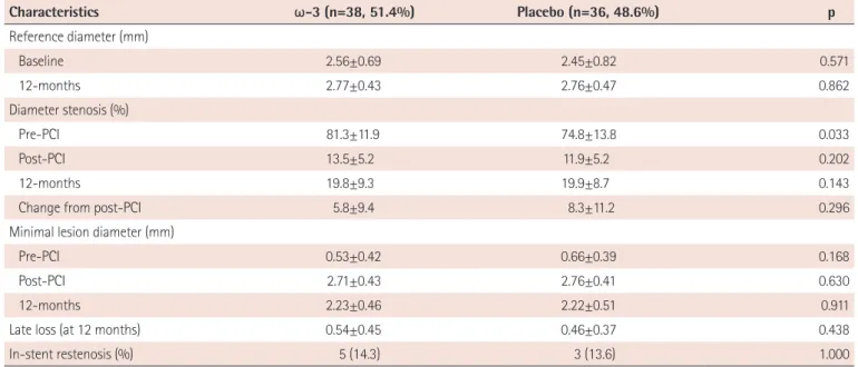
Quantitative coronary angiographic (QCA) data are presented in Table 3. Diameter stenosis at baseline was more significant in the n-3 group than in the placebo group (81.3% vs. 74.8%); however, all other post-procedural parameters were similar. No differences in late loss (0.54 mm, n-3 group; 0.46 mm, placebo group; p=0.438), or rate of in-stent restenosis (14.3%, n-3 group; 13.6%, placebo group; p>0.999), were observed between the two groups.

Intravascular ultrasound analyses at baseline and at the 12-month follow-up

The results of the IVUS analysis at the 12-month follow-up are shown in Table 4. No significant decrease in atheroma volume was observed in the n-3 group compared with the placebo group, measured in terms of percent change in atheroma volume index (-12.65% vs. -8.51%, p=0.768) and change in percent atheroma volume (-4.36% vs. -9.98%, p=0.526). Similarly, neointimal volume (neointimal volume index; 7.84 mm

3

/mm, n-3 group vs. 4.94 mm

3

/ mm, placebo group, p=0.154) did not differ between the two groups.

Changes in lipid profiles from baseline

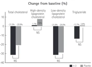
As shown in Table 5, lipid profiles at baseline were similar between the two groups. At the 12-month follow-up, the total cholesterol and LDL cholesterol levels significantly decreased in both groups (p<0.001). In addition, although HDL cholesterol levels increased significantly compared with the respective baseline values in the n-3 group (p<0.001), no significant differences in these levels were observed between the two groups. Triglyceride levels did not show a significant reduction between the two groups (Fig. 2) at the 12-month follow-up.

Total atheroma volume

=Σ(vessel-lumen area)

Atheroma area Lumen area Vessel area

Fig. 1. Intravascular ultrasound image for measurement of total atheroma

volume. A cross sectional image of stenosed vessel (white circle) is shown

in the inner picture. Total atheroma volume was calculated as a sum of the

differences between vessel and lumen area throughout the lesion by

Simpson’s method.

(4)

Discussion

The main results of this study can be summarized as follows:

addition of ω-3 PUFA to statin therapy in CAD patients requiring stent implantation did not have any effect on atheroma volume index, change in percent atheroma volume, neointimal volume index, late loss, and in-stent restenosis rates. These findings suggest that the additive benefits of ω-3 PUFA on atherosclerosis regression are questionable.

Atherosclerosis, a major cause of CAD, is characterized by a series of processes which include endothelial dysfunction, lipoprotein accumulation, leukocyte recruitment by receptors or adhesion molecules such as vascular cell adhesion molecule (VCAM) intercellular adhesion molecule (ICAM), or p-selectin, and macrophage differentiation to lipid-laden foam cells that then form the atheroma. Furthermore, smooth muscle cell proliferation

and migration, induced by pro-inflammatory cytokines such as tumor necrosis factor (TNF)-α, interleukin (IL)-1, or interferon (IFN)-γ, complicate atheroma evolution and increase the risk of CAD.

9)

Several studies

1-5)10-13)

have attempted to clarify the mechanisms of the atheroprotective effects of ω-3 PUFA. Apart from lowering the serum triglyceride level (a marker for several atherogenic lipoproteins or proinflammatory proteins promoting atherogenesis),

9-11)

ω-3 PUFA improves the endothelial function and displays antithrombogenic effects.

1)2)11)

Moreover, it reduces the levels of adhesion molecules and inflammatory mediators by the regulation of gene expression or transformation to downstream metabolites.

12)

It is believed that through these mechanisms, ω-3 PUFA increases the atherosclerotic plaque stability.

2)3)13)

This view is supported by results from a previous study using IVUS analysis by Amano et al.,

4)

in which a low serum ω-3 PUFA level was associated with the development of vulnerable plaques, suggesting a higher Table 1. Baseline clinical characteristics

Characteristics n-3 (n=38, 51.4%) Placebo (n=36, 48.6%) p

Age (years) 59.6±9.1 60.7±0.8 0.621

Male gender 24 (63.2) 26 (72.2) 0.405

BMI (kg/m

2

) 24.8±2.4 24.5±2.5 0.612

Hypertension 19 (50.0) 18 (50.0) 0.947

Diabetes mellitus 8 (21.1) 9 (23.7) 0.512

Current smoker 14 (36.8) 21 (58.3) 0.064

Dyslipidemia 25 (71.4) 14 (48.5) 0.059

Prior PCI 0 (0.0) 3 (8.3) 0.110

Clinical diagnosis

Stable AP 17 (44.7) 12 (33.3) 0.315

Unstable AP 5 (13.2) 11 (30.6) 0.069

NSTEMI 14 (36.8) 6 (16.7) 0.051

Medications at discharge

Aspirin 38 (100) 36 (100) 1.000

Clopidogrel 38 (100) 36 (100) 1.000

Cilostazol 21 (55.3) 15 (41.7) 0.242

ACEi/ARB 35 (92.1) 32 (88.9) 0.707

Beta blocker 22 (57.9) 26 (72.2) 0.230

Calcium antagonists 7 (18.4) 6 (16.7) 1.000

Nitrates 2 (5.3) 3 (8.3) 0.670

Statin

Atorvastatin 26 (68.4) 29 (80.5) 0.120

Rosuvastatin 12 (31.6) 7 (19.5) 0.211

LVEF (%) 59.3±9.9 56.8±7.2 0.493

Data are shown as mean ± standard deviation or number (%) as appropriate. BMI: body mass index, PCI: percutaneous coronary intervention, AP: angina

pectoris, NSTEMI: non-ST-segment elevation myocardial infarction, ACE: angiotensin-converting enzyme, ARB: angiotensin receptor blocker, LVEF: left ven-

tricular ejection fraction

(5)

Table 2. Baseline angiographic characteristics

Characteristics n-3 (n=38, 51.4%) Placebo (n=36, 48.6%) p

Angiographic vessel disease 0.987

1-VD 11 (28.9) 10 (27.8)

2-VD 16 (42.1) 15 (41.7)

3-VD 11 (28.9) 11 (30.6)

Culprit vessel 0.315

LAD 19 (50.0) 18 (50.0)

LCX 4 (10.5) 8 (22.2)

RCA 15 (39.5) 10 (27.8)

ACC/AHA lesion type 0.575

B1 3 (8.3) 4 (11.8)

B2 23 (63.9) 24 (70.6)

C 10 (27.8) 6 (17.6)

Stent diameter (mm) 3.0 (2.75, 3.5) 3.0 (2.75, 3.5) 0.325

Stent length (mm) 28.0 (23.0, 31.5) 28.0 (23.0, 32.3) 0.514

Stent type 0.426

BMS 0 (0.0) 1 (2.8)

ZES 20 (52.7) 16 (44.4)

EES 15 (39.4) 16 (44.5)

Others 3 (8.9) 3 (8.3)

Result of PCI 38 (100) 36 (100) .

Data are shown as number (%) or median (IQR), as appropriate. VD: vessel disease, LAD: left anterior descending artery, LCX: left circumflex artery, RCA:

right coronary artery, ACC: American College of Cardiology, AHA: American Heart Association, BMS: bare-metal stent, ZES: zotarolimus-eluting stent, EES:

everolimus-eluting stent, PCI: percutaneous coronary intervention

Table 3. Quantitative coronary angiographic analysis of the culprit lesion

Characteristics ω-3 (n=38, 51.4%) Placebo (n=36, 48.6%) p

Reference diameter (mm)

Baseline 2.56±0.69 2.45±0.82 0.571

12-months 2.77±0.43 2.76±0.47 0.862

Diameter stenosis (%)

Pre-PCI 81.3±11.9 74.8±13.8 0.033

Post-PCI 13.5±5.2 11.9±5.2 0.202

12-months 19.8±9.3 19.9±8.7 0.143

Change from post-PCI 5.8±9.4 8.3±11.2 0.296

Minimal lesion diameter (mm)

Pre-PCI 0.53±0.42 0.66±0.39 0.168

Post-PCI 2.71±0.43 2.76±0.41 0.630

12-months 2.23±0.46 2.22±0.51 0.911

Late loss (at 12 months) 0.54±0.45 0.46±0.37 0.438

In-stent restenosis (%) 5 (14.3) 3 (13.6) 1.000

Data are shown as mean ± standard deviation or number (%), as appropriate. PCI: percutaneous coronary intervention

(6)

risk of acute coronary syndrome.

4)

Other study

5)

demonstrated that low serum ω-3 PUFA was associated with atheroma progression in patients who achieved very low levels of LDL cholesterol during

statin therapy.

5)

In contrast, in this study we hypothesized that elevation of serum ω-3 PUFA by ω-3 PUFA supplementation could induce the regression of coronary atherosclerosis. Nakajima et al.

14)

Table 4. Intravascular ultrasound analysis on neointima and atheroma of the culprit lesion

Characteristics ω-3 (n=38, 51.4%) Placebo (n=36, 48.6%) p

Vessel volume index (mm

3

/mm)

Baseline (mm

3

/mm) 10.92±7.17 12.97±5.54 0.318

12-months (mm

3

/mm) 9.90±6.43 12.61±5.50 0.163

Change in volume (%) -9.36±16.24 -0.60±39.71 0.358

Lumen volume index (mm

3

/mm)

Baseline (mm

3

/mm) 5.41±2.99 5.88±2.48 0.594

12-months (mm

3

/mm) 5.57±3.19 6.20±2.88 0.518

Change in volume (%) 1.76±26.60 7.25±38.53 0.600

Atheroma volume index (mm

3

/mm)

Baseline (mm

3

/mm) 5.51±5.50 7.09±4.83 0.339

12-months (mm

3

/mm) 4.32±3.43 6.41±4.83 0.122

Change in volume (%) -12.65±30.19 -8.51±55.5 0.768

Percent atheroma volume (%)

Baseline (%) 45.24±14.70 51.13±14.69 0.209

12-months (%) 41.03±10.52 46.97±19.95 0.240

Change in volume (%) -4.36±26.77 -9.98±28.75 0.526

Neointimal volume index (mm

3

/mm) 7.84±4.90 4.94±5.23 0.087

Data are shown as mean±standard deviation

Table 5. Laboratory findings

ω-3 (n=38, 51.4%) Placebo (n=36, 48.6%) p

Total cholesterol (mg/dL)  

Baseline 203.53±42.09 183.50±47.21 0.059

12-month 139.29±28.73 145.17±25.99 0.364

p <0.001 <0.001

LDL cholesterol (mg/dL)

Baseline 127.00±29.42 113.80±35.62 0.100

12-month 86.81±51.77 80.77±21.87 0.525

p <0.001 <0.001

HDL cholesterol (mg/dL)

Baseline 45.82±12.04 41.06±13.58 0.130

12-month 46.31±12.13 44.29±12.77 0.497

p <0.001 0.202

Triglyceride cholesterol (mg/dL)

Baseline 131 (90.3, 158.8) 131 (86.5, 194.0) 0.883

12-month 113 (75.8, 150.8) 129 (94.0, 176.8) 0.367

p 0.287 0.674 0.871

Data are shown as mean±standard deviation or median (IQR), as appropriate. LDL: low-density lipoprotein, HDL: high-density lipoprotein

(7)

reported that orally administrated EPA induces the regression of atherosclerosis in LDL receptor-deficient mice, and that this effect was possibly mediated by alteration of dendritic cell functions and decrease in circulating T-lymphocytes. In our study, CAD patients received 3 g of ω-3 PUFA in addition to statin therapy; however, we did not observe regression of coronary atherosclerosis or prevention of neointimal proliferation as assessed by IVUS. Although this finding suggests that supplementation with 3g of ω-3 PUFA is not associated with regression of coronary atherosclerosis, ω-3 PUFA serum levels prior to ω-3 PUFA supplementation, were not measured in this study.

As no data on the optimal level of serum ω-3 PUFA necessary for regression of coronary atherosclerosis exists, we must consider the possibility that the 3 g ω-3 PUFA dose chosen was not adequate.

The cardiovascular protective roles of ω-3 PUFA can also be explained by mechanisms other than the above mentioned atheroprotective effects, including the suppression of cardiac arrhythmias, reduction of myocardial oxygen consumption, improvement of cardiac filling and myocardial efficiency, regulation of insulin resistance, and decrease in heart rate or blood pressure.

15–17)

Based on this information, results from meta-analyses have demonstrated a significant decrease or trend toward a decrease in mortality from CAD in patients with or without underlying cardiovascular disease.

3)18)19)

However, in contrast to earlier studies, no significant positive additive effects of ω-3 PUFA on atheroma regression were observed by IVUS analysis in the present study. Possible explanations for this discrepancy are as follows: first, medication usage, especially statins, differed between studies. As statins are known to significantly decrease the relative risk of morbidity and mortality from cardiovascular diseases,

20)

they are considered the

first-line therapy for primary or secondary prevention. However, although results from the GISSI-Prevenzione trial

21)

conducted in the mid-1990s showed benefits from statin treatment, only 5%

of the eligible patients received statin therapy, whereas no benefit was seen in two recently conducted landmark trials, i.e. the ORIGIN and the OMEGA trials,

8)9)

in which more than 50% and 80% of the patients received statin therapy, respectively. Moreover, while the effects of ω-3 PUFA on lipid profiles are mainly associated with triglycerides, a previous study showed that atheroma regression was related to LDL cholesterol levels.

22)

Considering the effects of statins, we hypothesized that adding ω-3 PUFA to statin therapy could have additive benefits. However, IVUS analysis showed that the use of ω-3 PUFA, compared to placebo, did not affect the regression of atherosclerosis, and only caused a slight decrease in triglyceride levels. Second, dietary differences in the consumption of fish containing abundant ω-3 PUFA may have an influence on serum ω-3 PUFA levels. For instance, the omega-3 index is relatively high in Korea because of a fish-rich diet.

23)

Furthermore, as the validity of a dose-response model for studying the cardiovascular effects of ω-3 PUFA remains controversial even in meta-analyses, the cut-off dose is currently undefined.

19)24)

In the present study, we hypothesized that a high dose (3 g/day) of ω-3 PUFA could have additive effects, but the possibility that patients in the placebo group had higher ω-3 PUFA baseline levels because of higher fish consumption, may have prevented induction of additive effects by the combined treatment. Third, the study populations differed between the studies. Previous trials, in which significant cardiovascular benefits of ω-3 PUFA were reported, included patients with a high risk of cardiovascular disease. Moreover, the GISSI-Prevenzione trial

21)

enrolled patients with MI within the previous 3 months, the GISSI-Heart Failure trial

25)

included patients with chronic congestive heart failure, and the ESPRIT trial

26)

included patients with traditional cardiovascular risk factors and hypertriglyceridemia>200 mg/dL. However, in this study, we excluded patients with acute MI, left main coronary artery disease, severe heart failure with LVEF<40%, uncontrolled hypertension, or diabetes. Therefore, our patients had a relatively low risk of future cardiovascular events, and the serum triglyceride level was an average of 150 mg/dL at baseline. Despite results from previous studies,

27)28)

the cardiovascular benefits of ω-3 PUFA in patients with relatively low risk of cardiovascular disease appears difficult to prove. Furthermore, one previous study reported that therapy with ω-3 PUFA did not reduce in-stent restenosis.

29)

In our study, ω-3 PUFA had no effect on late loss, in-stent restenosis, and neointimal volume index. Therefore, we consider that the anti- inflammatory effects of ω-3 PUFA are not strong enough to reduce neointimal proliferation.

10 0 -10 -20 -30 -40

NS Total cholesterol High-density lipoprotein

cholesterol

Low-density lipoprotein cholesterol

Triglyceride

NS NS

NS

-31.5% -31.6% -13.7%

n-3 Placebo

-20.9% 1.1% 7.8% -29.0% 2.8%

Change from baseline (%)

Fig. 2. Changes in lipid profiles from baseline. There were no significant

reductions in levels of total cholesterol, high density lipoprotein

cholesterol, low density lipoprotein cholesterol, and triglyceride betwen

patients with or without omega-3 polyunsaturated fatty acids.

(8)

Limitations

First, although we designed this study to investigate the benefits of adding ω-3 PUFA to statin therapy, we did not compare the differences between the statin group and the placebo group in the same patient population. Moreover, statins prescribed in this study were limited to only atorvastatin and rosuvastatin, since in actual clinical settings, these are the most commonly used statins.

Second, we did not measure ω-3 PUFA level in serum, red blood cells, or some other long-term marker of its enrichment prior to supplementation and did not control for dietary fish consumption.

Therefore, the optimal levels of ω-3 PUFA needed to induce regression of coronary atherosclerosis are unknown. Third, as the serum triglyceride level at baseline was relatively low (average of 150 mg/dL), no significant reduction in serum triglyceride level was observed after 3g of ω-3 PUFA supplementation. Therefore, the additive effect of ω-3 PUFA was not definite in our study.

Fourth, this study was single-blinded. However, this study design did not seem to affect the outcomes since all the patients in both groups completed a follow-up period, and a follow-up study was performed at the same time.

Despite these limitations, this study provides unique information, in that we used IVUS to examine the potential additive effects of ω-3 PUFA on atherosclerotic plaques in CAD patients receiving statin therapy.

Conclusions

This study suggests that adding ω-3 PUFA to statin therapy might not have additional effects on regression of coronary atherosclerosis in CAD patients requiring stent implantation.

Further, large-scale studies are warranted to investigate the role of ω-3 PUFA in this population.

Acknowledgements

This work was supported by clinical research grant from Pusan National University Hospital in 2016.

References

1. Kris-Etherton PM, Harris WS, Appel LJ; American Heart Association.

Nutrition Committee. Fish consumption, fish oil, omega-3 fatty acids, and cardiovascular disease. Circulation 2002;106:2747-57.

2. Cawood AL, Ding R, Napper FL, et al. Eicosapentaenoic acid (EPA) from highly concentrated n-3 fatty acid ethyl esters is incorporated into advanced atherosclerotic plaques and higher plaque EPA is

associated with decreased plaque inflammation and increased stability. Atherosclerosis 2010;212:252-9.

3. Mozaffarian D, Wu JH. Omega-3 fatty acids and cardiovascular disease:

effects on risk factors, molecular pathways, and clinical events. J Am Coll Cardiol 2011;58:2047-67.

4. Amano T, Matsubara T, Uetani T, et al. Impact of omega-3 polyunsaturated fatty acids on coronary plaque instability: An integrated backscatter intravascular ultrasound study. Atherosclerosis 2011;218:110-6.

5. Nozue T, Yamamoto S, Tohyama S, et al. A predictor of atheroma progression in patients achieving very low levels of low-density lipoprotein cholesterol. Am J Cardiovasc Dis 2013;3:255-63.

6. ORIGIN Trial Investigators, Bosch J, Gerstein HC. N-3 Fatty acids and cardiovascular outcomes in patients with dysglycemia. N Engl J Med 2012;367:309-18.

7. Rauch B, Schiele R, Schneider S, et al. OMEGA, a randomized, placebo-controlled trial to test the effect of highly purified omega-3 fatty acids on top of modern guideline-adjusted therapy after myocardial infarction. Circulation 2010;122:2152-9.

8. Wu JH, Mozaffarian D. ω-3 fatty acids, atherosclerosis progression and cardiovascular outcomes in recent trials: new pieces in a complex puzzle. Heart 2014;100:530-3.

9. Longo DL, Fauci AS, Kasper DL, Hauser SL, Jameson JL, Loscalzo J.

Harrison’s principles of Internal Medicine. 18th ed. New York:

McGrawHill; 2012. p.1983-91.

10. Chang CL, Seo T, Du CB, Accili D, Deckelbaum RJ. n-3 fatty acids decrease arterial low-density lipoprotein cholesterol delivery and lipoprotein lipase levels in insulin-resistant mice. Arterioscler Thromb Vasc Biol 2010;30:2510-7.

11. Talayero BG, Sacks FM. The role of triglycerides in atherosclerosis.

Curr Cardiol Rep 2011;13:544-52.

12. Calder PC. n-3 fatty acids and cardiovascular disease: evidence explained and mechanisms explored. Clin Sci (Lond) 2004;107:1-11.

13. Massaro M, Scoditti E, Carluccio MA, De Caterina R. Nutraceuticals and prevention of atherosclerosis: focus on omega-3 polyunsaturated fatty acids and Mediterranean diet polyphenols. Cardiovasc Ther 2010;28:e13-9.

14. Nakajima K, Yamashita T, Kita T, et al. Orally administered eicosapentaenoic acid induces rapid regression of atherosclerosis via modulating the phenotype of dendritic cells in LDL receptor-deficient mice. Arterioscler Thromb Vasc Biol 2011;31:1963-72.

15. O’Keefe JH Jr, Abuissa H, Sastre A, Steinhaus DM, Harris WS. Effects of omega-3 fatty acids on resting heart rate, heart rate recovery after exercise, and heart rate variability in men with healed myocardial infarctions and depressed ejection fractions. Am J Cardiol 2006;97:1127-30.

16. Charnock JS, McLennan PL, Abeywardena MY. Dietary modulation of

lipid metabolism and mechanical performance of the heart. Mol Cell

Biochem 1992;116:19-25.

(9)

17. Djoussé L, Gaziano JM, Buring JE, Lee IM. Dietary omega-3 fatty acids and fish consumption and risk of type 2 diabetes. Am J Clin Nutr 2011;93:143-50.

18. León H, Shibata MC, Sivakumaran S, Dorgan M, Chatterley T, Tsuyuki RT. Effect of fish oil on arrhythmias and mortality: systematic review.

BMJ 2008;337:a2931.

19. Marik PE, Varon J. Omega-3 dietary supplements and the risk of cardiovascular events: a systematic review. Clin Cardiol 2009;32:365-72.

20. Baigent C, Keech A, Kearney PM, et al. Efficacy and safety of cholesterol-lowering treatment: prospective meta-analysis of data from 90,056 participants in 14 randomised trials of statins. Lancet 2005; 366:1267-78.

21. Dietary supplementation with n-3 polyunsaturated fatty acids and vitamin E after myocardial infarction: results of the GISSI- Prevenzione trial. Gruppo Italiano per lo Studio della Sopravvivenza nell’Infarto miocardico. Lancet 1999;354:447-55.

22. Nissen SE, Nicholls SJ, Sipahi I, et al. Effect of very high-intensity statin therapy on regression of coronary atherosclerosis: the ASTEROID trial. JAMA 2006;295:1556-65.

23. Lee E, Lee S, Park Y. n-3 polyunsaturated fatty acids and trans fatty acids in patients with the metabolic syndrome: a case-control study

in Korea. Br J Nutr 2008;100:609-14.

24. Casula M, Soranna D, Catapano AL, Corrao G. Long-term effect of high dose omega-3 fatty acid supplementation for secondary prevention of cardiovascular outcomes: a meta-analysis of randomized, placebo controlled trials. Atherosclerosis Suppl 2013;14:243-51.

25. GISSI-HF investigators, Tavazzi L, Maggioni AP, et al. Effect of n-3 polyunsaturated fatty acids in patients with chronic heart failure (the GISSI-HF trial): a randomised, double-blind, placebo-controlled trial.

Lancet 2008;372:1223-30.

26. Maki KC, Orloff DG, Nicholls SJ, et al. A highly bioavailable omega-3 free fatty acid formulation improves the cardiovascular risk profile in high-risk, statin-treated patients with residual hypertriglyceridemia (the ESPRIT trial). Clin Ther 2013;35:1400-11.e1-3.

27. Burr ML, Ashfield-Watt PA, Dunstan FD, et al. Lack of benefit of dietary advice to men with angina: results of a controlled trial. Eur J Clin Nutr 2003;57:193-200.

28. Kromhout D, Giltay EJ, Geleijnse JM; Alpha Omega Trial Group. n-3 fatty acids and cardiovascular events after myocardial infarction. N Engl J Med 2010;363:2015-26.

29. Arnesen H. n-3 fatty acids and revascularization procedures. Lipids

2001;36 Suppl:S103-6.

수치

Fig. 1. Intravascular ultrasound image for measurement of total atheroma  volume. A cross sectional image of stenosed vessel (white circle) is shown  in the inner picture
Table 3. Quantitative coronary angiographic analysis of the culprit lesion
Table 5. Laboratory findings
Fig. 2. Changes in lipid profiles from baseline. There were no significant  reductions in levels of total cholesterol, high density lipoprotein  cholesterol, low density lipoprotein cholesterol, and triglyceride betwen  patients with or without omega-3 pol

참조

관련 문서