Original Article pISSN: 1011-8942 eISSN: 2092-9382
Korean J Ophthalmol 2013;27(4):235-242 http://dx.doi.org/10.3341/kjo.2013.27.4.235
© 2013 The Korean Ophthalmological Society
This is an Open Access article distributed under the terms of the Creative Commons Attribution Non-Commercial License (http://creativecommons.org/licenses /by-nc/3.0/) which permits unrestricted non-commercial use, distribution, and reproduction in any medium, provided the original work is properly cited.
The Effect of Bevacizumab versus Ranibizumab in the Treatment of Corneal Neovascularization: A Preliminary Study
Jin-Hyoung Kim, Hae-Won Seo, Hyun-Cheol Han, Jong-Hyun Lee, Suk-Kyue Choi, Doh Lee
Department of Ophthalmology, Ilsan Paik Hospital, Inje University College of Medicine, Goyang, Korea
Vascular endothelial growth factor (VEGF) plays a key role in vasculogenesis and the pathologic neovascular- ization (NV) associated with eye disease [1-4]. Although anti-VEGF therapy for ocular disease has been principally directed at retinal vascular conditions, it is widely accepted that anti-VEGF therapy is also effective when used to treat corneal NV, with results from animal and human studies supporting this suggestion [5-11]. Targeted corneal NV has been induced in animals by chemical burning, suturing,
application of a VEGF/bFBF pellet, or limbal injury, and anti-VEGF therapy has been used to treat human eyes with corneal NV associated with chemical burns, graft-versus- host disease, herpetic keratitis, limbal damage, ocular cica- tricial pemphigoid, use of penetrating keratoplasty, and Ste- vens-Johnson syndrome. Various reports show that the use of anti-VEGF therapy is not restricted by disease etiology.
Currently, popular anti-VEGF agents include bevaci- zumab (Avastin; Genentech Inc., San Francisco, CA, USA;
Roche, Basel, Switzerland) and ranibizumab (Lucentis;
Genentech Inc.). These materials are humanized monoclo- nal antibodies directed toward VEGF. No Food and Drug Administration (FDA)-approved drug is yet available for treatment of corneal NV. Most prior studies have investi- gated the effects of bevacizumab rather than ranibizumab.
However, it is notable that ranibizumab is the only agent Purpose: To compare the short term effects of bevacizumab and ranibizumab injections on the regression of
corneal neovascularization (NV).
Methods: Sixteen eyes of 16 patients with corneal NV were randomly assigned for an injection with 2.5 mg of bevacizumab (group 1, n = 8) or 1 mg of ranibizumab (group 2, n = 8) through subconjunctival and intrastromal routes. The patients were prospectively followed-up for one month after the injections. Corneal NV areas, as shown on corneal slit-lamp photographs stored in JPEG format, were calculated using Image J software before the injection, one week after the injection, and one month after the injection. The corneal NV areas were compared before and after the injections.
Results: Seven women and nine men, with an average age of 51 years, presented with corneal NV secondary to herpetic keratitis (7 cases), graft rejection (6), chemical burn (1), pemphigoid (1), and recurrent ulcer (1). In group I, the preoperative corneal NV area (8.75 ± 4.33%) was significantly decreased to 5.62 ± 3.86% one week after the injection and to 6.35 ± 3.02% one month after the injection (p = 0.012, 0.012, respectively). The corneal NV area in group 2 also exhibited a significant change, from 7.37 ± 4.33% to 6.72 ± 4.16% one week after the injection (p = 0.012). However, no significant change was observed one month after the injection. The mean decrease in corneal NV area one month after injection in group 1 (28.4 ± 9.01%) was significantly higher than in group 2 (4.51 ± 11.64%, p = 0.001).
Conclusions: Bevacizumab injection resulted in a more effective and stable regression of corneal NV compared to the ranibizumab injection. The potency and dose of these two drugs for the regression of corneal NV require further investigation.
Key Words: Bevacizumab, Corneal neovascularization, Ranibizumab
Received: June 2, 2012 Accepted: September 4, 2012
Corresponding Author: Doh Lee, MD, PhD. Department of Ophthal- mology, Ilsan Paik Hospital, Inje University College of Medicine, #170 Juhwa-ro, Ilsanseo-gu, Goyang 411-706, Korea. Tel: 82-31-910-7240, Fax:
82-31-911-7241, E-mail: [email protected]
approved by the FDA for intraocular use even though it is costly, especially considering that anti-VEGF therapy may need to be repeated [12,13]. Therefore, comparative work using both bevacizumab and ranibizumab therapy is neces- sary.
In the present study, we evaluated the short term effects of bevacizumab and ranibizumab on the regression of cor- neal NV and compared these effects in patients with corne- al NV caused by various ocular surface disorders.
Materials and Methods
Sixteen eyes of 16 patients with chronic corneal NV greater than six months in duration were enrolled in this prospective study. The eyes were randomly divided into a group of eight eyes of eight patients treated by bevaci- zumab injection (group 1) and eight eyes of eight patients receiving ranibizumab injection (group 2). The patients included in this study had no other ocular diseases and no history of prior treatment for corneal NV within three months prior to the study. Written informed consent was obtained from all the patients prior to study enrollment.
The work was approved by the institutional review board of Inje University and was conducted in accordance with the Declaration of Helsinki.
Under topical anesthesia, subconjunctival and intrastro- mal injections, delivered adjacent to the areas of the corne- al NV, were administered to both groups. Group 1 patients received 0.1 mL (2.5 mg equivalent to the intravitreal dose) of bevacizumab (Avastin), while 0.1 mL (1 mg being the same dose approved for intravitreal usage) of ranibizumab (Lucentis) was administered to the patients in group 2. No other medication, except for topical antibiotics, was given over the next three days.
The means and standard deviations of uncorrected visual acuity (UCVA) and best-corrected visual acuity (BCVA)
in logarithm of minimum angle resolution (logMAR) were measured before and after the injection in all patients.
Preoperative corneal photographs of each eye were taken at 1 : 1.8 magnification using a digital camera (×4.0 zoom;
E4500, Nikon, Tokyo, Japan) under slit-lamp examination at ×10 magnification and were stored in JPEG format (4.0 megapixels/image). The corneal NV area (as a percentage of the entire corneal area) was calculated using Image J software (Wayne Rasband of the Research Services Branch, National Institute of Mental Health, Bethesda, MD, USA).
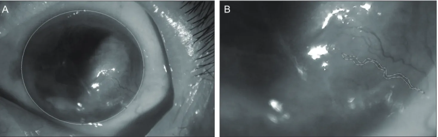
For objectivity, a double-blind method using two observers was used to measure the areas and verify the interobserver agreement. One observer measured the area three times and verified the intraobserver agreement. To calculate the increase or decrease in NV area, we measured the entire cornea area from a picture taken under the same digital camera settings and slit lamp magnification. Under the con- dition of higher slit lamp magnification, we measured the NV area by tracing the boundaries of the vessels including all visible branches (Fig. 1). The length of corneal NV was considered to be the distance from the limbal vascular plexus to the leading point of the new vessel. The width of corneal NV was considered to be the diameter including sprouting vessels around the suture. We then calculated the ratio between the entire cornea and NV area using slit lamp magnification. We converted the area of the entire cornea and NV into pixel counts using Image J software. The menu option ‘Analyze→Measure’ was selected, the values were recorded, and the process was repeated on subsequent images. The ‘Set Scale’ function in Image J was used to convert pixel numbers to standard units [14].
The corneal NV areas one week and one month after injection were estimated using the same method and were compared to the preoperative NV areas. Each decrease in NV area from baseline was calculated as a percentage.
Routine clinical examination was performed to detect
A B
Fig. 1. The measurement of corneal neovascularization (NV) area using the Image J program. We measured the area of the entire cornea (A) and the area of the new corneal vessels with its branches (B). The ratio of NV area was then calculated as a percentage of the entire
JH Kim, et al. Effect of Bevacizumab vs. Ranibizumab on Corneal NV
any side-effects occurring after the procedure. Statisti- cal analysis was performed using SPSS version 19.0 for Windows (SPSS Inc., Chicago, IL, USA). Comparisons between groups 1 and 2 were achieved with the use of the Mann-Whitney U-test. The change of corneal NV area with time in each group was analyzed using the Friedman test and the Wilcoxon signed-rank test. A p-value ≤0.05 was considered statistically significant.
Results
The patients included seven women and nine men, and each group was composed of eight patients that were ran- domly assigned. The average patient age was 51.06 ± 15.55 years (mean ± standard deviation; range, 21 to 82 years), and there was no age difference between groups 1 and 2 (p
= 0.328; Mann-Whitney U-test). Seven eyes had corneal NV secondary to herpetic keratitis, six eyes had graft rejec- tion, and one eye each experienced chemical burn, pemphi- goid, and recurrent sterile ulcers from secondary Sjogren’s syndrome (Table 1). The visual acuity in logMAR was measured in both groups before and after the injections.
The UCVA and BCVA were 0.79 ± 0.51, 0.64 ± 0.40 in group 1 and 0.81 ± 0.76, 0.78 ± 0.79 in group 2. There was no difference in UCVA or BCVA between groups 1 and 2 (p = 0.721, p = 0.878; Mann-Whitney U-test). The mean change of visual acuity did not show an improvement in either group after the injections (p > 0.05 in all tests; Wil- coxon signed-rank test) (Table 2).
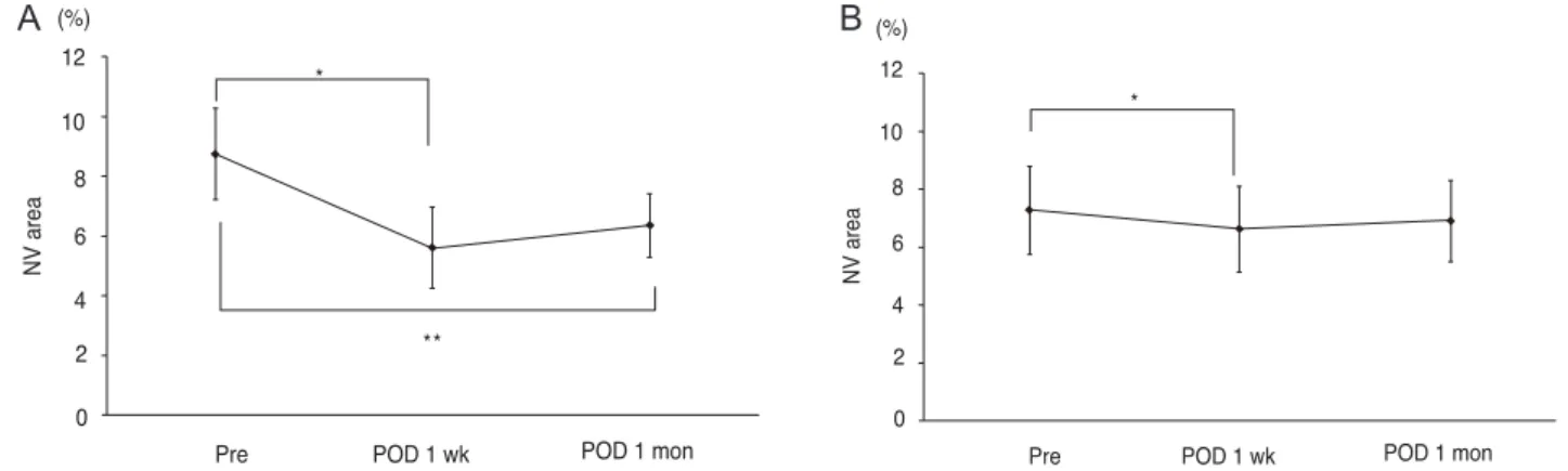
The areas of corneal NV did not differ between the two groups before the injections (p = 0.505; Mann-Whitney U-test). In both groups 1 and 2, significant differences were seen in the average areas of corneal NV with time (p = 0.002, p = 0.01, respectively; Friedman’s test). However, the group responses differed in detail. In group 1, the pre- operative corneal NV area (8.75 ± 4.33%, mean ± standard deviation) decreased significantly to 5.62 ± 3.86% one week after the injection and to 6.35 ± 3.02% one month after the injection (p = 0.012, p = 0.012, respectively; Wil- coxon signed-rank test) (Fig. 2A). The corneal NV area of group 2 patients also exhibited a significant change from 7.37 ± 4.33% preoperatively to 6.72 ± 4.16% one week after the injection (p = 0.012; Wilcoxon signed-rank test).
However, no significant change was evident one month af- ter the injection (7.02 ± 3.99%, p = 0.327, Wilcoxon signed- rank test) compared to the pre-injection area (Fig. 2B). The corneal NV area in group 1 decreased by 36.97 ± 27.34%
(mean ± standard deviation) one week after the injection and by 28.4 ± 9.01% after one month. In group 2, decreases of 10.40 ± 4.83% and 4.51 ± 11.64% were apparent one week and one month after the injection, respectively. The average decreases were significantly greater in group 1 than in the group 2 patients at one week and one month after the injection (p = 0.003, p = 0.001, respectively; Mann-Whit-
ney test) (Fig. 3).
After providing informed consent, five patients in group 2 (numbers 9, 11, 12, 13, and 16) (Table 1) received bevaci- zumab injections two months after completion of the pres- ent study because they had experienced no notable regres- sion of corneal NV, while one patient’s condition worsened (patient 12) despite the ranibizumab injections one month after initial therapy. All the patients exhibited good respons- es to the bevacizumab injection. Fig. 4 shows data from pa- tient 12 and highlights the effect of bevacizumab injection on the regression of corneal NV. Two patients with limbal insufficiency (patients 11 and 13) showed persistent epi- thelial defects with corneal melting after the bevacizumab
Table 1. Baseline characteristics of patients receiving a beva- cizumab or ranibizumab injection for corneal neovasculariza- tion
Group Patient Age Sex Etiology of corneal neovascularization
1 1 43 F Lipid keratopathy
2 44 M Rejection after limbal transplantation
3 82 M Pemphigoid
4 60 F Herpetic keratitis 5 70 M Herpetic keratitis
6 60 M Rejection after lamellar keratoplasty : Interface neovascularization
2 7 39 M Herpetic keratitis
8 49 F Sjogren syndrome 9 72 F Herpetic keratitis
10 62 F Rejection after lamellar keratoplasty : interface neovascularization 11 40 F Rejection after limbal ransplantation
due to chemical burn 12 21 F Lipid keratopathy
13 45 M Limbal insufficiency due to chemical burn
14 50 M Lipid keratopathy 15 43 M Herpetic keratitis 16 37 M Herpetic keratitis
Table 2. The visual acuity of patients receiving a bevacizum- ab or ranibizumab injection for corneal neovascularization
Group 1 Group 2 Before Injection UCVA 0.79 ± 0.51 0.81 ± 0.76
BCVA 0.64 ± 0.40 0.78 ± 0.79 One week after UCVA 0.73 ± 0.36 0.76 ± 0.79 injection BCVA 0.59 ± 0.35 0.75 ± 0.80 One month after UCVA 0.60 ± 0.45 0.80 ± 0.77 injection BCVA 0.50 ± 0.38 0.76 ± 0.81 Mean ± standard deviation of visual acuity in logarithm of mini- mum angle resolution.
UCVA = uncorrected visual acuity; BCVA = best-corrected visu- al acuity.
Korean J Ophthalmol Vol.27, No.4, 2013
injection even though they had a remarkable regression of corneal NV. No other complications were seen.
Discussion
The present study showed that subconjunctival and in- trastromal injections of either bevacizumab or ranibizumab were effective in achieving regression of corneal NV short-
bevacizumab injections, exhibited greater regression (a 36.97% decrease) one week after injection. This improve- ment was relatively long-lasting, up to one month, despite no repeat injections, considering the recommendation from the manufacturer that additional intraocular injections should be given at one-month intervals for the treatment of retinal disease. In contrast, the ranibizumab injections that were given to patients in group 2 were less effective and shorter acting. The decrease in the extent of corneal NV one week after the injection was significantly lower (10.40%) than in group 1 and was not maintained up to one month, at which time the extent of corneal NV did not dif- fer from that at baseline.
Both bevacizumab and ranibizumab were constructed from full-length murine anti-VEGF IgG A.4.6.1, sharing many essential properties with anti-VEGF agents [15,16].
However, differences in clinical efficacy can arise from variations in molecular size, drug development in different cell lines, drug affinity for VEGF, and drug formulation [12,13,15]. Bevacizumab is a 149 kDa full-length IgG1 antibody composed of two 214-residue light chains and two 453-residue heavy chains. Ranibizumab is a 48 kDa Fab from the expression plasmid pY0317 [17]. Ranibizum- ab and bevacizumab are both able to bind to all human VEGF-A isoforms. On a molar basis, ranibizumab was de- termined to be five- to 20-fold more potent than full-length bevacizumab with respect to binding to VEGF-A [15,18].
Affinity-matured ranibizumab may provide better VEGF inhibition compared with bevacizumab through stronger molecular binding. On the other hand, the half-life of bev- acizumab, estimated to be approximately 20 days, is longer than ranibizumab, estimated to be approximately six hours.
Therefore, bevacizumab may remain in the eye for a longer period of time as a result of its larger molecular size [19].
Decrement
Group 1 (bevacizumab) Group 2 (ranibizumab) 40
30
20
10
0
POD 1 wk POD 1 mon
(%)50
*
*
Fig. 3. A histogram showing a significant difference in the decrease in corneal neovascularization between group 1 (bev- acizumab) and group 2 (ranibizumab) patients after treatment.
postoperative day (POD) 1 week, one week after injection; POD 1 month, one month after injection. *p = 0.003 in POD 1 week, *p = 0.001 in POD 1 month; Mann-Whitney test. *A p-value <0.05 was considered statistically significant.
NV area
Pre
**
10 8 6 4 2 0
POD 1 wk POD 1 mon
Pre POD 1 wk POD 1 mon
NV area
12 *
10 8 6 4 2 0 (%)
NV area
Pre
12 *
**
10 8 6 4 2 0
POD 1 wk POD 1 mon
Pre POD 1 wk POD 1 mon
NV area
12 10 *
8 6 4 2 0 (%)
(%)
A B
Fig. 2. Areas of corneal neovascularization (NV; mean ± standard error, % value) in group 1 patients who received bevacizumab injections (A) and group 2 patients who received ranibizumab injections (B) before and after treatment. Pre, before injection; post- operative day (POD) 1 week, one week after injection; POD 1 month, one month after injection. (A) Group 1, *p = 0.012, **p = 0.012 by the Wilcoxon signed-rank test. (B) Group 2, *p = 0.012 by the Wilcoxon signed-rank test. A p-value <0.05 was considered statistically significant.
JH Kim, et al. Effect of Bevacizumab vs. Ranibizumab on Corneal NV
thelium (RPE)-choroid organ culture and RPE cell culture or human endothelial cell culture compared the in vitro effects of these two drugs [20,21]. It has been demonstrated that, at clinical doses, ranibizumab and bevacizumab are equally potent in neutralizing VEGF. In addition, a ran- domized clinical study showed no difference in the efficacy
of ranibizumab versus that of bevacizumab. Therefore, the potency of both drugs was comparable in regressing cho- roidal neovascularization in pathologic myopia (CNVM) [22]. Since there has been no comparative study about the potency of the two drugs for the regression of corneal NV, this study used the same dose for the intravitreal injections.
NV area
Pre-ranibizumab injection (A) 7
6 5 4 3 2 1
0 POD 1 wk POD 1 mon (B) Pre-bevacizumab
injection (C) POD 1 wk POD 1 mon (D)
(%) Fig. 4. Slit-lamp photographs of the cornea of patient 12, who
failed to show regression of corneal neovascularization (NV) after ranibizumab injection but demonstrated measurable regression after bevacizumab injection. The associated graph and photo- graphs show changes in the area of corneal neovascularization (%
values) after each treatment. Pre-ranibizumab injection (A): before ranibizumab injection ([A] shows the slit-lamp image obtained at that time). Postoperative day (POD) 1 week: one week after ranibi- zumab injection. POD 1 month (B): one month after ranibizumab injection ([B] shows the slit-lamp image obtained at that time).
Pre-bevacizumab injection (C): before bevacizumab injection, thus three months after the prior ranibizumab injection ([C] shows the aggravation of corneal NV in the slit-lamp image obtained at that time). POD 1 week: one week after bevacizumab injection. POD 1 month (D): one month after bevacizumab injection ([D] shows the remarkable regression of corneal NV in the slit-lamp image obtained at that time). (E) The graph shows changes in the area of corneal neovascularization (% values) after each treatment.
A B
C D
E
However, from this study, the potency of the two drugs does not seem to have the same effect in regressing corneal NV compared to CNVM.
The routes of injection may be a reason for this contrary result. Avisar et al. [23] compared the effect of bevacizum- ab in terms of the injection route and found that intracam- eral and intravitreal routes of injection were more effective than the subconjunctival route, but that the subconjunctival injection was associated with the earliest response peak.
In the vitreal cavity, the vitreous body is in contact with many retinal and choroidal vessels. However, the cavity is a closed space and may trap an agent prior to its leakage out of the eye into circulation. Therefore, the need for diffusion through the ciliary body epithelium means that the region of corneal NV is not immediately accessed. The subcon- junctival application of an anti-VEGF agent affords good proximity to any region of corneal NV and to a character- istic space containing abundant subconjunctival vessels, lymphatics, and a great deal of areolar tissue. As a result, clearance from the subconjunctival space may occur much faster than after injection into the intravitreal cavity. As a result, it is possible that a higher dose is required when ad- ministering through the subconjunctival space compared to an intravitreal injection. This is especially important if the drug is of low molecular weight or has a short half-life, like ranibizumab.
In addition, the shorter-acting effect of ranibizumab in the present study may be explained by the difference in half-life. The half-life of bevacizumab following intravitre- al injection in an animal model is 4.32 days, while the half- life of ranibizumab is 2.88 days [24,25].
Two previous studies using ranibizumab with different results suggest that the injection dose is a main factor in achieving a good result [11,26]. Mansour [11] described one patient exhibiting a prompt regression of conjunctival microvessels in the pterygial bed after a single subconjunc- tival injection of ranibizumab (1 mg) and no recurrence even after 13 months. However, Mandalos et al. [26] found that subconjunctival ranibizumab at a single dose of 0.3 mg had no effect on the vascularization extent of the primary pterygium. This suggests that the use of an insufficient dose resulted in a failure of vascularization regression even though no comparative study regarding the dose-related effect of ranibizumab has been performed. Ahmed et al.
[7] reported a dose-dependent effect in a study where two groups were treated with 5 or 10 mg of subconjunctival bevacizumab in rabbits. You et al. [27] compared the ef- fects of 1.25 mg/0.05 mL, 2.5 mg/0.1 mL, and 5.0 mg/0.2 mL of bevacizumab in patients with corneal NV. The effi- cacy of corneal NV regression correlated with the injection dose, and there were no significant changes in the areas of the eyes injected with 1.25 mg bevacizumab. Although the suggested dose used by You et al. [27] was not always
gest that a higher injection dose of ranibizumab needs to be considered to acquire a satisfactory result [12,16,28]. In addition, the effect of the anti-VEGF agents was dependent on time of treatment after the onset of NV. Papathanassiou et al. [5] concluded that early subconjunctival bevacizumab administration (one day after injury) inhibited corneal NV more effectively in a rabbit experimental model of limbal insufficiency than when treatment was performed on day 14. Recently, Lin et al. [29] demonstrated that the earlier the time of treatment with subconjunctival bevacizumab, the better the result in rabbit eyes with limbal insufficiency.
In the cited study, treatment was performed immediately (early group), at one week (mid-group), and one month after injury (late group). Early and mid-point bevacizumab injections inhibited epithelial alteration, the development of stromal opacity, and corneal NV manifestation as limbal insufficiency, while late treatment had no inhibitory effect.
The cited authors suggested that vessels may mature in chronic NV, and pericytes may be recruited to the region around the area of pathologic NV [30]. Such coverage may decrease the effect of bevacizumab on the regression of newly formed immature vessels. However, although we assessed eyes that had chronic corneal NV for longer than six months in duration, we found measurable regression.
Nevertheless, the mechanism suggested by the cited authors may explain why no complete resolution of corneal NV has been reported to date.
Five eyes refractory to ranibizumab therapy showed remarkable reductions in the extent of corneal NV after subsequent bevacizumab injections. The results shown in Fig. 4 indicate that the different effects are not due to the different nature of corneal NV in each case or to the unique characteristics of each patient. Therefore, in cases refrac- tory to ranibizumab, altering the drug to bevacizumab or increasing the dose of ranibizumab is a good alternative.
From the viewpoint of safety, as seen in the present study, complications of ranibizumab injections have been rare. Galor et al. [31] found that subconjunctival injection of ranibizumab in conjunction with pterygium surgery was well tolerated. However, use of topical bevacizumab may negatively affect the epithelium and delay wound healing [32,33]. Clinically, the potential side effects of topical bev- acizumab use including epitheliopathy, the development of epithelial defects, and stromal melting have been reported [9,34]. In the present study, subconjunctival bevacizumab injections resulted in the development of persistent epi- thelial defects with corneal melting in two patients within two weeks. The common feature of the four patients who suffered devastating side effects, in this and previous re- ports, was limbal deficiency induced by a chemical burn [9,34]. Therefore, the application of bevacizumab should be carefully considered, especially in patients with chemical burn-induced diffuse limbal deficiency, because such treat-
JH Kim, et al. Effect of Bevacizumab vs. Ranibizumab on Corneal NV
deficiencies.
The limitation of this work is that only a small number of patients were enrolled. Therefore, the effect of preoc- cupying disease entities on the response to the different anti-VEGF drugs have yet to be verified even though five eyes in this study, which were refractory to ranibizumab but showed good response to bevacizumab, did not have com- mon preoccupying pathology. Our results must therefore be regarded as preliminary. Additional larger studies including the evaluation of factors affecting the different responses of anti-VEGF drugs such as the preoccupying disease entity or onset of new vessels should be performed. Our compar- ative data indicates that subconjunctival and intrastromal bevacizumab injections result in a more effective and stable regression of corneal NV compared to ranibizumab. This implies that further research about the potency of the two drugs in regressing corneal NV is needed, and that the dos- age adjustment for each drug is important in order to obtain good clinical results.
Conflict of Interest
No potential conflict of interest relevant to this article was reported.
Acknowledgements
This study was supported by an Inje University research grant.
References
1. Chang JH, Gabison EE, Kato T, Azar DT. Corneal neovas- cularization. Curr Opin Ophthalmol 2001;12:242-9.
2. Philipp W, Speicher L, Humpel C. Expression of vascular endothelial growth factor and its receptors in inflamed and vascularized human corneas. Invest Ophthalmol Vis Sci 2000;41:2514-22.
3. Cursiefen C, Rummelt C, Kuchle M. Immunohistochemical localization of vascular endothelial growth factor, trans- forming growth factor alpha, and transforming growth fac- tor beta1 in human corneas with neovascularization. Cornea 2000;19:526-33.
4. Chen WL, Lin CT, Lin NT, et al. Subconjunctival injection of bevacizumab (avastin) on corneal neovascularization in different rabbit models of corneal angiogenesis. Invest Oph- thalmol Vis Sci 2009;50:1659-65.
5. Papathanassiou M, Theodossiadis PG, Liarakos VS, et al.
Inhibition of corneal neovascularization by subconjunc- tival bevacizumab in an animal model. Am J Ophthalmol 2008;145:424-31.
6. Yoeruek E, Ziemssen F, Henke-Fahle S, et al. Safety, pen- etration and efficacy of topically applied bevacizumab:
evaluation of eyedrops in corneal neovascularization after chemical burn. Acta Ophthalmol 2008;86:322-8.
7. Ahmed A, Berati H, Nalan A, Aylin S. Effect of bevaci- zumab on corneal neovascularization in experimental rabbit model. Clin Experiment Ophthalmol 2009;37:730-6.
8. Carrasco MA. Subconjunctival bevacizumab for corneal neovascularization in herpetic stromal keratitis. Cornea 2008;27:743-5.
9. Kim SW, Ha BJ, Kim EK, et al. The effect of topical bev- acizumab on corneal neovascularization. Ophthalmology 2008;115:e33-8.
10. Awadein A. Subconjunctival bevacizumab for vascularized re- jected corneal grafts. J Cataract Refract Surg 2007;33:1991-3.
11. Mansour AM. Treatment of inflamed pterygia or residual pterygial bed. Br J Ophthalmol 2009;93:864-5.
12. Steinbrook R. The price of sight: ranibizumab, bevacizum- ab, and the treatment of macular degeneration. N Engl J Med 2006;355:1409-12.
13. Rosenfeld PJ. Intravitreal avastin: the low cost alternative to lucentis? Am J Ophthalmol 2006;142:141-3.
14. Tang XN, Berman AE, Swanson RA, Yenari MA. Digitally quantifying cerebral hemorrhage using Photoshop and Im- age J. J Neurosci Methods 2010;190:240-3.
15. Ferrara N, Damico L, Shams N, et al. Development of ran- ibizumab, an anti-vascular endothelial growth factor antigen binding fragment, as therapy for neovascular age-related macular degeneration. Retina 2006;26:859-70.
16. Ferrara N, Hillan KJ, Gerber HP, Novotny W. Discovery and development of bevacizumab, an anti-VEGF antibody for treating cancer. Nat Rev Drug Discov 2004;3:391-400.
17. Presta LG, Chen H, O’Connor SJ, et al. Humanization of an anti-vascular endothelial growth factor monoclonal antibody for the therapy of solid tumors and other disorders. Cancer Res 1997;57:4593-9.
18. Chen Y, Wiesmann C, Fuh G, et al. Selection and analysis of an optimized anti-VEGF antibody: crystal structure of an affinity-matured Fab in complex with antigen. J Mol Biol 1999;293:865-81.
19. Stewart MW. Aflibercept (VEGF Trap-eye): the newest an- ti-VEGF drug. Br J Ophthalmol 2012;96:1157-8.
20. Klettner A, Roider J. Comparison of bevacizumab, ranibi- zumab, and pegaptanib in vitro: efficiency and possible addi- tional pathways. Invest Ophthalmol Vis Sci 2008;49:4523-7.
21. Carneiro A, Falcao M, Pirraco A, et al. Comparative effects of bevacizumab, ranibizumab and pegaptanib at intravitreal dose range on endothelial cells. Exp Eye Res 2009;88:522-7.
22. Gharbiya M, Giustolisi R, Allievi F, et al. Choroidal neovas- cularization in pathologic myopia: intravitreal ranibizumab versus bevacizumab: a randomized controlled trial. Am J Ophthalmol 2010;149:458-64.e1.
23. Avisar I, Weinberger D, Kremer I. Effect of subconjunctival and intraocular bevacizumab injections on corneal neovas- cularization in a mouse model. Curr Eye Res 2010;35:108- 24. Mordenti J, Cuthbertson RA, Ferrara N, et al. Comparisons 15.
of the intraocular tissue distribution, pharmacokinetics, and safety of 125I-labeled full-length and Fab antibodies in rhe- sus monkeys following intravitreal administration. Toxicol Pathol 1999;27:536-44.
25. Gaudreault J, Fei D, Rusit J, et al. Preclinical pharmacoki- netics of Ranibizumab (rhuFabV2) after a single intravitreal administration. Invest Ophthalmol Vis Sci 2005;46:726-33.
26. Mandalos A, Tsakpinis D, Karayannopoulou G, et al. The effect of subconjunctival ranibizumab on primary pterygi- um: a pilot study. Cornea 2010;29:1373-9.
27. You IC, Kang IS, Lee SH, Yoon KC. Therapeutic effect of sub- conjunctival injection of bevacizumab in the treatment of cor- neal neovascularization. Acta Ophthalmol 2009;87:653-8.
28. Bahar I, Kaiserman I, McAllum P, et al. Subconjunctival bevacizumab injection for corneal neovascularization in re-
current pterygium. Curr Eye Res 2008;33:23-8.
29. Lin CT, Hu FR, Kuo KT, et al. The different effects of early and late bevacizumab (Avastin) injection on inhib- iting corneal neovascularization and conjunctivalization in rabbit limbal insufficiency. Invest Ophthalmol Vis Sci 2010;51:6277-85.
30. Cursiefen C, Hofmann-Rummelt C, Kuchle M, Schlot- zer-Schrehardt U. Pericyte recruitment in human corneal an- giogenesis: an ultrastructural study with clinicopathological correlation. Br J Ophthalmol 2003;87:101-6.
31. Galor A, Yoo SH, Piccoli FV, et al. Phase I study of sub- conjunctival ranibizumab in patients with primary pte-
rygium undergoing pterygium surgery. Am J Ophthalmol 2010;149:926-31.e2.
32. Kim TI, Chung JL, Hong JP, et al. Bevacizumab application delays epithelial healing in rabbit cornea. Invest Ophthalmol Vis Sci 2009;50:4653-9.
33. Kim EC, Lee WS, Kim MS. The inhibitory effects of beva- cizumab eye drops on NGF expression and corneal wound healing in rats. Invest Ophthalmol Vis Sci 2010;51:4569-73.
34. Galor A, Yoo SH. Corneal melt while using topical bevaci- zumab eye drops. Ophthalmic Surg Lasers Imaging 2010 Mar 9 [Epub]. DOI: 10.3928/15428877-20100215-07.