WWW.KJOG.ORG 395
PORT-SITE METASTASIS OF ADVANCED PRIMARY
PERITONEAL CANCER AFTER LAPAROSCOPIC PERITONEAL BIOPSY: A CASE REPORT
Jin-Young Park, MD, Jung-Joo An, MD, Tae-Joong Kim, MD, Byoung-Gie Kim, MD, PhD, Duk-Soo Bae, MD, PhD
Department of Obstetrics and Gynecology, Samsung Medical Center, Sungkyunkwan University School of Medicine, Seoul, Korea
Due to the advancement in laparoscopic surgery in gynecology, laparoscopic surgery of patients with gynecologic malignancy is widely used. A 69-year-old woman who had elevated serum level of CA-125 and malignant ascites was transferred from general surgery after laparoscopic peritoneal biopsy. She complained about 2.5 cm sized abdominal wall mass which developed only 1week after surgery. With the impression of ovarian cancer with port-site metastasis, we performed debulking operation and abdominal wall mass excision. The final pathology confirmed primary peritoneal serous papillary adenocarcinoma. Whenever a female patient has elevated serum level of CA-125 and ascites cytology shows adenocarcinoma, she should be referred to gynecologic oncologists fi rst. And considering the primary peritoneal or ovarian cancer, primary debulking operation by gynecologic oncologist is recommended.
Keywords:
Port-site metastasis; Primary peritoneal cancer; Ovarian cancer
CASE REPORT
Received: 2011. 4.21. Revised: 2011. 5.23. Accepted: 2011. 6. 8.
Corresponding author: Duk-Soo Bae, MD, PhD
Department of Obstetrics and Gynecology, Samsung Medical Center, Sungkyunkwan University School of Medicine, 50 Ilwon- dong, Gangnam-gu, Seoul 135-710, Korea
Tel: +82-2-3410-3511 Fax: +82-2-3410-0630 E-mail: [email protected]
Th is is an Open Access article distributed under the terms of the Creative Commons Attribution Non-Commercial License (http://creativecommons.org/licenses/
by-nc/3.0/) which permits unrestricted non-commercial use, distribution, and reproduction in any medium, provided the original work is properly cited.
Copyright © 2011. Korean Society of Obstetrics and Gynecology Korean J Obstet Gynecol 2011;54(7):395-397
doi: 10.5468/KJOG.2011.54.7.395 pISSN 2233-5188 · eISSN 2233-5196
Owing to advancement of laparoscopic surgical technique, lapa- roscopic approach is now considered as a standard care for many benign gynecologic diseases. But in case of advanced malignant ovarian or peritoneal cancer, we should concern about port-site metastases after laparoscopic procedures. According to some gy- necologic oncology literature the reported incidence of port-site metastases varies from less than 1% to up to 19.6% depending on primary malignancy, stage of disease and indication for the laparoscopic procedure [1-5].
We report a patient who developed port-site metastasis of primary peritoneal cancer at left lower quadrant of abdomen only 1 week after laparoscopic biopsy of peritoneal seeding mass.
Case Report
A 69-year-old woman visited our hospital, diagnosed as malignant ascites at local hospital. She visited local clinic due to dyspepsia and abdominal discomfort which developed several weeks ago.
Results of esophagogastroduodenoscopy and colonoscopy were normal. Computerized tomography (CT) scanning of the abdomen and pelvis demonstrated moderate amount of ascites and omental
infiltration but bilateral normal sized ovaries. 18 F-fluorodeoxy-
glucose positron emission tomography-CT scan showed extensive
peritoneal carcinomatosis. Serum level of the tumor-associated
protein CA-125 was elevated over 600 U/mL. Ascites cytology
revealed metastatic adenocarcinoma. She visited outpatient de-
partment of general surgery of our hospital. Laparoscopic biopsy
of peritoneum was performed by general surgeon on Feburary
12, 2011. The 10 mm trocar was applied at umbilicus and 5 mm
trocars were applied at left and right lower quadrant of abdomen.
WWW.KJOG.ORG 396
KJOG Vol. 54, No. 7, 2011
Extensive peritoneal carcinomatosis and about 300 mL of ascites were found. The pathologic examination established histologic diagnosis of papillary serous adenocarcinoma. She was referred to gynecologic oncologist and debulking operation was scheduled.
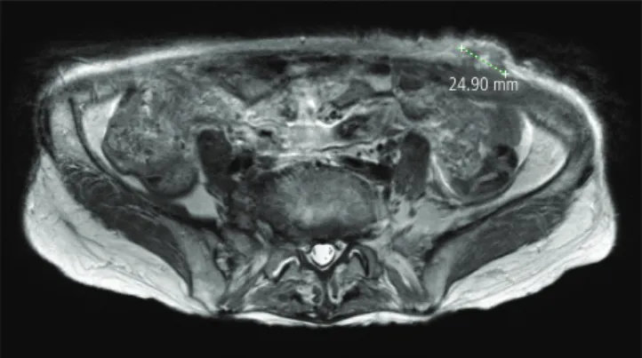
When she admitted for operation, she complained about 2.5 cm sized hard palpable mass at her left lower quadrant of abdomen where the laparoscopic trocar was inserted. Follow-up magnetic resonance imaging of the abdomen and pelvis demonstrated ex- trafascial 2.5 cm sized newly developed metastatic abdominal wall mass after laparoscopic surgery (Fig. 1). Serum level of the tumor- associated protein CA-125 was elevated over 4,009.7 U/mL. On March 8, 2011, she underwent a total abdominal hysterectomy with bilateral salpingo-oophrectomy, bilateral pelvic and para-aor- tic lymph node dissection, total omentectomy, and abdominal wall mass removal (Fig. 2). The pathologist diagnosed abdominal wall mass as grade 2 primary peritoneal serous papillary adenocarci- noma, and found the same pathologic feature on other specimens.
The patient discharged after combination chemotherapy of pacli- taxel and carboplatin.
Discussion
The first detailed descriptions of laparoscopic management and treatment of patients with gynecologic cancers were reported ap- proximately 30 years ago [6]. Since that time, an increasing num- ber of advanced laparoscopic techniques have been used in the management of gynecologic malignancies including second-look laparoscopy, laparoscopic lymphadenectomy, laparoscopically as- sisted vaginal hysterectomy, and laparoscopically assisted radical vaginal hysterectomy [7].
In case of ovarian malignancies, the advancement in laparoscopic surgery offered some advantages over previous laparotomy includ- ing smaller incisions, improved visualization, less blood loss, re- duction in the need for analgesics, decreased morbidity and rapid recovery.
But increasing amount of literature describes postoperative tumor growth at the specifi c puncture sites associated with trocar place- ment [1,3].
The true incidence of port-site metastases in patients with gyne- cologic malignancies has not been clearly defi ned and wide range of incidence was reported by several studies. Childers et al. [3]
reported port-site metastases in 1 of 88 patients (1.4% per proce- dure) undergoing a laparoscopic procedure for ovarian cancer and 1% per procedure for gynecologic cancers in general, while Kruit- wagen et al. [1] reported the incidence of port-site metastases to be 16% in patients with ovarian cancer undergoing laparoscopic procedures 9 to 35 days prior to the initial debulking procedure.
And van Dam et al. [8] reported port-site metastases in 9% of pa- tients (9/104) undergoing laparoscopic procedures for primary or recurrent ovarian cancer.
Although the concept of tumor cell contamination of the surgical wound was described over 40 years ago [9], the mechanism by which wound metastases occurs is still not completely understood.
Hypotheses specifi c to laparoscopy include exfoliation and spread of tumor cells by laparoscopic instruments, direct implantation at the trocar site by frequent changes of instruments, direct im- plantation from the passage of the specimen, the presence of the pneumoperitoneum, which can create a “chimney effect” that causes an increase in the passage of tumor cells at port-sites, and preferential growth of malignant cells at areas of laparoscopic peritoneal perforation [10].
The prognostic effect of port-site metastases is not fully under- stood yet, but Huang et al. [4] reported it is associated with poor outcome. Therefore it is of worthy to prevent port-site metastases, and whenever a female patient has elevated serum level of CA-
Fig. 2. Post debulking surgery including abdominal wall mass removal.Fig. 1. The MRI fi nding of extrafascial metastatic mass (green line mea- sured) after laparoscopic operation.
24.90 mm
WWW.KJOG.ORG 397
Jin-Young Park, et al. Port-site metastasis of advanced peritoneal cancer
125 and ascites cytology shows adenocarcinoma, we should con- sider the possibility of primary peritoneal cancer or normal sized ovarian cancer [11] in case of imaging studies show normal sized ovaries. To prevent the laparoscopic port-site metastases of these patients, all the suspicious patients must be referred to gynecolog- ic oncologists fi rst. If the gynecologic oncologist decided to do the laparoscopic biopsy rather than debulking surgery, the plastic re- trieval bag should be used when removing the specimen through 11 mm or 12 mm trocar to prevent cancer cell implantation. But in case of large amount of ascites are present, it would be better to do the primary debulking surgery rather than to do laparoscopic biopsy.
References
1. Kruitwagen RF, Swinkels BM, Keyser KG, Doesburg WH, Schijf CP. Incidence and effect on survival of abdominal wall me- tastases at trocar or puncture sites following laparoscopy or paracentesis in women with ovarian cancer. Gynecol Oncol 1996;60:233-7.
2. Abu-Rustum NR, Rhee EH, Chi DS, Sonoda Y, Gemignani M, Barakat RR. Subcutaneous tumor implantation after laparo- scopic procedures in women with malignant disease. Obstet Gynecol 2004;103:480-7.
3. Childers JM, Aqua KA, Surwit EA, Hallum AV, Hatch KD.
Abdominal-wall tumor implantation after laparoscopy for ma- lignant conditions. Obstet Gynecol 1994;84:765-9.
4. Huang KG, Wang CJ, Chang TC, Liou JD, Hsueh S, Lai CH, et al. Management of port-site metastasis after laparoscopic surgery for ovarian cancer. Am J Obstet Gynecol 2003;189:16- 21.
5. Chung MJ, Lee JM, Kim YJ, Park IS, Cho YL, Lee YS. Trocar-site metastasis after laparoscopic gynecologic oncologic surgery.
Korean J Obstet Gynecol 2007;50:117-25.
6. Rosenoff SH, DeVita T Jr, Hubbard S, Young RC. Peritoneoscopy in the staging and follow-up of ovarian cancer. Semin Oncol 1975;2:223-8.
7. Jennings TS, Dottino P, Rahaman J, Cohen CJ. Results of selec- tive use of operative laparoscopy in gynecologic oncology.
Gynecol Oncol 1998;70:323-8.
8. van Dam PA, DeCloedt J, Tjalma WA, Buytaert P, Becquart D, Vergote IB. Trocar implantation metastasis after laparoscopy in patients with advanced ovarian cancer: can the risk be re- duced? Am J Obstet Gynecol 1999;181:536-41.
9. Thomas CG. Tumor Cell Contamination of the Surgical Wound: Experimental and Clinical Observations. Ann Surg 1961;153:697-704.
10. Wang PH, Yuan CC, Lin G, Ng HT, Chao HT. Risk factors con- tributing to early occurrence of port site metastases of laparo- scopic surgery for malignancy. Gynecol Oncol 1999;72:38-44.
11. Choi CH, Kim TJ, Kim WY, Ahn GH, Lee JW, Kim BG, et al.
Papillary serous carcinoma in ovaries of normal size: a clini- copathologic study of 20 cases and comparison with extra- ovarian peritoneal papillary serous carcinoma. Gynecol Oncol 2007;105:762-8.
진행된 원발성 복막암의 복강경하 복막생검 후 투관 침 삽입부 전이
성균관대학교 의과대학 산부인과학교실 박진영, 안정주, 김태중, 김병기, 배덕수
부인과 복강경수술 기술의 발달로 인하여 부인과 악성종양 환자들에게도 복강경수술이 널리 시행되고 있다. CA-125의 혈중 수치가 상승 되어 있었으며, 다량의 악성복수가 있었던 69세 여자 환자가 일반외과에서 복강경하 복막생검 후 산부인과로 전원되었다. 환자는 수술 1 주일 후부터 만져진 2.5 cm 크기의 복벽종괴를 호소하였다. 난소암 및 투관 침 삽입부 전이 의심하에 우리는 종양감축술 및 복벽종괴제 거술을 시행하였다. 최종 병리 결과 원발성 복막 유두상 장액성 선암으로 진단되었다. 혈중 CA-125 수치가 상승되어 있으며, 복수천자 결과상 선암을 진단받은 여성 환자는 반드시 부인 종양학 전문의에게 전원되어야 한다. 그리고 부인종양학 전문의는 원발성 복막암이나 원발성 난소암 의심하에 일차적 종양감축술을 시행하여야 한다.
중심단어: 투관침 삽입부 전이, 원발성 복막암, 원발성 난소암