http://dx.doi.org/10.5468/ogs.2016.59.2.130 pISSN 2287-8572 · eISSN 2287-8580
Introduction
Inflammation of the vulvar and vaginal epithelium, vulvo- vaginitis, is frequently reported in prepubertal girls. Patients present with signs and symptoms such as vaginal discharge, redness of the vulva, and genital soreness. Archetypal low es- trogen milieu, as well as a proclivity for deprived local hygiene, in young prepubertal girls are the main causes of susceptibility to non-specific pediatric vulvovaginitis [1,2]. In addition, bub- ble bath use, and a medical history of upper respiratory tract infection (URI) or vulvovaginitis have been reported as risk fac-
Clinical and microbiologic characteristics of
vulvovaginitis in Korean prepubertal girls, 2009–2014:
a single center experience
Hounyoung Kim, Sun Myung Chai, Eun Hee Ahn, Mee-Hwa Lee
Department of Obstetrics and Gynecology, CHA Bundang Medical Center, CHA University, Seongnam, Korea
Objective
To update information on the clinical and microbiologic characteristics of pediatric vulvovaginitis in Korean prepubertal girls.
Methods
A total of 120 girls (aged 0 to 9 years) with culture-confirmed pediatric vulvovaginitis, diagnosed between 2009 and 2014, were enrolled in the study. The epidemiologic and microbiologic characteristics, and clinical outcomes were assessed. Patients with sexual precocity, as well as those who were referred for suspected sexual abuse, were excluded.
Results
Girls aged 4 to 6 years were at the highest risk of pediatric vulvovaginitis. Seasonal distribution indicated obvious peaks in summer and winter. Of the 120 subjects, specific pathogens were identified in the genital specimens in only 20 cases (16.7%). Streptococcus pyogenes (n=12, 60%) was the leading cause of specific vulvovaginitis. Haemophilus influenzae was isolated in one patient. No cases presented with enteric pathogens, such as Shigella or Yersinia. A history of recent upper respiratory tract infection, swimming, and bubble bath use was reported in 37.5%, 15.8%, and 10.0% of patients, respectively. Recent upper respiratory tract infection was not significantly correlated with the detection of respiratory pathogens in genital specimens (P>0.05). Of 104 patients who underwent perineal hygienic care, 80 (76.9%) showed improvement of symptoms without antibiotic treatment. Furthermore, the efficacy of hygienic care was not significantly different between patients with or without specific pathogens (P>0.05).
Conclusion
Specific pathogens were only found in 16.7% of pediatric vulvovaginitis cases. Our results indicate an excellent outcome with hygienic care, irrespective of the presence of specific pathogens.
Keywords: Childhood; Hygienic care; Vaginal flora; Vulvitis; Vulvovaginitis
Articles published in Obstet Gynecol Sci are open-access, distributed under the terms of the Creative Commons Attribution Non-Commercial License (http://creativecommons.
org/licenses/by-nc/3.0/) which permits unrestricted non-commercial use, distribution, and reproduction in any medium, provided the original work is properly cited.
Copyright © 2016 Korean Society of Obstetrics and Gynecology Received: 2015.7.31. Revised: 2015.9.25. Accepted: 2015.10.20.
Corresponding author: Mee-Hwa Lee
Department of Obstetrics and Gynecology, CHA Bundang Medical Center, CHA University, 59 Yatap-ro, Bundang-gu, Seongnam 13496, Korea
Tel: +82-31-780-5290 Fax: +82-31-780-1947 E-mail: [email protected]
http://orcid.org/0000-0001-9159-3961
tors for pediatric vulvovaginitis [1,3].
In 25% to 75% of prepubertal girls with symptoms of vulvo- vaginitis, genital microbiologic investigations indicate the pres- ence of normal vaginal microflora or non-pathogenic bacteria [4]. Specific pathogens are reported in <50% of patients, and are typically respiratory in origin [5-8]. Streptococcus pyogenes is the leading cause of specific pediatric vulvovaginitis [5,7].
Although there are numerous reports on the causative mi- croorganisms and clinical features of pediatric vulvovaginitis, data from Korea is very limited. A solitary Korean study, pub- lished by our institute in 1999, described some clinical aspects of pediatric vulvovaginitis using data from a small number of cases [6]. In that study, Haemophilus influenzae had been reported as the most common pathogenic bacteria, unlike the results of western studies in recent years. The present study aimed to reinforce the information on clinical characteristics of pediatric vulvovaginitis in Korean prepubertal girls with data from participants of larger studies as well as to elucidate con- temporary changes in microbiologic etiology since 1999.
Materials and methods
1. Study samples
This was a retrospective analysis of data from 120 prepubertal girls, aged 0 to 9 years, who presented to the outpatient clinic for pediatric and adolescent gynecology at the CHA Bundang Medical Center between January 2009 and December 2014.
Cases were defined as prepubertal girls who visited a clinic with typical genital symptoms or physical findings consistent with vulvovaginitis, either vaginal discharge and/or genital redness. Children who were diagnosed without culture were excluded. Patients with a record of Tanner stage ≥2 for breast development were excluded from this study. In addition, pa- tients who were referred to the clinic for the evaluation of genital trauma or child sexual abuse were also excluded.
2. Clinical characteristics
Patient demographics, presenting symptoms, objective find- ings of external genitalia, and clinical outcomes were assessed.
The patient history for URI in the previous month, bubble bath use, and swimming were analyzed as associated factors.
3. Microbiologic data
All subjects had undergone vaginal (n=108) or vulvar (n=12)
swabs for culture of aerobic bacteria or yeasts using conven- tional culture methods. Vulvar swabs had been selectively performed in subjects with small hymenal opening inade- quate for vaginal swab (n=8) or subjects with severe vulvar in- flammation without vaginal discharge (n=4). The swabs were transported in Amies transport medium, and cultured on 5%
sheep blood agar, chocolate agar, and MacConkey agar. Ad- ditionally, 7 subjects with abnormal hymenal configuration had undergone Chlamydia antigen polymerase chain reac- tion from genital specimens. An early morning cellophane tape test had been selectively performed on 10 subjects with genital pruritus to confirm the presence of pinworm eggs. No cases had undergone anaerobic culture tests. Among 6 cases presenting with urinary symptoms, 2 cases with moderate to severe urinary symptoms underwent further investigation by urine culture. In contrast, 4 cases with a definite finding of vaginal discharge accompanied with mild urinary symptoms did not undergo urine microbiologic study.
4. Hygienic care
Subjects were routinely provided instructions for perineal hy- gienic care at their first visit. The instructions included use of frequent sitz baths, genital drying, and front-to-back wiping, along with avoidance of genital irritants and tight underpants.
Treatment success was defined as the complete or partial im- provement of genital symptoms without the requirement for antibiotics.
5. Statistical analysis
Various characteristics were compared between the subgroups
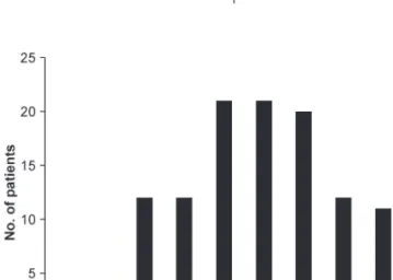
Fig. 1. Age distribution of study participants (n=120).
using analysis of variance or the chi-square test. A P-value of
<0.05 was regarded as statistically significant in all analyses.
Results
1. Demographics and clinical characteristics
The age of the patients at presentation is illustrated in Fig. 1.
The peak age at presentation was 4 to 6 years, and the mean (±standard deviation) age was 5.1 (±2.1) years. The seasonal distribution at presentation demonstrated an obvious bimodal trait with peaks in summer and winter (Fig. 2A). However, in terms of the time of symptom onset, the seasonal distribution represented a less apparent bimodal trait (Fig. 2B).
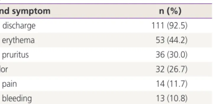
The frequency of signs and symptoms are shown in Table 1. Vaginal discharge was the most frequent symptom in this patient group; it was reported for 92% of patients as a sub- jective symptom or an objective finding. Genital erythema and pruritus were reported in 44% and 30% of subjects, respec-
tively. Genital bleeding or a bloody discharge was reported in 11% of subjects. As a matter of course, a thorough history had been taken and careful genital examination had been performed for these patients to exclude other causes of pedi- atric vaginal bleeding.
Fig. 2. Seasonal distribution of (A) presentation to clinic, and (B) time of symptom onset in prepubertal girls with vulvovaginitis (n=120).
Table 1. Signs and symptoms of vulvovaginitis in prepubertal girls (n=120)
Sign and symptom n (%)
Vaginal discharge 111 (92.5)
Genital erythema 53 (44.2)
Genital pruritus 36 (30.0)
Foul odor 32 (26.7)
Genital pain 14 (11.7)
Genital bleeding 13 (10.8)
Urinary symptoms 6 (5.0)
Table 2. Organisms isolated from the genital specimens of prepu- bertal girls with vulvovaginitis (n=120)
Organism isolated No. of cases (%)
Normal flora
a)61 (50.8)
Non-pathogenic bacteria 39
b)(32.5)
Escherichia coli 30 (25.0)
Coagulase-negative staphylococci 7 (5.8)
Klebsiella pneumoniae 3 (2.5)
Citrobacter freundii 2 (1.7)
Enterobacter aerogenes 2 (1.7)
Klebsiella ozaenae 1 (0.8)
Morganella morganii 1 (0.8)
Stenotrophomonas maltophilia 1 (0.8)
Pathogenic organisms 20 (16.7)
Streptococcus pyogenes 12 (10.0)
Staphylococcus aureus 3 (2.5)
Haemophilus influenzae 1 (0.8)
Chlamydia trachomatis 2 (1.7)
Candida albicans 1 (0.8)
Pinworms 1 (0.8)
Total 120 (100)
a)
Includes lactobacilli, α-hemolytic streptococcus, Enterococcus sp.,
and Neisseria sp.;
b)Seven cases represented more than one species of
non-pathogenic bacteria.
2. Microbiology
Microbiologic investigation revealed normal flora (n=61, 50.8%) or non-pathogenic bacteria (n=39, 32.5%) in the majority of patients (n=100, 83.3%) (Table 2). Escherichia coli were the most commonly identified non-pathogenic bacteria (n=30, 25%).
Pathogenic organisms were isolated in 16.7% of the pa- tients (n=20). The respiratory pathogens Streptococcus pyo- genes (n=12, 10.0%), Staphylococcus aureus (n=3, 2.5%), and Haemophilus influenzae (n=1, 0.8%) were specifically identified. Candida albicans was detected in 1 patient (0.8%), a 2-year-old girl who was not yet toilet trained. Chlamydial antigen polymerase chain reaction tests were selectively per- formed in 7 children with abnormal hymenal configuration;
a positive result was identified in 2 children (1.7%). Pinworm was reported in a 5-year-old girl with symptoms of genital itching and vaginal discharge. There were no cases of Shigella or Yersinia reported, even in one patient with accompanying diarrhea.
Fig. 3 illustrates the seasonal distribution of genital microor- ganisms in 116 patients. The 4 cases that had presented with Chlamydia trachomatis, Candida albicans, and pinworm were excluded. There were no obvious seasonal traits found in any microorganism subgroup; however, the vulvovaginitis cases that were associated with respiratory pathogens seemed to occur more frequently in summer and winter.
3. Associated factors
A history of URI in the previous month was reported in 45 (37.5%) patients with vulvovaginitis (data not shown). In ad-
Table 3. Frequency of upper respiratory tract infection and recent antibiotic use in prepubertal patients with or without respiratory patho- gens
Parameter Patients with normal flora or
non-pathogenic bacteria
Patients with respiratory
bacteria
a)P-value
Upper respiratory tract infection 36 (36.0) 7 (43.8) NS
Recent antibiotic use 40 (40.0) 5 (31.3) NS
No. of participants 100 16
Values are presented as n (%).
NS, not significant.
a)
Includes Streptococcus pyogenes (n=12), Staphylococcus aureus (n=3), and Haemophilus influenzae (n=1).
Table 4. Efficacy of perineal hygienic care in prepubertal girls with vulvovaginitis (n=104) Efficacy of hygienic care Total Patients with normal flora or
non-pathogenic bacteria
Patients with specific
bacteria
a)P-value
Effective 80 (76.9) 71 (78.0) 9 (69.2) NS
Ineffective 12 (11.5) 9 (9.9) 3 (23.1) NS
Lost to follow-up 12 (11.5) 11 (12.1) 1 (7.7) NS
Total 104 (100) 91 (100) 13 (100)
Values are presented as number (%).
NS, not significant.
a)