CT Evaluation of Vocal Cord Paralysis due to Thoracic Diseases:
A 10-Year Retrospective Study
Sun Wha Song,
1Beom Cho Jun,
2Kwang Jae Cho,
2Sungwon Lee,
1Young Joo Kim,
1and Seog Hee Park
31Departments of Radiology and 2Otolaryngology-Head and Neck Surgery, Uijeongbu St. Mary’s Hospital, The Catholic University of Korea College of Medicine, Uijeongbu;
3Department of Radiology, Seoul St. Mary’s Hospital, The Catholic University of Korea College of Medicine, Seoul, Korea.
Received: September 28, 2010 Revised: November 6, 2010 Accepted: December 2, 2010
Corresponding author: Dr. Beom Cho Jun, Department of Otolaryngology-Head and Neck Surgery, Uijeongbu St. Mary’s Hospital, The Catholic University of Korea
College of Medicine,
65-1 Geumo-dong, Uijeongbu 480-821, Korea.
Tel: 82-31-820-3657, Fax: 82-31-847-0038 E-mail: [email protected]
∙ The authors have no financial conflicts of interest.
© Copyright:
Yonsei University College of Medicine 2011 This is an Open Access article distributed under the terms of the Creative Commons Attribution Non- Commercial License (http://creativecommons.org/
licenses/by-nc/3.0) which permits unrestricted non- commercial use, distribution, and reproduction in any medium, provided the original work is properly cited.
Purpose: To discuss computed tomography (CT) evaluation of the etiology of vo- cal cord paralysis (VCP) due to thoracic diseases. Materials and Methods: From records from the past 10 years at our hospital, we retrospectively reviewed 115 cases of VCP that were evaluated with CT. Of these 115 cases, 36 patients (23 M, 13 F) had VCP due to a condition within the thoracic cavity. From these cases, we collected the following information: sex, age distribution, side of paralysis, symp- tom onset date, date of diagnosis, imaging, and primary disease. The etiology of VCP was determined using both historical information and diagnostic imaging.
Imaging procedures included chest radiograph, CT of neck or chest, and esopha- gography or esophagoscopy. Results: Thirty-three of the 36 patients with thoracic disease had unilateral VCP (21 left, 12 right). Of the primary thoracic diseases, malignancy was the most common (19, 52.8%), with 18 of the 19 malignancies presenting with unilateral VCP. The detected malignant tumors in the chest con- sisted of thirteen lung cancers, three esophageal cancers, two metastatic tumors, and one mediastinal tumor. We also found other underlying etiologies of VCP, in- cluding one aortic arch aneurysm, five iatrogenic, six tuberculosis, one neurofibro- matosis, three benign nodes, and one lung collapse. A chest radiograph failed to de- tect eight of the 19 primary malignancies detected on the CT. Nine patients with lung cancer developed VCP between follow-ups and four of them were diagnosed with a progression of malignancy upon CT evaluation of VCP. Conclusion: CT is helpful for the early detection of primary malignancy or progression of malignancy between follow-ups. Moreover, it can reveal various non-malignant causes of VCP.
Key Words: Vocal cord paralysis, thorax, CT
INTRODUCTION
Vocal cord paralysis may arise from neurogenic paralysis or mechanical fixation.1 It is sometimes the only sign of an underlying disease.2 Thus, it is clinically impor- tant to diagnose the primary disease in cases of vocal cord paralysis (VCP) be- cause many of its potential causes, such as symptom-free malignant tumors, can
VCP was caused by chest diseases, and in 34 (29.6%) VCP was caused by neck diseases. A summary of the identified underlying etiologies of VCP is shown in Table 2.
Of the 36 patients whose VCP had a thoracic pathology, nine patients underwent only a neck CT, seven had only a chest CT, and 20 underwent both neck and chest CT scans.
Of the 36 patients with thoracic disease, the majority were in their 6th decade of life, 23 (63.9%) were men, and 13 (36.1%) were women. The sex and age distributions of the VCP patients with thoracic disease are shown in Table 3.
The duration of VCP symptoms varied widely, ranging from two days to 30 years.
The diagnostic categories of the primary thoracic diseases be fatal or cause serious morbidity if detected late.2,3 Radio-
logic evaluation is often useful for determining the etiology of VCP, especially for conditions within the thoracic cavity.
However, chest radiographs can sometimes miss small le- sions in the mediastinum.2 In these cases, computed tomog- raphy (CT) can be a valuable diagnostic tool. We have found only a few original reports on CT evaluation of VCP for thoracic disease.2,4 In this article, we performed a retrospec- tive CT analysis of our patients in order to review cases of VCP with an underlying etiology of thoracic disease.
MATERIALS AND METHODS
The study was approved by the institutional review board.
We retrospectively reviewed 115 cases of VCP with CT evaluation from patient records at our hospital among cases occurring any time from January 2000 to March 2010. We separated out the cases where VCP was caused by an un- derlying thoracic disease (a total of 36 patients; 23 male and 13 female) and patient data such as age, distribution, side of paralysis, symptom onset date, date of diagnosis, di- agnostic imaging studies, and primary disease was extract- ed from each case.
After the general and otolaryngologic examinations, la- ryngeal fiberscopy was performed for all new patients with VCP to examine the state of paralysis and to look for the presence of a primary lesion in the larynx or hypopharynx.
The etiology of VCP was determined using both historical information and diagnostic imaging. Imaging procedures included a chest radiograph, a CT of the neck or chest, and esophagography or esophagoscopy.
RESULTS
CT was used in 115 patients with VCP. In the majority of cases, VCP was unilateral (Table 1). One hundred and five (91.3%) patients were identified as having unilateral VCP, of whom 59 were male and 46 were female. Of the 105 unilateral cases of VCP, 73 cases had left-side and 32 had right-side paralysis. Ten (8.7%) patients were identified as having bilateral VCP, of whom four were male and six were female.
Our analysis found that 70 (60.9%) of the 115 patients with VCP had an identified cause, and 45 (39.1%) were id- iopathic. Furthermore, in 36 (31.3%) of the 115 patients,
Table 1. Sex and Side Distributions of the Primary Diseases of 115 VCP Patients
Unilateral (105) Bilateral R L (10)
Chest (36) M (23) 5 16 2
F (13) 4 8 1
Neck (34) M (15) 4 10 1
F (19) 6 10 3
Idiopathic (45) M (25) 8 16 1
F (20) 5 13 2
Total (115) 32 73 10
VCP, vocal cord paralysis; R, right vocal cord paralysis; L, left vocal cord paralysis.
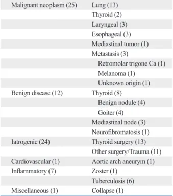
Table 2. The Identified Diagnostic Categories of 70 VCP Pa- tients
Malignant neoplasm (25) Lung (13) Thyroid (2) Laryngeal (3) Esophageal (3) Mediastinal tumor (1) Metastasis (3)
Retromolar trigone Ca (1) Melanoma (1)
Unknown origin (1) Benign disease (12) Thyroid (8)
Benign nodule (4) Goiter (4) Mediastinal node (3) Neurofibromatosis (1) Iatrogenic (24) Thyroid surgery (13)
Other surgery/Trauma (11) Cardiovascular (1) Aortic arch aneurym (1) Inflammatory (7) Zoster (1)
Tuberculosis (6) Miscellaneous (1) Collapse (1) VCP, vocal cord paralysis; Ca, carcinoma.
The causes of bilateral VCP (n=3) were caused by lung cancer (n=1), pulmonary tuberculosis (n=1) and injury from a tracheostomy procedure (n=1).
The malignant tumor types detected by CT and side dis- tribution of the VCP are shown in Table 5. Of the detected malignant tumors in the chest, lung cancer was the most identified among patients presenting with VCP are shown in
Table 4. Thirty-three of 36 patients with thoracic diseases had unilateral VCP, 21 (63.6%) had left-side paralysis, and 12 (36.4%) had right-side paralysis. Of the primary thoracic dis- eases, malignancy was the most common (19 cases, 52.8%).
Eighteen (94.7%) cases of 19 malignant diseases had unilat- eral paralysis, 12 (66.7%) had left-side paralysis, and six (33.3%) had right-side paralysis (Fig. 1).
Seventeen (47.2%) of the 36 patients with thoracic dis- ease were found to have a non-malignant condition within the thoracic cavity that accounted for their VCP. Tuberculo- sis was the second most common primary chest disease causing VCP (six cases, 16.7%). We found five cases of in- active tuberculosis and one case of active apical tuberculo- sis (Fig. 2).
One case of VCP was caused by aortic arch aneurysm.
One case involved neurofibromatosis associated with left VCP, and five cases of VCP were caused by iatrogenic injury (Fig. 3).
Table 3. Sex and Age Distributions of 36 VCP Patients with Thoracic Disease
Age (yrs) Male Female Total
20-29 0 0 0
30-39 1 0 1
40-49 3 0 3
50-59 5 2 7
60-69 6 4 10
70-79 4 5 9
80-89 3 2 5
90-99 1 0 1
Total 23 13 36
VCP, vocal cord paralysis.
Fig. 1. A 72-year-old woman with a paralyzed left vocal cord. Contrast-enha- nced CT demonstrates a large lung cancer in the left upper lobe with inva- sion in the aorticopulmonary window (arrows). CT, computed tomography.
Fig. 2. A 61-year-old woman with a paralyzed left vocal cord for 11 years. (A and B) Chest radiograph (A) and contrast-enhanced CT (B) show the left lung destroyed from pulmonary tuberculosis with a marked decrease in lung volume and a mediastinal shift to the left. Fibrocalcific tuberculous scars can be seen in the right upper lobe. CT, computed tomography.
Table 4. The Identified Diagnostic Categories and Side Distributions of 36 VCP Patients with Thoracic Disease
R L B Total
Malignant neoplasm 6 12 1 19
Benign disease 0 4 0 4
Mediastinal node 0 3 0 3
Neurofibromatosis 0 1 0 1
Iatrogenic 2 2 1 5
Esophageal Ca surgery 0 1 0 1
Lung Ca surgery 0 1 0 1
Tracheostomy 1 0 1 2
Chemo port insertion 1 0 0 1
Cardiovascular disease 0 1 0 1
Tuberculosis 4 1 1 6
Miscellaneous
Collapse 0 1 0 1
Total 12 21 3 36
VCP, vocal cord paralysis; R, right vocal cord paralysis; L, left vocal cord paralysis; B, bilateral vocal cord paralysis; Ca, carcinoma.
A B
common (13, 68.4%). The other malignancies were three (15.8%) cases of esophageal carcinoma, two (10.5%) cases of metastatic tumors, and one (5.3%) case of mediastinal tumor.
Of the 12 patients diagnosed with a malignancy who pre- sented with left VCP, we discovered direct cancer invasion into the left recurrent laryngeal nerve in three cases, all of whom were diagnosed with lung cancer, and metastasis to the lymph nodes in nine cases (six cases of lung carcinoma, two cases of esophageal carcinoma, and one case of un- known metastasis). Of the six patients diagnosed with a malignancy who presented with right VCP, we found direct cancer invasion into the right recurrent laryngeal nerve in two cases; among these, one case was diagnosed as apical lung carcinoma, and the other case was diagnosed as a su- perior mediastinal tumor. In the other four patients, metas- tasis to the either the right highest mediastinal or the right supraclavicular lymph node was found in three cases, and metastasis was found in both sites in one case. In the only patient diagnosed with a malignancy who presented with bilateral VCP, lung cancer was detected in the right upper lobe, with metastasis to the bilateral lymph nodes.
Nine patients with lung cancer developed VCP between follow-ups and four of them were diagnosed with progres- sion of lung cancer after CT evaluation of VCP (Fig. 4).
One of the patients with lung cancer who presented with VCP had a left lung collapse due to an enlarged mass in the left hilum, but there was no evidence of metastatic nodes, or of a mass in the aortopulmonary window or near the pathway of the left recurrent laryngeal nerve. So we con- cluded that VCP was probably caused by the effect of pres- sure on the nerve from the collapsed lung and classified the patient under the miscellaneous group instead of the lung cancer group in Table 4.
The malignant chest lesions were clearly detectable with contrast-enhanced chest CT or neck CT in all 19 cases. How- ever, a chest radiograph failed to detect the primary lesions in
Fig. 5. A 61-year-old man with a paralyzed right vocal cord. (A) Contrast- enhanced CT demonstrates a small irregular lung cancer in the right apex (white arrow) and a metastatic node in the right upper paratracheal region (black arrow). (B) Retrospective review of chest radiograph suggests a hid- den mass being overlapped by the medial end of the right first rib (arrow).
CT, computed tomography.
A
Fig. 3. A 73-year-old woman with a paralyzed right vocal cord after failure of a chemo port insertion. (A and B) Contrast-enhanced CT scans demon- strate cellulitis in the right anterior chest wall along the tract of a previous chemo port (arrows) (A) and right upper mediastinitis (arrows) (B). CT, com- puted tomography.
A B
Fig. 4. A 73-year-old man with paralyzed left vocal cord. (A) Contrast- enhanced CT prior to vocal cord paralysis demonstrates an irregular lung cancer (short arrow) in the apicoposterior segment of the left upper lobe and conglomerated metastatic nodes (long arrow) in the aortopulmonary window. (B) Follow-up CT after development of left vocal cord paralysis shows enlargement of a lung cancer (short arrow) and metastatic nodes in the aortopulmonary window (long arrow). CT, computed tomography.
A B
B
Table 5. Nineteen Malignant Tumors Detected in the Chests of VCP Patients and Side Distribution
Diagnosis R L B Total
Lung cancer 3 9 1 13
Esophageal cancer 1 2 0 3
Mediastinal tumor 1 0 0 1
Metastatic node 1 1 0 2
Retromolar trigone Ca 0 1 0 1
Unknown origin 1 0 0 1
Total 6 12 1 19
VCP, vocal cord paralysis; R, right vocal cord paralysis; L, left vocal cord paralysis; B, bilateral vocal cord paralysis; Ca, carcinoma.
and nine of 13 VCP patients with lung carcinoma had left- side VCP.
Left VCP associated with a mass in the lung is usually of sinister significance, implying a lung carcinoma with subaor- tic lymph node metastasis.10 In our patients with left-side pa- ralysis, six of nine patients with lung carcinoma showed me- tastasis to a subaortic lymph node, and three of them showed direct tumor invasion of the aortopulmonary window.
However, right-side mediastinal adenopathy extending cephalad to the region of the right subclavian artery can also involve the right recurrent laryngeal nerve. In addition, patients with a superior sulcus tumor can develop right-side VCP when the tumor extends into the supraclavicular space.4 The right recurrent laryngeal nerve hooks around the first part of the subclavian artery and is related only to the apex of the right lung.5 Of the six patients with malignant tumors and right-side VCP, we found direct tumor invasion of the nerve in two and metastasis to the highest mediastinal or supraclavicular lymph node in three cases.
The diagnostic approach used to evaluate patients pre- senting with VCP differs among otolaryngologists, and no consensus has been achieved regarding the appropriate ini- tial workup of these patients. Although no precise evalua- tion sequence exists, a thorough history and physical exami- nation reveal the etiology in the majority of cases. Previously, radiologic examination of patients with extralaryngeal causes of VCP included chest and skull radiographs, barium esopha- gography, and a thyroid scan.4 With the advent of advanced studies of the head and neck [CT and magnetic resonance imaging (MRI)], it is now possible to image the entire course of the laryngeal nerves. These imaging methods obviate the need for tests such as thyroid scans, thyroid function tests, skull radiographs, and barium esophagography.9 The proba- bility of an isolated laryngeal motor nerve encased by an intracranial lesion is highly unlikely due to the relationship of the corticobulbar fibers to the surrounding structures.
Thus, brain MRI or CT is not recommended unless signs or symptoms suggest an intracranial lesion.11
The most important objective in evaluating a patient with VCP is to exclude the existence of a treatable and potential- ly life-threatening primary disease as the cause of VCP.9 Chest radiograph remains a useful screening study, but, it is not sufficient for the full evaluation of VCP.11 Because it is very important to scrutinize the mediastinum, particularly the aortopulmonary window and the paratracheal region in pa- tients with VCP,2 further diagnostic imaging may be needed.
The CT images analyzed in this study demonstrated that eight cases (Fig. 5).
Of the 36 patients with chest diseases, nine patients un- derwent an esophagography or esophagoscopy. Of these nine patients, seven were male, and two were female; four had right-side VCP, and five had left-side VCP. Of the nine patients with an esophagography or esophagoscopy, six had no abnormalities, and three had esophageal cancer (two left- sided VCP cases and one right-sided VCP case). The esopha- gography was able to detect all three esophageal cancers, and the CT scans also revealed them in all three cases.
DISCUSSION
The recurrent laryngeal nerves innervate the vocal cords.
The right recurrent laryngeal nerve leaves the vagus nerve at the anterior surface of the right subclavian artery and runs inferiorly, looping around the subclavian artery, then as- cends medially in the tracheoesophageal groove. The left recurrent nerve branches more caudally from the vagus nerve on the anterior surface of the aortic arch. It passes in- feriorly around the arch through the aortopulmonary win- dow to ascend in the tracheoesophageal groove. Both recur- rent laryngeal nerves are intimately associated with the thyroid gland.4
In the thorax, the left recurrent laryngeal nerve is more vulnerable than the right because it pursues a longer intra- thoracic course, coming into contact with the mediastinal surface of the left lung, continuing along the mediastinal lymph nodes, and finally looping around the aortic arch.5 VCP has been reported to be about 1.4-2.5 times more fre- quent on the left side than on the right.3 In our cases diag- nosed with a thoracic disease, left-side VCP was 1.75 times more frequent than right-side VCP.
The function of the recurrent laryngeal nerve can be dis- rupted by pressure or by injury of the nerve caused by dis- ease pathology invading the nerve.5 Malignant invasion of the vagus or recurrent laryngeal nerves can also occur with thyroid neoplasms, carcinoma of the lung, esophageal carci- noma, and metastases to the mediastinum.6,7 Earlier studies found that tumors were the most frequent cause, and of these tumors, bronchogenic carcinoma was the most common cause of unilateral VCP, as can be seen from our results.7-9
In patients with lung carcinoma, the left recurrent larynge- al nerve is most frequently involved as it courses through the aortopulmonary window.4 In our study, 12 of 19 VCP pa- tients with a malignant chest neoplasm had left-side VCP,
the cause of VCP in females.3 Our study revealed similar results in that all three esophageal carcinoma patients were male. A marked sex difference exists in the origin of malig- nant tumors. In addition to esophageal carcinoma, the inci- dence of lung cancer is higher in males with VCP.3 In our study, we also found similar results, with 11 of 13 lung can- cers being found in men.
Acquired cardiovascular disease such as cardiomegaly, pulmonary hypertension, aortic arch aneurysm, right sub- clavian aneurysm, ductus arteriosus aneurysm, or an en- larged left atrium may compress or stretch the left recurrent laryngeal nerve.12-17 We experienced a case of VCP caused by aortic arch aneurysm.
Left recurrent laryngeal nerve palsy in a patient with me- diastinal lymphadenopathy caused by tuberculosis, pneu- moconiosis, or sarcoidosis has rarely been reported.18 In chronic pulmonary disease, paralysis may be caused by three possible mechanisms that exert effects on the nerve: 1) the nerve is passing through or is adjacent to a mass of caseat- ing nodes, 2) the nerve is trapped in the dense fibrous pleu- ral thickening or in the chronic fibrosing mediastinitis that may occur, and 3) the nerve is being stretched due to retrac- tion of the left upper lobe bronchus pulled towards the apex.19 We found five cases of patients with tuberculosis- destroyed lungs or tuberculous pleurisy scars including the upper mediastinal pleura who had a history of VCP for 4-30 years (mean 14 years). The probable mechanism in these cases would be mechanisms 2) and 3) noted above. We also found a patient with active tuberculosis in the apex. The mechanism in this case was uncertain, but other factors are possible, such as the direct spread of infection, damaging the nerve. Tuberculosis, with six cases, was the second most common cause of VCP in our study, following the most common cause, malignant neoplasm, with 19 cases.
These results are similar to those of a report by Titche.5 Intrathoracic vagus nerve sheath tumors have also been reported.20 We also encountered a case of neurofibromatosis with multiple neurofibroma masses, including in the aorto- pulmonary window, causing left-side VCP.
Vocal cord paralysis can occur after resection of a thorac- ic aortic aneurysm, carotid endarterectomy, parathyroidec- tomy, radical neck dissection for thyroid carcinoma, thorac- ic esophagectomy, mitral valve replacement, tracheostomy or intubation.21,22 In our cases, VCP associated with surgery or trauma occurred only after tracheostomy (n=2), chemo port insertion (n=1), surgery for lung cancer (n=1), and sur- gery for esophageal cancer (n=1).
in a patient presenting with VCP, CT can more accurately detect the extent and location of the responsible pathology than a chest radiograph. We found that of the 19 CT-detect- ed malignant neoplasms, eight were not detected on the chest radiograph. Five of them were associated with left-side pa- ralysis and showed a metastatic node in the aortopulmonary window on CT. The other three were associated with right- side paralysis, and the CT scan detected one case of right apical lung carcinoma and two cases of metastatic nodes in the right upper paratracheal region. Masaki, et al. suggested that neck ultrasonography and chest X-rays were a suffi- cient evaluation modality for patients with right VCP.3 But in our study, CT was needed in a patient with a small right apical lung carcinoma that was hidden behind rib shadows, and also in a patient with VCP caused by phlebitis and right upper mediastinitis during use of an intravenous chemo port (an implantable port for chemotherapy). We do not recom- mend a routine chest radiograph for evaluating the etiology of VCP caused by thoracic diseases.
The CT scan is a very sensitive study for lesions in the neck and upper chest. Furthermore, it is now available at a moderate cost.9 Nevertheless, a chest CT scan was not rou- tinely taken in this study. We discovered that when neck CT scans were taken for patients with right VCP, including an area of the skull base to the thoracic inlet, and for left VCP, including the area up to the aortic triangle, most primary diseases in the chest were detectable. Therefore, it might be cost effective to scan down to the level of the aortic triangle when taking neck CT scans for VCP patients, regardless of the paralytic side.
However, in certain situations, an additional chest CT is also required. When cancer is found in the neck CT, an addi- tional chest CT scan is needed to evaluate the extent and stage of the malignancy. An additional chest CT scan is also warranted when the neck CT shows an enlarged aortopulmo- nary window, supraclavicular nodes, or right highest medias- tinal nodes without a definite primary malignancy focus, such as mid to low esophageal cancer or lung cancers that are outside the boundaries of the neck CT scan. Thus, we recom- mend taking both neck and chest CT scans at the first visit.
For males, when no primary malignancy is found on the CT scans, contrast esophagography or esophagoscopy is rec- ommended to examine the possibility of esophageal cancer.3 Esophagoscopy has the advantage of tissue visualization and biopsy. It is not necessary to perform contrast esopha- gography or esophagoscopy as an initial diagnostic tool in female patients with VCP, as esophageal cancer is rarely
2. Bando H, Nishio T, Bamba H, Uno T, Hisa Y. Vocal fold paralysis as a sign of chest diseases: a 15-year retrospective study. World J Surg 2006;30:293-8.
3. Furukawa M, Furukawa MK, Ooishi K. Statistical analysis of ma- lignant tumors detected as the cause of vocal cord paralysis. ORL J Otorhinolaryngol Relat Spec 1994;56:161-5.
4. Glazer HS, Aronberg DJ, Lee JK, Sagel SS. Extralaryngeal causes of vocal cord paralysis: CT evaluation. AJR Am J Roentgenol 1983;141:527-31.
5. Titche LL. Causes of recurrent laryngeal nerve paralysis. Arch Otolaryngol 1976;102:259-61.
6. Evans JM, Schucany WG. Hoarseness and cough in a 67-year-old woman. Proc (Bayl Univ Med Cent) 2004;17:469-72.
7. Chen HC, Jen YM, Wang CH, Lee JC, Lin YS. Etiology of vocal cord paralysis. ORL J Otorhinolaryngol Relat Spec 2007;69:167- 71.
8. de Gandt JB. [Recurrent nerve compression]. Acta Otorhinolaryn- gol Belg 1970;24:520-54.
9. Terris DJ, Arnstein DP, Nguyen HH. Contemporary evaluation of unilateral vocal cord paralysis. Otolaryngol Head Neck Surg 1992;107:84-90.
10. Sherani TM, Angelini GD, Passani SP, Butchart EG. Vocal cord paralysis associated with coalworkers’ pneumoconiosis and pro- gressive massive fibrosis. Thorax 1984;39:683-4.
11. Altman JS, Benninger MS. The evaluation of unilateral vocal fold immobility: is chest X-ray enough? J Voice 1997;11:364-7.
12. Camishion RC, Gibbon JH Jr, Pierucci L Jr. Paralysis of the left re- current laryngeal nerve secondary to mitral valvular disease. Report of two cases and literature review. Ann Surg 1966;163:818-28.
13. Che GW, Chen J, Liu LX, Zhou QH. Aortic arch aneurysm rup- ture into the lung misdiagnosed as lung carcinoma. Can J Surg 2008;51:E91-2.
14. Bin HG, Kim MS, Kim SC, Keun JB, Lee JH, Kim SS. Intratho- racic aneurysm of the right subclavian artery presenting with hoarseness: a case report. J Korean Med Sci 2005;20:674-6.
15. Kokotsakis J, Misthos P, Athanassiou T, Skouteli E, Rontogianni D, Lioulias A. Acute Ortner’s syndrome arising from ductus arte- riosus aneurysm. Tex Heart Inst J 2008;35:216-7.
16. Gothi R, Ghonge NP. Case Report: Spontaneous aneurysm of ductus arteriosus: A rare cause of hoarseness of voice in adults. In- dian J Radiol Imaging 2008;18:322-3.
17. Morgan AA, Mourant AJ. Left vocal cord paralysis and dysphagia in mitral valve disease. Br Heart J 1980;43:470-3.
18. el-Kassimi FA, Ashour M, Vijayaraghavan R. Sarcoidosis present- ing as recurrent left laryngeal nerve palsy. Thorax 1990;45:565-6.
19. Fowler RW, Hetzel MR. Tuberculous mediastinal lymphadenopa- thy can cause left vocal cord paralysis. Br Med J (Clin Res Ed) 1983;286:1562.
20. Strickland B, Wolverson MK. Intrathoracic vagus nerve tumours.
Thorax 1974;29:215-22.
21. Barondess JA, Pompei P, Schley WS. A study of vocal cord palsy.
Trans Am Clin Climatol Assoc 1986;97:141-8.
22. Ellis PD, Pallister WK. Recurrent laryngeal nerve palsy and endo- tracheal intubation. J Laryngol Otol 1975;89:823-6.
23. Holinger LD, Holinger PC, Holinger PH. Etiology of bilateral ab- ductor vocal cord paralysis: a review of 389 cases. Ann Otol Rhi- nol Laryngol 1976;85:428-36.
The etiologies of bilateral VCP in adults are 1) following thyroidectomy, which is the most common reason; 2) vari- ous neurogenic diseases, such as poliomyelitis, Parkinson’s disease, cerebrovascular accident, multiple sclerosis, etc.; 3) malignancy of the neck and mediastinum, such as in our case diagnosed with right lung cancer with bilateral metastatic nodes; and 4) many others, including foreign bodies, intuba- tion, bilateral neck dissection, infection, congenital lesions, trauma, and idiopathic paralysis.23 In our study, we found a case of bilateral VCP caused by bilateral upper lobe tubercu- losis, another by tracheostomy, and a third by lung cancer.
Although a CT scan is important, the history of the patient is also essential under the following circumstances: 1) VCP in a patient during follow-up for a known malignancy, as it may indicate recurrence or aggravation of the primary lesion;
2) VCP after tracheostomy, chemo port insertion, or an oper- ation on the lung or esophagus, which suggests the possibili- ty of iatrogenic paralysis; and 3) a 10- to 30-year history of VCP, which may indicate VCP caused by tuberculosis.
A negative CT in both the chest and neck reassures clini- cians that the VCP is idiopathic in origin, thus obviating the need for aggressive therapeutic regimens or further inva- sive diagnostic testing.4 Among our cases, 39.1% were id- iopathic.
Among the patients who presented to an otolaryngologist with VCP as the initial sign, the frequency of detection of a malignant tumor as the primary lesion is relatively high.
Hence, it is clinically important to diagnose the primary dis- ease in cases of VCP.3 Identifying the cause of VCP can benefit the patient through early detection and treatment of a malignant mass, help recognize recurrence or aggravation of the primary malignancy, or identify metastasis. In our study, nine patients with lung cancer developed VCP during the periods between follow-up appointments and four of them were subsequently diagnosed with lung cancer pro- gression. Radiologic imaging is also needed to differentiate between various non-malignant causes of VCP. To determine the etiology of VCP caused by thoracic diseases, we recom- mend both neck and chest CT scans and in some cases, esophagography or esophagoscopy, especially for males.
REFERENCES
1. Benninger MS, Gillen JB, Altman JS. Changing etiology of vocal fold immobility. Laryngoscope 1998;108:1346-50.