INTRODUCTION
Estrogen and progesterone production is essential for normal proliferation of the endometrium. Previous studies have shown
that continuous and excessive exposure to estrogen increases the risk of endometrial cancer.1-4 However, there is a paradoxi- cal increase of estrogen-dependent endometrial cancer in postmenopausal women who have stopped producing estro- gen in the ovaries. Hence, intrinsic synthesis and metabolism of estrogen plays an important role in the prevalence and pro- gression of endometrial cancer.5 Inhibition of sex hormones in patients with endometrial cancer is thus an important ther- apeutic goal to reduce levels of biologically significant estro- gen and achieve better prognosis.6,7
Regulation of estrogen synthesis and action in the treatment of hormone-dependent breast cancer has been successful in the past.8 Tamoxifen, the estrogen receptor (ER) blocker has been standard treatment until now, and aromatase inhibitors (AIs) have been used as a first line therapy in metastatic and hormone-dependent breast cancer in postmenopausal wom- en. AIs are also widely used currently, as first line therapy in
The Role of Steroid Sulfatase as a Prognostic Factor in Patients with Endometrial Cancer
Won Moo Lee1, Ki-Seok Jang2, Jaeman Bae1, and A Ra Koh1
1Division of Gynecologic Oncology and Gynecologic Minimally Invasive Surgery, Department of Obstetrics and Gynecology, Hanyang University College of Medicine, Seoul, Korea;
2Department of Pathology, Hanyang University College of Medicine, Seoul, Korea.
Purpose: The aim of the study was to determine steroid sulfatase (STS) expression in endometrial cancer patients and its correla- tion with disease prognosis.
Materials and Methods: We conducted a retrospective study in 59 patients who underwent surgery with histologically confirmed endometrial cancer from January 2000 to December 2011 at Hanyang University Hospital. Immuno-histochemical staining of STS was performed using rabbit polyclonal anti-STS antibody.
Results: Sixteen of the 59 patients (27.1%) were positive for STS expression. Disease free survival (DFS) was 129.83±8.67 [95%
confidence interval (CI): 112.84–146.82] months in the STS positive group (group A) and 111.06±7.17 (95% CI: 97.01–125.10) months in the STS negative group (group B) (p=0.92). Overall survival (OS) was 129.01±9.38 (95% CI: 110.63–147.38) months and 111.16±7.10 (95% CI: 97.24–125.07) months for the groups A and B, respectively (p=0.45). Univariate analysis revealed that FIGO stage and adjuvant therapy are significantly associated with DFS and OS. However, in multivariate analysis, FIGO stage and adju- vant therapy did not show any statistical significance with DFS and OS. STS was also not significantly associated with DFS and OS in univariate and multivariate analysis.
Conclusion: STS expression was not significantly associated with DFS and OS, despite positive STS expression in 27% of endome- trial cancer patients. Therefore, the role of STS as a prognostic factor in patients with endometrial cancer remains unclear and re- quires further research.
Key Words: Steroid sulfatase, endometrial cancer, prognostic factor
pISSN: 0513-5796 · eISSN: 1976-2437
Received: February 25, 2015 Revised: July 13, 2015 Accepted: August 31, 2015
Corresponding author: Dr. Jaeman Bae, Division of Gynecologic Oncology and Gynecologic Minimally Invasive Surgery, Department of Obstetrics and Gynecology, Hanyang University College of Medicine, 222 Wangsimni-ro, Seongdong-gu, Seoul 04763, Korea.
Tel: 82-2-2290-8418, Fax: 82-2-2296-8472, E-mail: [email protected] We presented this work as an abstract at the Korean Society of Obstetrics and Gynecology meeting, which was held in Seoul, Korea in October 2014.
•The authors have no financial conflicts of interest.
© Copyright: Yonsei University College of Medicine 2016
This is an Open Access article distributed under the terms of the Creative Com- mons Attribution Non-Commercial License (http://creativecommons.org/licenses/
by-nc/3.0) which permits unrestricted non-commercial use, distribution, and repro- duction in any medium, provided the original work is properly cited.
Yonsei Med J 2016 May;57(3):754-760 http://dx.doi.org/10.3349/ymj.2016.57.3.754
early ER positive breast cancer in postmenopausal women.
Unfortunately, despite advances in therapeutic strategies, the majority of breast cancer patients who undergo these treat- ments have progressive disease after a few years of a complete response.
Androgen receptor (AR) is expressed in >80% of ER positive postmenopausal breast cancer women. However, AR is also expressed in ER negative patients. Androstenediol (Adiol) can bind to ER, despite being an androgen, and results in an in- crease in the number of ER positive breast cancer cells. Al- though Adiol has a weak affinity to ER, high concentrations of Adiol of >100 fold can have the same effect as estradiol (E2).9 Clinical treatment with AIs inhibits the synthesis of E2 by
>99%, but simultaneously sensitizes to very E2 low concentra- tions. Adiol-induced low E2 concentration potentially affects the progression of breast cancer.8 The androgen-induced acti- vation of estrogen through AR in ER-negative breast cancer patients is via steroid sulfatase (STS). Therefore, inhibition of STS can reduce estrogen synthesis by Adiol.
Most STS inhibitor clinical studies in breast cancer patients are in the clinical trial phase, and an ongoing study on hor- mone-dependent endometrial cancer that is similar to breast cancer is in the animal study phase. To the best of our knowl- edge, there is no study on STS with human endometrial can- cer tissue. Therefore, we investigated STS expression in hu- man endometrial cancer tissue and determined its correlation with prognosis in patients with or without expression of STS.
MATERIALS AND METHODS
Patient selection
We conducted a retrospective study in 59 patients who under- went surgery for endometrial cancer from January 2000 to December 2011 at Hanyang University Hospital. STS expres- sion was confirmed through immuno-histochemical staining of sections from paraffin-embedded endometrial cancer tissue.
We excluded patients who were diagnosed as recurrent endo- metrial cancer, transferred from another hospital after hyster- ectomy, and were lost to follow up after surgery. We evaluated patient characteristics, including age, parity, types of surgery, serum levels of CA125 and 19-9 before and after surgery, the International Federation of Gynecology and Obstetrics (FIGO) stage, histologic type and grade, and adjuvant therapies, such as chemotherapy, radiation therapy, and concurrent chemo- radiation therapy (CCRT). In survival analysis, last day of treat- ment and follow up, day of recurrence, and day of death were investigated. The study was approved by the Institutional Re- view Board (Study approval No.: HYUH 2012-R-03).
Patient follow up
Patients were followed up by physical examination and for tu- mor markers (CA125/19-9) every 3 months for 2 years, and ev-
ery 6 months for the next 3 years. Chest X-ray, abdomino-pel- vic computed tomography, and 18F-fluoro-D-glucose positron emission tomography-computed tomography (18FDG PET- CT) were performed annually. Recurrence of disease was de- fined as presence of tumor histologically or radiologically.
Any suspicious lesion on CT scan was followed-up with CT scans every 3 months until recurrence was confirmed clinically.
In cases of recurrence, 18FDG PET-CT was performed to lo- cate other sites of recurrence, and all sites of recurrence were documented after 2006.
Surgery and adjuvant treatment
We performed total hysterectomy with bilateral salpingo-oo- phorectomy without lymphadenectomy in cases where the tu- mor mass was confined to the endometrium and there was no significant lymphadenopathy. Para-aortic lymphadenectomy was performed in cases where the tumor mass was ≥2 cm in size, or there were more than 1/2 invasions of the myometrium or positive pelvic lymphadenopathy in frozen sections.
We did not perform adjuvant treatment in patients with stage IA and grade 1, 2, or vaginal brachytherapy in patients with stage IA and grade 3, or stage IB. Patients with stage II received external pelvic radiotherapy and chemotherapy, and extended radiotherapy was given in patients with stage III or stage IV.
Construction of tissue microarray
The most morphologically representative and non-necrotic area was carefully selected and marked on the hematoxylin-eo- sin stained slide. Two tissue cores (2 mm in diameter) were sampled from each paraffin block and assembled into a re- cipient paraffin block using a tissue microarray instrument (Ac- cuMax array, ISU Abix, Seoul, Korea).
Immunohistochemical staining
The 4-μm-thick tissue sections were cut from the tissue micro- array blocks and deparaffinized with xylene and rehydrated with graded alcohol. Antigen retrieval was performed by microwav- ing the samples for 12 min in preheated 10 mM sodium citrate buffer (pH 6.0). Endogenous peroxidase activity was blocked with peroxidase blocking solution (DAKO, Carpinteria, CA, USA) for 10 min. The rabbit polyclonal anti-STS antibody (ab62219, Abcam, Cambridge, MA, USA) was diluted 1:100 and incubated at 4°C for 16 h. The sections were then incubated with a peroxidase labeled anti-mouse/rabbit secondary antibody for 30 min (DAKO, Carpinteria, CA, USA). The samples were de- veloped with DAB substrate (DAKO, Carpinteria, CA, USA) for 2 min and counterstained with Mayer’s hematoxylin. Subse- quently, standard procedure was used to dehydrate the slides and seal with coverslips. Negative controls were performed by omitting STS antibody during the primary antibody incubation.
Normal placental tissue served as positive control.
Interpretation of steroid sulfatase immunostaining Immunohistochemical staining of STS was evaluated semi- quantitatively based on staining intensity and percentage of positive cells. The intensity score was based on the staining in- tensity as negative (0 point), weak (1 points), intermediate (2 points), and strong (3 points). A final score was then calculat- ed by a labeling index with percentage of positive cells.10 Ex- pression of STS was considered ‘positive’ when >5% of tumor cells had any cytoplasmic staining.10,11
Statistical analysis
Group wise comparisons of categorical clinical characteristics were by the chi-square and Fisher’s exact tests. For continuous variables, mean values were compared between the groups using the Mann-Whitney U-test. Disease-free survival (DFS) and overall survival (OS) were estimated using the Kaplan-
Meier method, and hazard ratio estimates and confidence in- tervals (CI) were generated using the Cox proportional hazard model. Statistical significance was defined as 2-sided p value of
<0.05. Data were analyzed using SPSS software for Windows (version 18.0; SPSS Inc., Chicago, IL, USA).
RESULTS
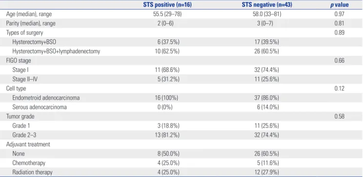
Fifty-nine patients were included in this study. Of these, 16 pa- tients (27.1%) were STS positive (group A) (Fig. 1A) and 43 pa- tients were STS negative (group B) (Fig. 1B). There was no sig- nificant difference in general characteristics between group A and group B. None of the patients received CCRT as an adju- vant treatment (Table 1). In the STS positive group, 8 patients were diagnosed as stage IA, and 3 patients were diagnosed as
Fig. 1. The results of immunohistochemical staining (×400) of steroid sulfatase. (A) Positive. (B) Negative.
A B
Table 1. General Characteristics in Patients with Endometrial Cancer
STS positive (n=16) STS negative (n=43) p value
Age (median), range 55.5 (29–78) 58.0 (33–81) 0.97
Parity (median), range 2 (0–6) 3 (0–7) 0.81
Types of surgery 0.89
Hysterectomy+BSO 6 (37.5%) 17 (39.5%)
Hysterectomy+BSO+lymphadenectomy 10 (62.5%) 26 (60.5%)
FIGO stage 0.66
Stage I 11 (68.6%) 32 (74.4%)
Stage II–IV 5 (31.2%) 11 (25.6%)
Cell type 0.12
Endometroid adenocarcinoma 16 (100%) 37 (86.0%)
Serous adenocarcinoma 0 (0%) 6 (14.0%)
Tumor grade 0.58
Grade 1 3 (18.8%) 11 (25.6%)
Grade 2–3 13 (81.2%) 32 (74.4%)
Adjuvant treatment
None 8 (50.0%) 26 (60.5%)
Chemotherapy 4 (25.0%) 5 (11.6%)
Radiation therapy 4 (25.0%) 12 (27.9%)
STS, steroid sulfatase; BSO, bilateral salpingo-oophorectomy; FIGO, International Federation of Gynecology and Obstetrics.
more than stage IB who were received adjuvant treatment;
three patients were grade 1, 5 patients were grade 2 and 8 pa- tients were grade 3. In the STS negative group, 26 patients were stage IA, and 6 patients were stage IB and IC who were re-
ceived adjuvant treatment; eleven patients were grade 1 and 15 patients were grade 2 and 6 patients were grade 3.
Two of the 16 patients in group A had recurrence and 1 pa- tient died. Six patients had recurrence and subsequent mor-
Fig. 2. Kaplan-Meier estimates of disease free survival between STS pos- itive and STS negative in patients with endometrial cancer. STS, steroid sulfatase.
1.0
0.8
0.6
0.4
0.2
0.0
0.0 25.0 50.0 75.0 100.0 125.0 Follow up time (months)
Disease free survival (%)
p=0.92 STS
STS negative STS positive 0-sensored 1-sensored
Fig. 3. Kaplan-Meier estimates of overall survival between STS positive and STS negative in patients with endometrial cancer. STS, steroid sulfa- tase.
1.0
0.8
0.6
0.4
0.2
0.0
0.0 25.0 50.0 75.0 100.0 125.0 Follow up time (months)
Overall survival (%)
p=0.45 STS
STS negative STS positive 0-sensored 1-sensored
Table 2. Association of Clinicopathologic Variables with Disease-Free Survival in Patients with Endometrial Cancer
Variable Univariate analysis Multivariate analysis
HR 95% CI p value HR 95% CI p value
Age 0.58 0.72
<50 1 1
≥50 1.0 0.92–1.05 1.4 0.22–9.0
Types of surgery 0.23 0.76
Hysterectomy+BSO 1 1
Hysterectomy+BSO+lymphadenectomy 0.6 0.27–1.36 1.3 0.21–9.03
FIGO stage 0.01 0.16
Stage I 1 1
Stage II–IV 9.6 1.8–49.9 4.7 0.6–41.4
Cell type 0.81 0.15
Endometroid 1 1
Serous 0.9 0.31–2.49 0.1 0.02–1.89
Tumor grade 0.54 0.96
Grade 1 1 1
Grade 2–3 1.9 0.2–16.2 1.1 1.2–9.4
Adjuvant treatment 0.03 0.35
None 1 1
Chemotherapy or radiation therapy 9.9 1.2–82.9 3.8 0.2–62.9
STS 0.47 0.24
Negative 1 1
Positive 0.5 0.06–3.8 0.3 0.3–2.4
STS, steroid sulfatase; HR, hazard ratio; CI, confidential interval; BSO, bilateral salpingo-oophorectomy; FIGO, International Federation of Gynecology and Obstetrics.
tality in group B. Using Kaplan-Meier survival analysis, DFS (mean±standard deviation) was 129.83±8.67 (95% CI: 112.84–
146.82) months in group A, and 111.06±7.17 (95% CI: 97.01–
125.10) months in group B (p=0.92) (Fig. 2). OS (mean±standard deviation) was 129.01±9.38 (95% CI: 110.63–147.38) months in group A, and 111.16±7.10 (95% CI: 97.24–125.07) months in group B (p=0.45) (Fig. 3).
In univariate Cox proportional hazard analysis, FIGO stage (p=0.007 in DFS, p=0.006 in OS) and adjuvant treatment (p=
0.034 in DFS, p=0.034 in OS) were significantly associated with DFS and OS. In multivariate analysis, however, FIGO stage and adjuvant treatment were not significantly associated with DFS and OS. The expression of STS was not statistically signifi- cant with DFS and OS (Table 2 and 3).
DISCUSSION
STS is a single enzyme that hydrolyses not only estrone sulfate (E1S) and estradiol sulfate (E2S), but also various steroid sul- fates, such as dehydroepiandrosterone sulfate (DHEA-S) and cholesterol sulfate. Synthesis of sex hormones involves the con- version of androstenedione or testosterone to E2 or estrone (E1) by aromatase, and these hormones are converted by 17β- hydroxysteroid dehydrogenase (17βHSD). The inhibition of aromatase or 17βHSD at this point would inhibit the synthesis of E2 or E1. However, Adiol, which is synthesized from DHEA
or DHEA-S via STS, is not inhibited and is eventually convert- ed into estrogen.
The activity of STS is 12 times higher in endometrial cancer tissue, as compared to normal endometrial tissue,12 and is ex- pressed up to 86% on immunohistochemical staining.13 Previ- ous studies showed that the STS pathway is an important source of estrogen and STS inhibitors are effective in breast cancer. These results have led to the clinical use of STS and STS inhibitors in patients with another hormone-dependent cancer (i.e., endometrial cancer).
STX 64 is the only STS inhibitor that completed the phase I clinical trial among the developed STS inhibitors. In various in vivo tumor models, STX 64 showed a great inhibitory effect of estrogen activity.14 STX 213 is a second generation STS in- hibitor that showed an eight times stronger effect than STX 64 in vitro to completely block estrogen activity. The most signifi- cant characteristic of second generation STS inhibitor is the long duration of STS inhibition. In the study with a mouse model, the time to recover of STS activity was 10 days in STX 213, as compared to 3 days in STX 64.15,16 However, the clinical application of STS inhibitors is limited. DFS was reported from 2.8 months to 7 months in phase I of clinical trial with 14 breast cancer patients using STX 64.17
Although limited, clinical studies were conducted in breast cancer patients, but only animal studies were conducted in en- dometrial cancer patients. When STX 64 and STX 213 were ad- ministered orally to ovariectomized rats with endometrial Table 3. Association of Clinicopathologic Variables with Overall Survival in Patients with Endometrial Cancer
Variable Univariate analysis Multivariate analysis
HR 95% CI p value HR 95% CI p value
Age 0.62 0.26
<50 1 1
≥50 0.6 0.14–2.76 0.5 0.03–2.61
Types of surgery 0.79 0.21
Hysterectomy+BSO 1 1
Hysterectomy+BSO+lymphadenectomy 0.8 0.18–3.63 0.2 0.02–2.46
FIGO stage 0.01 0.13
Stage I 1 1
Stage II–IV 9.9 1.9–51.2 10.35 0.49–2.18
Cell type 0.81 0.18
Endometroid 1 1
Serous 0.9 0.31–2.49 0.18 0.01–2.26
Tumor grade 0.53 0.99
Grade 1 1 1
Grade 2–3 1.9 0.24–16.3 1.0 0.1–9.2
Adjuvant treatment 0.03 0.35
None 1 1
Chemotherapy or radiation therapy 9.9 1.2–82.8 3.8 0.2–62.6
STS 0.46 0.18
Negative 1 1
Positive 0.5 0.05–3.7 0.2 0.03–1.9
STS, steroid sulfatase; HR, hazard ratio; CI, confidential interval; BSO, bilateral salpingo-oophorectomy; FIGO, International Federation of Gynecology and Obstetrics.
cancer xenograft, the cancer cell growth was inhibited by 48%
and 67%, respectively. Furthermore, cancer cell growth was in- hibited up to 59% and serum estradiol was significantly re- duced by STX 213 at 10 mg/kg daily.18
Unlike previous in vitro and in vivo studies, we used human endometrial cancer tissues. In this study, STS was expressed in sixteen patients out of 59 patients (27%), which was lower than the previous animal study with immuno-histochemical staining (86%). A possible explanation for this discrepancy might be the use of paraffin-embedded tissue post-formalin fixing vs. fresh endometrial cancer tissue. The use of rabbit polyclonal anti-STS antibody for STS detection might be an- other cause. Although previous study showed that the im- mune response was lower in the endometrium, as compared to human breast or hepatic tissue,19 there is no antibody with better response to STS detection.
Our results showed that the expression of STS did not affect DFS and OS in endometrial cancer patients. Although the ef- fect was not statistically significant, survival was better in the STS positive group, unlike in the previous study conducted in breast cancer patients. In addition, FIGO stage and adjuvant treatment, which are known prognostic factors, were not as- sociated with DFS and OS. The main reasons for this were the small sample size and the few events of recurrence and death.
Other reasons were subtle differences between the two groups in the proportions of the cell types and in the numbers of pa- tients receiving radiation therapy. Specifically, the STS nega- tive group contained a larger number of patients with papil- lary serous adenocarcinoma and undergoing radiation therapy, and a previous study has shown that papillary serous adeno- carcinoma has a poorer prognosis than endometroid adeno- carcinoma.20 Also, radiation therapy has been considered the standard of adjuvant treatment, but the treatment failure rate in advanced stage patients is reportedly up to 67%.21
Another consideration is the sex hormone inhibitor with different response in breast and endometrium tissue. Tamoxi- fen has an anti-estrogen effect on breast tissue, but has an es- trogen effect on endometrium tissue, which can cause endo- metrial hyperplasia or dysfunctional uterine bleeding and endometrial cancer.22-24 On the other hand, raloxifene, another selective ER active substance, has a treatment effect on breast cancer and neutral effect on endometrial tissue.25 AIs also showed excellent clinical results in breast cancer, but there is only a minimal inhibitory effect on endometrial cancer.26-28 These results can be explained by ER. The subtype of ER is di- vided into alpha receptor (ERα) and beta receptor (ERβ), both of which have a different distribution in each organ.29 ERα is dominant in breast tissue, while ERβ is dominant in endome- trial tissue.30 Therefore, the effect of the same sex hormone in- hibitor can differ in breast and endometrial cancer. Another study showed that the mutation of exon 5, 8, and 36 in ERα may lead to lower anti-cancer effects in endometrial cancer.31-33
Theoretically, the clinical effect of STS inhibitor in endome-
trial cancer is expected to be significant. The effect would be greater if there is concurrent inhibition of aromatase and 17βHSD in the main pathway of estrogen synthesis and STS. However, we did not show the effect of the expression of STS in endo- metrial cancer as a prognostic factor. Although these findings were rather disappointing, they warrant future endometrial cancer trials with STS including ER, more specific antibody and endometrial cell biological markers.
In conclusion, STS was detected in approximately 27% hu- man endometrial cancer tissue by immuno-histochemical staining. However, STS expression was not significantly asso- ciated with DFS and OS. Therefore, STS as a prognostic factor in patients with endometrial cancer is questionable, hence re- quires more study and a careful result-based approach.
ACKNOWLEDGEMENTS
This work was supported by the research fund of Hanyang Uni- versity (HY-2012-N).
REFERENCES
1. Sherman ME, Sturgeon S, Brinton L, Kurman RJ. Endometrial can- cer chemoprevention: implications of diverse pathways of carci- nogenesis. J Cell Biochem Suppl 1995;23:160-4.
2. Sherman ME. Theories of endometrial carcinogenesis: a multi- disciplinary approach. Mod Pathol 2000;13:295-308.
3. Thomas DB. Do hormones cause breast cancer? Cancer 1984;53(3 Suppl):595-604.
4. Kelsey JL, LiVolsi VA, Holford TR, Fischer DB, Mostow ED, Schwartz PE, et al. A case-control study of cancer of the endome- trium. Am J Epidemiol 1982;116:333-42.
5. Ryan KJ. Editorial: Cancer risk and estrogen use in the meno- pause. N Engl J Med 1975;293:1199-200.
6. Reed MJ, Purohit A, Woo LW, Newman SP, Potter BV. Steroid sul- fatase: molecular biology, regulation, and inhibition. Endocr Rev 2005;26:171-202.
7. Woo LW, Purohit A, Potter BV. Development of steroid sulfatase inhibitors. Mol Cell Endocrinol 2011;340:175-85.
8. Geisler J, Sasano H, Chen S, Purohit A. Steroid sulfatase inhibitors:
promising new tools for breast cancer therapy? J Steroid Biochem Mol Biol 2011;125:39-45.
9. Masamura S, Santner SJ, Heitjan DF, Santen RJ. Estrogen depriva- tion causes estradiol hypersensitivity in human breast cancer cells.
J Clin Endocrinol Metab 1995;80:2918-25.
10. Sasano H, Frost AR, Saitoh R, Harada N, Poutanen M, Vihko R, et al. Aromatase and 17 beta-hydroxysteroid dehydrogenase type 1 in human breast carcinoma. J Clin Endocrinol Metab 1996;81:
4042-6.
11. Utsunomiya H, Suzuki T, Kaneko C, Takeyama J, Nakamura J, Kimura K, et al. The analyses of 17beta-hydroxysteroid dehydroge- nase isozymes in human endometrial hyperplasia and carcino- ma. J Clin Endocrinol Metab 2001;86:3436-43.
12. Tokunaga K, Nakamura Y, Sakata K, Fujimori K, Ohkubo M, Sawa- da K, et al. Enhanced expression of a glyceraldehyde-3-phosphate dehydrogenase gene in human lung cancers. Cancer Res 1987;47:
5616-9.
13. Utsunomiya H, Ito K, Suzuki T, Kitamura T, Kaneko C, Nakata T, et al. Steroid sulfatase and estrogen sulfotransferase in human en-
dometrial carcinoma. Clin Cancer Res 2004;10:5850-6.
14. Purohit A, Woo LW, Potter BV, Reed MJ. In vivo inhibition of es- trone sulfatase activity and growth of nitrosomethylurea-induced mammary tumors by 667 COUMATE. Cancer Res 2000;60:3394-6.
15. Foster PA, Newman SP, Chander SK, Stengel C, Jhalli R, Woo LL, et al. In vivo efficacy of STX213, a second-generation steroid sulfatase inhibitor, for hormone-dependent breast cancer therapy. Clin Cancer Res 2006;12:5543-9.
16. Foster PA, Chander SK, Parsons MF, Newman SP, Woo LW, Potter BV, et al. Efficacy of three potent steroid sulfatase inhibitors: pre- clinical investigations for their use in the treatment of hormone- dependent breast cancer. Breast Cancer Res Treat 2008;111:129-38.
17. Stanway SJ, Purohit A, Woo LW, Sufi S, Vigushin D, Ward R, et al.
Phase I study of STX 64 (667 Coumate) in breast cancer patients:
the first study of a steroid sulfatase inhibitor. Clin Cancer Res 2006;
12:1585-92.
18. Foster PA, Woo LW, Potter BV, Reed MJ, Purohit A. The use of ste- roid sulfatase inhibitors as a novel therapeutic strategy against hor- mone-dependent endometrial cancer. Endocrinology 2008;149:
4035-42.
19. Selcer KW, Difrancesca HM, Chandra AB, Li PK. Immunohisto- chemical analysis of steroid sulfatase in human tissues. J Steroid Biochem Mol Biol 2007;105:115-23.
20. Greggi S, Mangili G, Scaffa C, Scala F, Losito S, Iodice F, et al. Uter- ine papillary serous, clear cell, and poorly differentiated endome- trioid carcinomas: a comparative study. Int J Gynecol Cancer 2011;
21:661-7.
21. Ayeni TA, Bakkum-Gamez JN, Mariani A, McGree ME, Weaver AL, Haddock MG, et al. Comparative outcomes assessment of uterine grade 3 endometrioid, serous, and clear cell carcinomas. Gynecol Oncol 2013;129:478-85.
22. Cohen I. Endometrial pathologies associated with postmenopaus- al tamoxifen treatment. Gynecol Oncol 2004;94:256-66.
23. Neven P, Vergote I. Controversies regarding tamoxifen and uterine carcinoma. Curr Opin Obstet Gynecol 1998;10:9-14.
24. Slomovitz BM, Sun CC, Ramirez PT, Bodurka DC, Diaz P, Lu KH.
Does tamoxifen use affect prognosis in breast cancer patients who
develop endometrial cancer? Obstet Gynecol 2004;104:255-60.
25. DeMichele A, Troxel AB, Berlin JA, Weber AL, Bunin GR, Turzo E, et al. Impact of raloxifene or tamoxifen use on endometrial cancer risk: a population-based case-control study. J Clin Oncol 2008;26:
4151-9.
26. Berstein L, Maximov S, Gershfeld E, Meshkova I, Gamajunova V, Tsyrlina E, et al. Neoadjuvant therapy of endometrial cancer with the aromatase inhibitor letrozole: endocrine and clinical effects.
Eur J Obstet Gynecol Reprod Biol 2002;105:161-5.
27. Barker LC, Brand IR, Crawford SM. Sustained effect of the aroma- tase inhibitors anastrozole and letrozole on endometrial thickness in patients with endometrial hyperplasia and endometrial carci- noma. Curr Med Res Opin 2009;25:1105-9.
28. Ma BB, Oza A, Eisenhauer E, Stanimir G, Carey M, Chapman W, et al. The activity of letrozole in patients with advanced or recurrent endometrial cancer and correlation with biological markers--a study of the National Cancer Institute of Canada Clinical Trials Group. Int J Gynecol Cancer 2004;14:650-8.
29. Couse JF, Curtis Hewitt S, Korach KS. Receptor null mice reveal contrasting roles for estrogen receptor alpha and beta in reproduc- tive tissues. J Steroid Biochem Mol Biol 2000;74:287-96.
30. Kuiper GG, Carlsson B, Grandien K, Enmark E, Häggblad J, Nilsson S, et al. Comparison of the ligand binding specificity and transcript tissue distribution of estrogen receptors alpha and beta. Endocri- nology 1997;138:863-70.
31. Bryant W, Snowhite AE, Rice LW, Shupnik MA. The estrogen re- ceptor (ER)alpha variant Delta5 exhibits dominant positive activ- ity on ER-regulated promoters in endometrial carcinoma cells. En- docrinology 2005;146:751-9.
32. Lin SL, Yan LY, Liang XW, Wang ZB, Wang ZY, Qiao J, et al. A novel variant of ER-alpha, ER-alpha36 mediates testosterone-stimulated ERK and Akt activation in endometrial cancer Hec1A cells. Reprod Biol Endocrinol 2009;7:102.
33. Ogawa S, Inoue S, Watanabe T, Orimo A, Hosoi T, Ouchi Y, et al.
Molecular cloning and characterization of human estrogen recep- tor betacx: a potential inhibitor ofestrogen action in human. Nucle- ic Acids Res 1998;26:3505-12.