INTRODUCTION
Fractional flow reserve (FFR) is currently the gold standard for detecting lesion-specific myocardial ischemia.1-3 Prior studies have demonstrated that ischemia-causing lesions detected us- ing FFR portend poor prognosis.1 Furthermore, FFR-guided percutaneous coronary intervention (PCI) has shown survival benefits over invasive coronary angiography (ICA)-guided PCI alone.2,3
Coronary computed tomography angiography (CTA) is a noninvasive method for accurate detection and exclusion of
Diagnostic Accuracy of a Novel On-site Virtual
Fractional Flow Reserve Parallel Computing System
Hyung-Bok Park1,2*, Yeonggul Jang1*, Reza Arsanjani3, Minh Tuan Nguyen4, Sang-Eun Lee1,5, Byunghwan Jeon1, Sunghee Jung1, Youngtaek Hong1, Seongmin Ha1,
Sekeun Kim1, Sang-Wook Lee4, and Hyuk-Jae Chang1,5
1Connect-AI Research Center, Yonsei University College of Medicine, Seoul, Korea;
2Department of Cardiology, International St. Mary’s Hospital, Catholic Kwandong University College of Medicine, Incheon, Korea;
3Mayo Clinic, Division of Cardiology, Department of Internal Medicine, Scottsdale, AZ, USA;
4School of Mechanical Engineering, University of Ulsan, Ulsan, Korea;
5Division of Cardiology, Severance Cardiovascular Hospital, Yonsei University Health System, Seoul, Korea.
Purpose: To evaluate the diagnostic accuracy of a novel on-site virtual fractional flow reserve (vFFR) derived from coronary com- puted tomography angiography (CTA).
Materials and Methods: We analyzed 100 vessels from 57 patients who had undergone CTA followed by invasive FFR during cor- onary angiography. Coronary lumen segmentation and three-dimensional reconstruction were conducted using a completely automated algorithm, and parallel computing based vFFR prediction was performed. Lesion-specific ischemia based on FFR was defined as significant at ≤0.8, as well as ≤0.75, and obstructive CTA stenosis was defined that ≥50%. The diagnostic performance of vFFR was compared to invasive FFR at both ≤0.8 and ≤0.75.
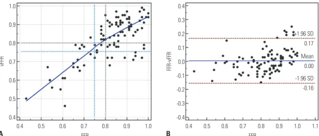
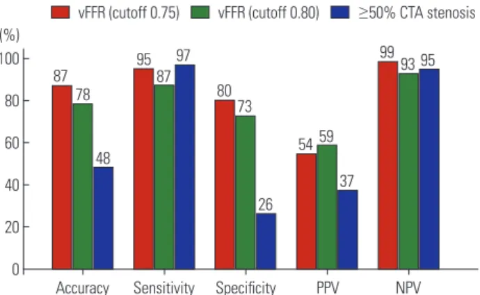
Results: The average computation time was 12 minutes per patient. The correlation coefficient (r) between vFFR and invasive FFR was 0.75 [95% confidence interval (CI) 0.65 to 0.83], and Bland-Altman analysis showed a mean bias of 0.005 (95% CI -0.011 to 0.021) with 95% limits of agreement of -0.16 to 0.17 between vFFR and FFR. The accuracy, sensitivity, specificity, positive predic- tive value, and negative predictive value were 78.0%, 87.1%, 72.5%, 58.7%, and 92.6%, respectively, using the FFR cutoff of 0.80.
They were 87.0%, 95.0%, 80.0%, 54.3%, and 98.5%, respectively, with the FFR cutoff of 0.75. The area under the receiver-operating characteristics curve of vFFR versus obstructive CTA stenosis was 0.88 versus 0.61 for the FFR cutoff of 0.80, respectively; it was 0.94 versus 0.62 for the FFR cutoff of 0.75.
Conclusion: Our novel, fully automated, on-site vFFR technology showed excellent diagnostic performance for the detection of lesion-specific ischemia.
Key Words: Fractional flow reserve, myocardial; computed tomography angiography; patient-specific computational modeling pISSN: 0513-5796 · eISSN: 1976-2437
Received: August 21, 2019 Revised: December 4, 2019 Accepted: December 19, 2019
Corresponding author: Sang-Wook Lee, PhD, School of Mechanical Engineering, University of Ulsan, 93 Daehak-ro, Nam-gu, Ulsan 44610, Korea.
Tel: 82-52-259-2765, Fax: 82-52-259-1680, E-mail: [email protected]
*Hyung-Bok Park and Yeonggul Jang contributed equally to this work.
•The authors have no potential conflicts of interest to disclose.
© Copyright: Yonsei University College of Medicine 2020
This is an Open Access article distributed under the terms of the Creative Com- mons Attribution Non-Commercial License (https://creativecommons.org/licenses/
by-nc/4.0) which permits unrestricted non-commercial use, distribution, and repro- duction in any medium, provided the original work is properly cited.
Yonsei Med J 2020 Feb;61(2):137-144 https://doi.org/10.3349/ymj.2020.61.2.137
high-grade coronary stenoses, hence it may serve as a gate- keeper for invasive catheterization.4-7 However, CTA tends to overestimate the severity of coronary artery stenosis, resulting in low specificity and positive predictive values (PPVs).8,9
Non-invasive CTA-derived FFR, which applies computa- tional fluid dynamics (CFD) onto three-dimensional (3D) cor- onary lumen geometry derived from CTA, has demonstrated high diagnostic accuracy, as well as improved specificity and PPVs, according to three prospective multicenter trials.10-12 Ul- timately, this novel technique allows for comprehensive ana- tomic and physiologic diagnosis of coronary artery disease (CAD).13 However, current non-invasive FFR techniques have several major limitations when applied to day-to-day clinical practice: The required simulation has to be performed outside of the hospital because it necessitates a supercomputer, there- fore requiring transfer of patient data potentially leading to ex- posure of personal patient information.14 Furthermore, the processing time can take several hours, and the service can be expensive.14-18
Therefore, we aimed to develop a new non-invasive on-site vFFR computing system derived from CTA that would allow for instantaneous utilization in clinical practice that is timelier and cost efficient. This technique uses a novel parallel com- puting method with a completely automated lumen segmen- tation algorithm without the need for a supercomputer.
MATERIALS AND METHODS
Study population
We consecutively enrolled clinically stable adult patients from September 2015 to February 2016 at Severance Cardiovascu- lar Hospital who underwent clinically indicated ICA within 30 days following CTA with no intervening coronary events. In- stitutional Review Board (Severance Hospital, IRB Number 1-2017-0031) approval was obtained for this retrospective study and informed consent was waived. Patients were ex- cluded if they met any of the following criteria: history of cor- onary artery bypass graft surgery; prior PCI with suspected in- stent restenosis; old myocardial infarction; complex congenital heart disease; prior pacemaker or defibrillator; prosthetic heart valve; significant arrhythmia; body mass index greater than 40; or evidence of active clinical instability or life-threat- ening disease.
CTA data acquisition
CTA images were acquired using two 64-slice multi-detector row computed tomography systems (Somatom Sensation 64, Siemens Medical Solutions, Forchheim, Germany) with pro- spective or retrospective electrocardiographic gating. All pa- tients with a heart rate of 65 beats per minute or higher received 100 mg of atenolol orally prior to the CT, unless contraindicat- ed. In addition, all patients received a 0.3-mg sublingual dose
of nitroglycerin just prior to scanning, unless contraindicated.
Bolus tracking was used for contrast injection. The scan pa- rameters for the machines were as follows: 64×0.6-mm section collimation and a 330-ms rotation time. Depending on body habitus, tube voltages and currents were adjusted as follows:
80, 100, or 120 kVp and 150–500 mAs. The median radiation dose was 3.92 mSv [95% confidence interval (CI) 2.56 to 5.65], with 9 patients receiving less than 1 mSv.
Invasive FFR measurement
Fractional flow reserve was measured in vessels deemed clini- cally indicated for evaluation. After administration of nitro- glycerin, a pressure-monitoring guidewire was advanced distal to a lesion. Hyperemia was induced by administration of intra- venous or intracoronary adenosine at a rate of 140 mg/kg/min, and FFR was calculated by dividing the mean distal coronary pressure by the mean aortic pressure during hyperemia. FFR at a threshold of ≤0.80 or ≤0.75 were considered hemodynam- ically significant leading to ischemia.
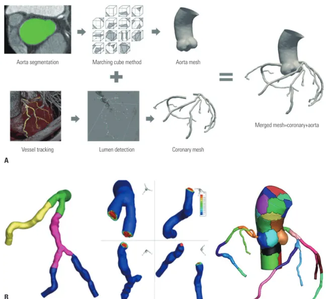
Fully automated lumen segmentation algorithm We reconstructed patient-specific coronary geometry auto- matically using in-house software on CTA images (Fig. 1A).
The fully automatic segmentation was performed with the fol- lowing four steps: at first, aorta and ostia were detected by our algorithm, which uses a Bayesian formulation in a pairwise fashion using anatomical and geometrical information.19 The detected points were used as seeds for coronary artery track- ing. Second, the centerlines of the left and right coronary arter- ies (RCA) were extracted starting from the detected ostia by our vessel tracking method based on stochastic geometric processes using an active branch search.20 The tracking meth- od could better find the branches, any stenotic lesions, and seemingly disconnected vessels that may be occluded by ath- erosclerotic plaque by modeling the statistical branch occur- rence and the vessel disconnection. Third, luminal boundaries for all vessels were automatically delineated every cross-sec- tional plane based on the image gradient and prior CT infor- mation on the number of plaques. Finally, we generated the surface mesh of the coronary structure with the estimated lu- minal boundaries. The connections of bifurcated vessels were processed smoothly using a mesh merging method that merged the vessel mesh and the divaricating branch mesh.21
Computational fluid dynamics for vFFR
To calculate vFFR, 3D blood flow dynamics were simulated us- ing the CFD technique for patient-specific coronary geometry (Fig. 1B). The continuity and Navier-Stokes equations for un- steady incompressible Newtonian fluid flow were solved with appropriate boundary conditions. A fully implicit four-step fractional method with a regular Crank-Nicolson scheme for time advancement and a P2P1 finite element method for spa- tial discretization were employed. In order to accommodate
the specific time-varying pressure-flow characteristics of cor- onary flow, which is affected by intramyocardial pressure sub- ject to cyclic contraction of the left ventricle and microcircula- tion in coronary distal networks, zero dimensional lumped parameter network (LPN) models were integrated into the 3D CFD model at all coronary outlets in a fully implicit manner.
The patient-specific total resistances and capacitance for the distal coronary bed in a resting condition, as well as a hyper- emic condition (i.e., the maximum flow condition resulting from the minimal microvascular resistance), were determined as de- scribed by Sankaran, et al.22 and Sharma, et al.23 The distribu- tion of flow rates between left anterior descending artery (LAD) and left circumflex artery (LCX) was determined according to the total volume of the corresponding vessel tree. Flow divi- sion into further distal branches was approximated by allome- tric scaling laws based on the coronary lumen cross-sectional area.24 The aortic pressure and flow rate were acquired by solv- ing the LPN models with constraints that matched the systolic
and the diastolic blood pressure of the individual patients.
To expedite simulation time, a parallel computing procedure based on a domain decomposition method was applied (Fig.
1B).25 The P2P1 unstructured mesh of the whole computation- al domain was decomposed into multiple subdomains using a k-way partition method,26 and the computing work for each subdomain was equally distributed to corresponding proces- sors. Simulations were carried out on a parallel cluster system (two nodes, 40 cores), where each node comprised two CPUs (E5-2680V2, 2.8 GHz, 25 MB L3 cache) and 64 GB RAM, with an Infiniband interconnect network between nodes.
Statistical analysis
Data are reported as a mean±SD for continuous variables and as proportions (%) for categorical variables. Pearson correla- tion coefficients using two-sided p-values, with p-values<0.05 considered statistically significant, and Fisher’s Z transforma- tion statistics were applied for comparing correlations. Bland–
Fig. 1. Workflow of the automated segmentation algorithm and the novel parallel computing method. (A) A fully automated lumen segmentation algorithm was applied to reconstruct patient-specific coronary geometry. (B) A novel parallel computing procedure based on a cluster with 40 cores decomposing the domain into 40 sub-domains and assigning a sub-domain to each computing core was applied.
Aorta segmentation
Vessel tracking
Marching cube method
Lumen detection
Aorta mesh
Coronary mesh
Merged mesh=coronary+aorta
A
B
Altman plots with 95% CIs for correlations were calculated.
FFRs were each dichotomized at a threshold of 0.80 and 0.75, with FFR values ≤0.80 and ≤0.75, respectively, considered he- modynamically significant and causal of ischemia. The diag- nostic accuracy, sensitivity, specificity, PPV, and negative pre- dictive value (NPV) between vFFR and FFR at 0.80 and 0.75 were presented as proportions and 95% CIs. The areas under receiver operating characteristic curves (AUC) were com- pared according to the method proposed by DeLong, et al.27 Statistical analyses were performed using Medcalc software (version 17.6; MedCalc Software, Mariakerke, Belgium).
RESULTS
Three out of 60 patients analyzed (5%) were excluded based on failure of lumen segmentation due to severely calcified lesions or motion artifacts. We subsequently analyzed 100 lesions from 57 patients (42 men and 15 women, mean age: 67.3±8.5 years).
Baseline patient characteristics are presented in Table 1. The
lesions were primarily located in the LAD (47%), followed by the RCA (23%), LCX (18%), diagonal branch (DG, 5%), obtuse marginal branch (OM, 4%), left main (LM, 2%), and ramus in- termedius (RI, 1%). The plaque composition profiles of the le- sions were as follows: mixed plaque (59%), non-calcified plaques (29%), and calcified plaques (12%). Forty percent of plaques were located in the proximal segments of coronary arteries.
The average time for generating patient-specific 3D coronary geometry was 233 seconds (min=175 s, Q1=196 s, median=212 s, Q3=221 s, max=386 s). CFD simulations were carried out up to two cardiac cycles to damp initial transients and most runs were able to be completed within 20 minutes (average=514.7 s, min= 232 s, Q1=380 s, median=439 s, Q3=544 s, max=1370 s).
The run time was highly dependent on the smoothness of cor- onary lumen surface and computational mesh quality. The representative simulation case of vFFR is shown in Fig. 2.
The correlation coefficient between vFFR and FFR was 0.75 (95% CI 0.65 to 0.83), and Bland-Altman analysis reavealed a mean bias of 0.005 (95% CI -0.011 to 0.021), with 95 % limits of agreement of -0.16 to 0.17 between vFFR and FFR (Fig. 3). Di- agnostic performance was evaluated separately using FFR cut- offs of 0.80 and 0.75. The accuracy, sensitivity, specificity, PPV, and NPV when the FFR cutoff of 0.80 was used were 78.0% (95%
CI 64.0–87.0), 87.1% (95% CI 70.2–96.4), 72.5% (95% CI 60.4–
82.5), 58.7% (95% CI 48.6–68.1), and 92.6% (95% CI 83.2–96.9), respectively. In addition, the AUC value for vFFR was signifi- cantly higher [0.88 (95% CI 0.80–0.94) vs. 0.61 (95% CI 0.51–
0.71)] than CTA ≥50% stenosis (Fig. 4A). Moreover, when a FFR cutoff of 0.75 was used the diagnostic performance signifi- cantly improved, with accuracy, sensitivity, specificity, PPV, Table 1. Baseline Characteristics of the Study Population
Baseline characteristics (n=57)
Mean age, yrs 67.3±8.5
Male, % 73.7
Mean body-mass index 25.1±2.7
Hypertension, % 56.1
Diabetes, % 31.6
Dyslipidemia, % 45.6
Family history, % 7
Current smoker, % 29.8
Vital signs
Systolic blood pressure, mm Hg 135.9±15.5 Diastolic blood pressure, mm Hg 77.0±10.0
Heart rate, beat/min 60.7±8.9
Laboratory measures
Hemoglobin, mg/dL 13.9±1.5
Hematocrit, % 41.0±4.5
Creatinine, mg/dL 0.89±0.22
Total cholesterol, mg/dL 159.5±39.8
LDL cholesterol, mg/dL 88.7±33.3
HDL cholesterol, mg/dL 44.6±11.7
Triglycerides, mg/dL 124.1±79.6
Medications, %
Aspirin 50.9
Clopidogrel 28.1
Beta-blocker 26.3
Nitrate 19.3
Statins 57.9
ACE inhibitors/ARB 22.8
Calcium channel blocker 36.8
LDL, low-density lipoprotein; HDL, high-density lipoprotein; ACE, angiotensin- converting enzyme; ARB, angiotensin II receptor blocker.
Fig. 2. Example simulation case of on-site virtual fractional flow reserve (vFFR). A noninvasive on-site vFFR simulation defined the distal portion of the right coronary artery (A) as an ischemic lesion (0.76) and the middle portion of the left anterior descending artery (B) as a non-ischemic lesion (0.84). These simulation derived values matched perfectly with the inva- sively measured FFR values.
1.0
0.9
0.8
vFFRA=0.76 0.7 (FFR=0.76)
vFFR vFFRB=0.84
(FFR=0.84) A
B
and NPV of 87.0% (95% CI 77.0–95.7), 95.0% (95% CI 75.1–
99.9), 80.0% (95% CI 69.6–88.1), 54.3% (95% CI 43.1–65.1), and 98.5% (95% CI 90.4–99.8), respectively. In addition, the AUC value for vFFR was excellent [0.94 (95% CI 0.88–0.98) vs.
0.62 (95% CI 0.52–0.71)], compared with CTA ≥50% stenosis (Fig. 4B). Fig. 5 shows the comparison of the diagnostic perfor- mances between vFFR and FFR at 0.8, between vFFR and FFR at 0.75, and between CTA ≥50% stenosis and FFR at 0.8.
The diagnostic performances according to vessel size were also evaluated by separating large vessels (n=70), such as the LM, LAD, and RCA, from small vessels (n=30), such as LCX, DG, OM, and RI. Large vessels showed higher accuracy than small vessels both with FFR cutoffs of 0.80 (80.5% vs. 73.3%) and 0.75
(88.9% vs. 83.3%).
DISCUSSION
In the present study, we developed a novel non-invasive CTA- derived on-site vFFR method and demonstrated its excellent correlation with invasively measured FFR. In addition, this method exhibited excellent diagnostic performance for de- tecting ischemia producing lesions, compared to invasive FFR as a reference standard. Furthermore, vFFR had significantly higher diagnostic performance and improved discriminatory power for the detection of lesion-specific ischemia, compared Fig. 3. Linear regression (A) and Bland-Altman analysis (B) between vFFR and FFR. Correlation coefficient (r) between vFFR and FFR was 0.75 (95% CI 0.65 to 0.83), and Bland-Altman analysis showed a mean bias of 0.005 (95% CI -0.011 to 0.021), with 95% limits of agreement of -0.16 to 0.17. vFFR, virtual frac- tional flow reserve; CI, confidence interval.
1.0
0.9
0.8
0.7
0.6
0.5
0.4
vFFR
0.4 0.5 0.6 0.7 0.8 0.9 1.0 A FFR
0.4 0.3 0.2 0.1 0.0 -0.1 -0.2 -0.3 -0.4
FFR-vFFR
0.4 0.5 0.6 0.7 0.8 0.9 1.0 1.1 -0.16
0.00 0.17
-1.96 SD +1.96 SD
Mean
B FFR
Fig. 4. ROC demonstrating AUCs for vFFR and obstructive (≥50%) CTA stenosis for the discrimination of lesion-specific ischemia using FFR cutoff values of 0.8 and 0.75. (A) The AUC for vFFR was significantly higher (0.88, 95% CI 0.80–0.94) than CTA ≥50% stenosis (0.61, 95% CI 0.51–0.71) when an FFR cutoff of 0.8 was used. (B) The AUC value for vFFR was excellent (0.94, 95% CI 0.88–0.98), compared to the CTA ≥50% stenosis (0.62, 95% CI 0.52–0.71), when an FFR cutoff of 0.75 was used. ROC, receiver operating characteristic curve; AUC, areas under receiver operating characteristic curve; vFFR, virtual frac- tional flow reserve; CTA, com-puted tomography angiography; CI, confidence interval.
100
80
60
40
20
0
Sensitivity
0 20 40 60 80 100 vFFR, AUC 0.88 CTA, AUC 0.61
100-specificity A
100
80
60
40
20
0
Sensitivity
0 20 40 60 80 100 vFFR, AUC 0.94 CTA, AUC 0.62
100-specificity B
to obstructive (≥50%) CTA stenosis alone. Moreover, we also discovered that when an FFR cutoff of 0.75 was used, the diag- nostic accuracy and discriminatory ability of vFFR for ischemia detection was greatly enhanced.
CTA is a robust noninvasive tool for ruling out obstructive or high-grade coronary artery stenosis, obviating unnecessary use of invasive catheterization.4-7 On the other hand, CTA’s tenden- cy to overestimate coronary artery stenosis can lead to addi- tional testing that is often unnecessary due to its low specificity and PPV for the detection of ischemia causing stenosis.8,9 Based on these limitations, FFR-CT was created, and several FFR-CT studies have shown great improvement in the diagnostic per- formance of ischemia detection by increasing specificity and PPV compared with other non-invasive imaging modalities, such as stress echocardiography, single photon emission com- puted tomography, or cardiac magnetic resonance.10-12,28 How- ever, FFR-CT has several major limitations, including high add- on costs (approximately $1500), potential risk of exposure of personal patient information based on need for off-site post- processing, and considerable processing time leading to a po- tential delay in patient care.14-18 Furthermore, a recently intro- duced on-site CTA derived cFFR (Siemens cFFR, version 1.4;
Siemens Healthcare, Forchheim, Germany) has been shown to have high diagnostic accuracy.29 However, this method re- quires a semi-automated coronary lumen segmentation pro- cess (–60 minutes per case), which might be a considerable limitation when considering its application to daily practice.30
On the other hand, the vFFR does not require the use of a su- percomputer to compute coronary flow. Instead, the imple- mentation of on-site CFD calculation is done using a novel parallel computing method. This method is based on a cluster with 40 cores, which then decomposes the domain into 40 sub- domains and assigns a sub-domain to each computing core.
Additionally, the coronary lumen segmentation and 3D recon- struction is conducted by a completely automated algorithm without any operator intervention, thus making the results available within 30 minutes, including segmentation and CFD
calculation. In addition, the median radiation dose of CTA in this study was 3.92 mSv (95% CI 2.56 to 5.65), and 9 patients (16%) received less than 1 mSv (using 80 kVp along prospec- tive electrocardiogram gating). To date, several studies have shown that CTA is able to visualize not only stenosis severity but also adverse plaque characteristics, which are closely re- lated with future adverse events, in addition to lesion specific ischemia.31-35 For example, Gaur, et al.36 demonstrated that ad- verse plaque characteristics provide additive valve in FFR-CT for prediction of ischemia. Based on these findings, CTA with the aid of on-site vFFR could potentially provide a comprehen- sive evaluation of coronary stenosis and adverse plaque fea- tures, as well as assessment of hemodynamically significant CAD, which could lead to a true “one-stop shop” with sub mil- li-Sievert radiation exposure.
This study is not without limitations. First, this was a retro- spective pioneering study with a limited number of cases. Pres- ently, vFFR requires further well-powered validation in pro- spective multicenter trials. Second, the vessels interrogated by FFR were limited to those clinically indicated, introducing po- tential selection bias. Lastly, we were unable to evaluate 5%
(3/60) of patients in whom vessels were heavily calcified or there were severe motion artifacts that made CTA-derived FFR meth- ods impossible. CTA image quality was also an important fac- tor for successful simulation in this study, which is the case in most other imaging studies.37,38 Recently developed high spa- tial resolution and wider CTA detector coverage may provide a solution for this issue in the near future.39
In conclusion, a novel on-site vFFR computing system em- ploying a fully automated segmentation algorithm using a par- allel computation method showed excellent diagnostic perfor- mance for the detection of lesion-specific ischemia and was significantly faster and cheaper.
ACKNOWLEDGEMENTS
This work was supported by the Institute for Information &
Communications Technology Promotion (IITP) grant funded by the Korea government (MSIP) (No. R0101-16-0171, Devel- opment of Multi-modality Imaging and 3D Simulation-Based Integrative Diagnosis-Treatment Support Software System for Cardiovascular Diseases).
AUTHOR CONTRIBUTIONS
Conceptualization: Hyung-Bok Park, Sang-Wook Lee, and Hyuk-Jae Chang. Data curation: Hyung-Bok Park, Yeonggul Jang, Sang-Wook Lee, and Hyuk-Jae Chang. Formal analysis: Hyung-Bok Park, Yeonggul Jang, and Sang-Wook Lee. Funding acquisition: Hyuk-Jae Chang. In- vestigation: Hyung-Bok Park, Yeonggul Jang, Sang-Wook Lee, Reza Arsanjani, Minh Tuan Nguyen, Sang-Eun Lee, Byunghwan Jeon, Sunghee Jung, and Seongmin Ha. Methodology: Hyung-Bok Park, Sang-Wook Lee, and Hyuk-Jae Chang. Project administration: Hyuk- Jae Chang. Resources: Hyuk-Jae Chang and Sang-Wook Lee. Software:
100 80 60 40 20 0
87 87
8073
54 59 37
9993 95
26 95 97
78
48
Accuracy Sensitivity Specificity PPV NPV vFFR (cutoff 0.75) vFFR (cutoff 0.80) ≥50% CTA stenosis
Fig. 5. Diagnostic performance of vFFR using a cutoff of 0.75 (red), vFFR us- ing a cutoff of 0.8 (green), and obstructive (≥50%) CTA stenosis (blue) for lesion-specific ischemia detection. vFFR, virtual fractional flow reserve;
CTA, computed tomography angiography.
(%)
Yeonggul Jang, Reza Arsanjani, Minh Tuan Nguyen, and Youngtaek Hong. Supervision: Sang-Wook Lee and Hyuk-Jae Chang. Validation:
Yeonggul Jang, Reza Arsanjani, Minh Tuan Nguyen, and Sang-Eun Lee. Visualization: Seongmin Ha and Sekeun Kim. Writing—original draft: Hyung-Bok Park, Yeonggul Jang, Sang-Wook Lee, and Hyuk-Jae Chang. Writing—review & editing: Hyung-Bok Park, Yeonggul Jang, Sang-Wook Lee, and Hyuk-Jae Chang. Approval of final manuscript:
All authors.
ORCID iDs
Hyung-Bok Park https://orcid.org/0000-0002-3773-2665 Yeonggul Jang https://orcid.org/0000-0002-5805-7494 Reza Arsanjani https://orcid.org/0000-0001-7081-4286 Minh Tuan Nguyen https://orcid.org/0000-0002-2915-9133 Sang-Eun Lee https://orcid.org/0000-0001-6645-4038 Byunghwan Jeon https://orcid.org/0000-0002-0414-1762 Sunghee Jung https://orcid.org/0000-0002-8042-457X Youngtaek Hong https://orcid.org/0000-0003-2104-5905 Seongmin Ha https://orcid.org/0000-0002-0731-2301 Sekeun Kim https://orcid.org/0000-0003-4196-6242 Sang-Wook Lee https://orcid.org/0000-0002-8600-9991 Hyuk-Jae Chang https://orcid.org/0000-0002-6139-7545
REFERENCES
1. Tonino PA, De Bruyne B, Pijls NH, Siebert U, Ikeno F, van’ t Veer M, et al. Fractional flow reserve versus angiography for guiding per- cutaneous coronary intervention. N Engl J Med 2009;360:213-24.
2. Pijls NH, Fearon WF, Tonino PA, Siebert U, Ikeno F, Bornschein B, et al. Fractional flow reserve versus angiography for guiding per- cutaneous coronary intervention in patients with multivessel cor- onary artery disease: 2-year follow-up of the FAME (Fractional Flow Reserve Versus Angiography for Multivessel Evaluation) study. J Am Coll Cardiol 2010;56:177-84.
3. De Bruyne B, Pijls NH, Kalesan B, Barbato E, Tonino PA, Piroth Z, et al. Fractional flow reserve-guided PCI versus medical therapy in stable coronary disease. N Engl J Med 2012;367:991-1001.
4. Budoff MJ, Dowe D, Jollis JG, Gitter M, Sutherland J, Halamert E, et al. Diagnostic performance of 64-multidetector row coronary com- puted tomographic angiography for evaluation of coronary artery stenosis in individuals without known coronary artery disease: re- sults from the prospective multicenter ACCURACY (Assessment by Coronary Computed Tomographic Angiography of Individuals Undergoing Invasive Coronary Angiography) trial. J Am Coll Car- diol 2008;52:1724-32.
5. Miller JM, Rochitte CE, Dewey M, Arbab-Zadeh A, Niinuma H, Gottlieb I, et al. Diagnostic performance of coronary angiography by 64-row CT. N Engl J Med 2008;359:2324-36.
6. Meijboom WB, Meijs MF, Schuijf JD, Cramer MJ, Mollet NR, van Mieghem CA, et al. Diagnostic accuracy of 64-slice computed to- mography coronary angiography: a prospective, multicenter, mul- tivendor study. J Am Coll Cardiol 2008;52:2135-44.
7. Dewey M, Rief M, Martus P, Kendziora B, Feger S, Dreger H, et al.
Evaluation of computed tomography in patients with atypical an- gina or chest pain clinically referred for invasive coronary angiog- raphy: randomised controlled trial. BMJ 2016;355:i5441.
8. Raff GL, Gallagher MJ, O’Neill WW, Goldstein JA. Diagnostic accu- racy of noninvasive coronary angiography using 64-slice spiral computed tomography. J Am Coll Cardiol 2005;46:552-7.
9. Leber AW, Knez A, von Ziegler F, Becker A, Nikolaou K, Paul S, et al. Quantification of obstructive and nonobstructive coronary le-
sions by 64-slice computed tomography: a comparative study with quantitative coronary angiography and intravascular ultrasound. J Am Coll Cardiol 2005;46:147-54.
10. Koo BK, Erglis A, Doh JH, Daniels DV, Jegere S, Kim HS, et al. Di- agnosis of ischemia-causing coronary stenoses by noninvasive fractional flow reserve computed from coronary computed tomo- graphic angiograms. Results from the prospective multicenter DISCOVER-FLOW (Diagnosis of Ischemia-Causing Stenoses Ob- tained Via Noninvasive Fractional Flow Reserve) study. J Am Coll Cardiol 2011;58:1989-97.
11. Min JK, Leipsic J, Pencina MJ, Berman DS, Koo BK, van Mieghem C, et al. Diagnostic accuracy of fractional flow reserve from ana- tomic CT angiography. JAMA 2012;308:1237-45.
12. Nørgaard BL, Leipsic J, Gaur S, Seneviratne S, Ko BS, Ito H, et al. Di- agnostic performance of noninvasive fractional flow reserve de- rived from coronary computed tomography angiography in sus- pected coronary artery disease: the NXT trial (analysis of coronary blood flow using CT angiography: next steps). J Am Coll Cardiol 2014;63:1145-55.
13. Ding A, Qiu G, Lin W, Hu L, Lu G, Long X, et al. Diagnostic perfor- mance of noninvasive fractional flow reserve derived from coro- nary computed tomography angiography in ischemia-causing coronary stenosis: a meta-analysis. Jpn J Radiol 2016;34:795-808.
14. Min JK, Taylor CA, Achenbach S, Koo BK, Leipsic J, Nørgaard BL, et al. Noninvasive fractional flow reserve derived from coronary CT angiography: clinical data and scientific principles. JACC Car- diovasc Imaging 2015;8:1209-22.
15. Kimura T, Shiomi H, Kuribayashi S, Isshiki T, Kanazawa S, Ito H, et al. Cost analysis of non-invasive fractional flow reserve derived from coronary computed tomographic angiography in Japan. Car- diovasc Interv Ther 2015;30:38-44.
16. Douglas PS, De Bruyne B, Pontone G, Patel MR, Norgaard BL, By- rne RA, et al. 1-year outcomes of FFRCT-guided care in patients with suspected coronary disease: the PLATFORM study. J Am Coll Cardiol 2016;68:435-45.
17. Rajani R, Modi B, Ntalas I, Curzen N. Non-invasive fractional flow reserve using computed tomographic angiography: where are we now and where are we going? Heart 2017;103:1216-22.
18. Otake H, Taylor CA, Matsuo H, Tanaka N, Akasaka T. Noninvasive fractional flow reserve derived from coronary computed tomog- raphy angiography-is this just another new diagnostic test or the long-awaited game changer? Circ J 2017;81:1085-93.
19. Jeon B, Hong Y, Han D, Jang Y, Jung S, Hong Y, et al. Maximum a posteriori estimation method for aorta localization and coronary seed identification. Pattern Recognition 2017;68:222-32.
20. Han D, Shim H, Jeon B, Jang Y, Hong Y, Jung S, et al. Automatic cor- onary artery segmentation using active search for branches and seemingly disconnected vessel segments from coronary CT angi- ography. PLoS One 2016;11:e0156837.
21. Jang Y, Kim DH, Jeon B, Han D, Shim H, Chang HJ. Generation of triangular mesh of coronary artery using mesh merging. Journal of KIISE 2016;43:419-29.
22. Sankaran S, Esmaily Moghadam M, Kahn AM, Tseng EE, Guccione JM, Marsden AL. Patient-specific multiscale modeling of blood flow for coronary artery bypass graft surgery. Ann Biomed Eng 2012;40:2228-42.
23. Sharma P, Itu L, Zheng X, Kamen A, Bernhardt D, Suciu C, et al. A framework for personalization of coronary flow computations during rest and hyperemia. Conf Proc IEEE Eng Med Biol Soc 2012;
2012:6665-8.
24. Murray CD. The physiological principle of minimum work: I. The vascular system and the cost of blood volume. Proc Natl Acad Sci U S A 1926;12:207-14.
25. Nguyen MT, Jeon BJ, Chang HJ, Lee SW. Domain decomposition based parallel computing for multi-scale coronary blood flow sim- ulations. Computers & Fluids 2019;191:104254.
26. Karypis G, Kumar V. A fast and high quality multilevel scheme for partitioning irregular graphs. SIAM J Sci Comput 1998;20:359-92.
27. DeLong ER, DeLong DM, Clarke-Pearson DL. Comparing the ar- eas under two or more correlated receiver operating characteristic curves: a nonparametric approach. Biometrics 1988;44:837-45.
28. Nørgaard BL, Jensen JM, Leipsic J. Fractional flow reserve derived from coronary CT angiography in stable coronary disease: a new standard in non-invasive testing? Eur Radiol 2015;25:2282-90.
29. Renker M, Schoepf UJ, Wang R, Meinel FG, Rier JD, Bayer RR 2nd, et al. Comparison of diagnostic value of a novel noninvasive coro- nary computed tomography angiography method versus standard coronary angiography for assessing fractional flow reserve. Am J Cardiol 2014;114:1303-8.
30. Yang DH, Kim YH, Roh JH, Kang JW, Ahn JM, Kweon J, et al. Diag- nostic performance of on-site CT-derived fractional flow reserve versus CT perfusion. Eur Heart J Cardiovasc Imaging 2017;18:432- 40.
31. Motoyama S, Kondo T, Sarai M, Sugiura A, Harigaya H, Sato T, et al. Multislice computed tomographic characteristics of coronary lesions in acute coronary syndromes. J Am Coll Cardiol 2007;50:
319-26.
32. Motoyama S, Sarai M, Harigaya H, Anno H, Inoue K, Hara T, et al.
Computed tomographic angiography characteristics of athero- sclerotic plaques subsequently resulting in acute coronary syn- drome. J Am Coll Cardiol 2009;54:49-57.
33. Motoyama S, Ito H, Sarai M, Kondo T, Kawai H, Nagahara Y, et al.
Plaque characterization by coronary computed tomography angi- ography and the likelihood of acute coronary events in mid-term follow-up. J Am Coll Cardiol 2015;66:337-46.
34. Park HB, Heo R, Ó Hartaigh B, Cho I, Gransar H, Nakazato R, et al.
Atherosclerotic plaque characteristics by CT angiography identify coronary lesions that cause ischemia: a direct comparison to frac- tional flow reserve. JACC Cardiovasc Imaging 2015;8:1-10.
35. Nakazato R, Park HB, Gransar H, Leipsic JA, Budoff MJ, Mancini GB, et al. Additive diagnostic value of atherosclerotic plaque char- acteristics to non-invasive FFR for identification of lesions causing ischaemia: results from a prospective international multicentre trial. EuroIntervention 2016;12:473-81.
36. Gaur S, Øvrehus KA, Dey D, Leipsic J, Bøtker HE, Jensen JM, et al.
Coronary plaque quantification and fractional flow reserve by cor- onary computed tomography angiography identify ischaemia- causing lesions. Eur Heart J 2016;37:1220-7.
37. Leipsic J, Yang TH, Thompson A, Koo BK, Mancini GB, Taylor C, et al. CT angiography (CTA) and diagnostic performance of noninva- sive fractional flow reserve: results from the Determination of Frac- tional Flow Reserve by Anatomic CTA (DeFACTO) study. AJR Am J Roentgenol 2014;202:989-94.
38. Nørgaard BL, Gaur S, Leipsic J, Ito H, Miyoshi T, Park SJ, et al. Influ- ence of coronary calcification on the diagnostic performance of CT angiography derived FFR in coronary artery disease: a sub- study of the NXT trial. JACC Cardiovasc Imaging 2015;8:1045-55.
39. Nerlekar N, Ko BS, Nasis A, Cameron JD, Leung M, Brown AJ, et al.
Impact of heart rate on diagnostic accuracy of second generation 320-detector computed tomography coronary angiography. Car- diovasc Diagn Ther 2017;7:296-304.