http://dx.doi.org/10.5534/wjmh.2012.30.2.141
Case Report
Received: Jun 4, 2012; Revised: Jul 20, 2012; Accepted: Jul 26, 2012 Correspondence to: Jae Young Park
Department of Urology, Korea University Ansan Hospital, 123, Jeokgeum-ro, Danwon-gu, Ansan 425-707, Korea.
Tel: +82-31-412-6545, Fax: +82-31-412-2370, E-mail: [email protected] Copyright © 2012 Korean Society for Sexual Medicine and Andrology
This is an Open Access article distributed under the terms of the Creative Commons Attribution Non-Commercial License (http://creativecommons.
org/licenses/by-nc/3.0) which permits unrestricted non-commercial use, distribution, and reproduction in any medium, provided the original work is properly cited.
Concurrent Bladder Lymphoma and Bladder Cancer Presenting as Metastatic Bladder Cancer
Jae Heon Kim1, Ji Sung Shim2, Tae Il Noh2, Hong Jae Ahn2, Jae Hyun Bae2, Jae Young Park2
1Department of Urology, Soon Chun Hyang University Seoul Hospital, Soon Chun Hyang University College of Medicine, Seoul,
2Korea University Ansan Hospital, Korea University College of Medicine, Ansan, Korea
Malignant lymphoma of the bladder is a rare lesion, representing approximately 0.2% of the primary lesions and approximately 1.8% of the secondary lesions. A disseminated lymphoma presenting as a bladder mass is an infrequent phenomenon. The authors report the case of a 71-year-old patient with concurrent bladder lymphoma and bladder cancer presenting as metastatic bladder cancer. To the best of our knowledge, this is the first report of concurrent bladder lymphoma and bladder cancer.
Key Words: Urinary bladder neoplasms; Carcinoma, transitional cell; Lymphoma, non-Hodgkin
Malignant lymphoma of the bladder is a rare lesion, rep- resenting approximately 0.2% of primary neoplastic le- sions and approximately 1.8% of secondary lesions.1 Patients with bladder lymphoma can be classified into 3 groups according to their clinical presentation: primary cases in the bladder, cases occurring in the bladder as a manifestation of systemic disease, and secondary cases with a clinical history of malignant lymphoma recurring in the bladder.2 Although urogenital involvement by malig- nant lymphoma has been demonstrated in approximately 7% of autopsies, a disseminated lymphoma clinically pre- senting as a bladder mass is a rare phonomenon.3 The au- thors report the case of a 71-year-old patient with con- current bladder lymphoma and bladder urothelial carci- noma (UC) presenting as metastatic bladder UC. Bladder lymphoma in this patient was a manifestation of systemic
disseminated disease.
CASE REPORT
A 71-year-old man presented to our urologic depart- ment with a history of gross hematuria and dysuria for 1 month. Initial laboratory findings showed normochromic normocytic anemia without thrombocytopenia and an elevated level of lactate dehydrogenase at 798 IU/L.
Abdominal computed tomography (CT) revealed a large bladder mass in the base and the left lateral wall of the bladder associated with perivesical infiltration and region- al lymphadenopathy (Fig. 1). Abdominal CT also revealed a low attenuated, homogeneous mass in the distal ileum.
Homogeneous, enlarged lymph nodes in the right peri- cardiophrenic angle, and around the suprahepatic inferior
142 World J Mens Health Vol. 30, No. 2, August 2012
Fig. 1. Abdominal computed tomography features of the bladder tumor. (A) A large bladder mass (black arrow) found in the base and (B) right lateral wall (white arrow) of the bladder with perivesical infiltration (asterix) and enlarged regional lymph nodes (black arrow) around the bladder.
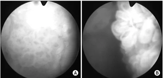
Fig. 2. Cystoscopic findings of a papillary mass in the trigone (A) and left lateral wall of the bladder (B).
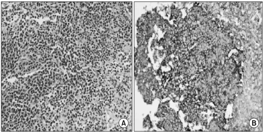
vena cava, aortocaval, right lower quadrant mesenteric, and bilateral inguinal areas were also seen. Cystoscopy re- vealed a large papillary mass in the trigone and left lateral wall of the bladder (Fig. 2). For histologic confirmation, the patient underwent transurethral resection of the blad- der tumor. Histopathologic examination of specimens from the trigone specimen and the lateral wall confirmed superficial low-grade UC. In the subepithelial connective tissue, infiltration of atypical medium to large lympho- cytes showing vesicular nuclei, prominent multiple nucle- oli, and an appreciable amount of basophilic cytoplasm was seen (Fig. 3). For the stage work-up, positron emission tomography (PET), and ultrasonography-guided percuta- neous biopsy of the right inguinal and axillary regions were performed, in addition to bone marrow biopsy bi- laterally in the superior iliac crest. PET showed abnormal
uptake in the bladder, small bowel mesentery, cervical spine, and the lymph nodes in the right pericardiophrenic angle, around the suprahepatic inferior vena cava, aorto- caval, and both superior inguinal lesions. The right in- guinal lymph node specimen showed a vague follicular growth pattern with relatively monotonous small lympho- cytes, plasma cells, and plasmacytoid lymphocytes. They were positive for immunohistochemical stains for CD20, CD79a, Bcl-2, CD43, and immunoglobulin M (IgM). The VS38 immunohistochemical stain was positive (Fig. 4).
Magnetic resonance imaging of the spine revealed an in- tradural mass at the 7th cervical level of the spine.
Malignant lymphoma was found to be marginal zone B-cell lymphoma (MZBCL) based on the normal serum monoclonal IgM level and non-involvement of the bone marrow and spleen.
Fig. 3. Histologic features of a resection specimen of the bladder obtained from transurethral resection of the bladder tumor. The specimen showed low grade, non-invasive papillary urothelial carcinoma in the epithelium (arrow), while infiltration of atypical medium-sized to large lymphocytes showing vesicular nuclei, prominent multiple nucleoli, and an appreciable amount of basophilic cytoplasm were seen in the subepithelial connective tissue (asterisk). The atypical lymphoid cells showed more aggressive and dedifferentiated features than those of the inguinal lymph nodes (A: H&E, ×100, B: H&E, ×400).
Fig. 4. Histologic features of the right inguinal lymph node specimen. (A) The specimen showed a vague follicular growth pattern with relatively monotonous small lymphocytes, plasma cells, and plasmacytoid lymphocytes (H&E, ×400). (B) They were positive for immunohistochemical stains for CD20, CD79a, Bcl-2, CD43, and immunoglobulin M (IgM), but negative for CD3, CD5, Cyclin D1, CD23, and Bcl-6. The VS38 immunohistochemical stain was positive but was negative for CD138 and CD10 (Immunohistochemical examination stain or IgM, ×400).
During preparation for chemotherapy, the patient un- derwent surgical exploration of the abdomen due to perfo- ration of a lymphoma lesion in the distal ileum. At six months after transurethral resection of the bladder tumor, cystoscopy was performed for follow-up of the bladder tu- mor, which revealed a papillary tumor on the trigone and
subsequent transurethral resection was conducted. The patient underwent 6 cycles of cyclophosphamide, doxor- ubicin, vincristine, and prednisolone chemotherapy but died from pneumonia and heart failure at 1 year after the end of chemotherapy.
144 World J Mens Health Vol. 30, No. 2, August 2012
DISCUSSION
Most tumors of the bladder are derived from the epi- thelium, and non-epithelial tumors are extremely rare.3 Metastatic tumors represent approximately 15% of all known bladder malignancies.4 In our case, the tumors of the bladder were demonstrated to be UC involving the ep- ithelium and as B cell lymphoma in a subepithelial lesion.
This is the first case of concurrent bladder tumors involv- ing two different malignancies.
Usually, the diagnosis of bladder lymphoma is one of exclusion. It is made in the absence of any other nodal or extranodal involvement after biopsy with immunohisto- chemical study, and after a negative study of disease ex- tension, which includes bone marrow biopsy and CT.4 This approach is changing with the introduction of PET to search for other nodal or extranodal involvement.
The most frequent symptoms of bladder lymphoma are gross hematuria followed by concomitant urinary tract in- fection, dysuria, and other lower urinary tract symptoms.4 The cystoscopic appearance of bladder lymphoma is a large, multinodular mass which has a characteristic sub- mucosal pattern with minimal or no ulceration.5 The CT appearances of bladder lymphoma are multinodular thick- ening of the bladder wall, a lobulated mass involving the bladder wall, and intermittent extravesical extension.6 In our case, the cystoscopic appearance was a multiple papillary mass, which is a typical finding of bladder UC, and the CT showed multinodular thickening with extra- vesical extension and regional lymph node enlargement.
Our first impression after cystoscopy and CT was meta- static bladder cancer with regional lymph node and small intestine metastasis. However, by comparing the CT find- ings of the bladder mass with other reported cases, it be- came clear that the multinodular pattern was a classic find- ing of bladder lymphoma.
MZBCL, previously known as monocytoid B-cell lym- phoma in the lymph nodes, has been an evolving concept since these neoplasms were first described in the 1980s. It was only in the World Health Organization classification that extranodal, nodal, and splenic types of MZBCL be- came recognized as three distinct entities. Lymphoplas- macytic lymphoma (LPL) is characterized by a mono- clonal expansion of predominantly small B-lymphocytes
with variable plasmacytoid differentiation; small B-lym- phocytes are usually CD5-, CD10-, and CD23-, and they are associated with serum IgM paraprotein.7 Although LPL is an indolent tumor in most cases, it also involves extra- medullary sites. Lin et al8 reported 44 patients with LPL in- volving extramedullary sites, and LPL was subclassifed in- to lymphoplasmacytic, lymphoplasmacytioid, polymor- phous, DLBCL transformed from LPL, and MZBCL. This subclassification in LPL with extranodal involvement sug- gested the possibility that these tumors are part of the spec- trum of LPL. MZBCL is indistinguishable from LPL by his- tologic appearance and only distinguishable by elevation of serum monoclonal IgM and the involvement of the bone marrow.7
In conclusion, we describe an extremely infrequent in- stance of concurrent bladder tumors, which were demon- strated to be UC and MZBCL. Although the incidence of bladder lymphoma is low, in cases with gross hematuria, regional lymph node enlargement, and a multinodular pattern with bladder wall thickening in abdominal CT findings, the diagnosis of bladder lymphoma should be considered.
ACKNOWLEDGEMENTS
This work was supported by a National Research Foundation of Korea (NRF) grant funded by the Korean government (MEST) (No. 2011-0020128).
REFERENCES
1. Bates AW, Norton AJ, Baithun SI. Malignant lymphoma of the urinary bladder: a clinicopathological study of 11 cases. J Clin Pathol 2000;53:458-61
2. Kempton CL, Kurtin PJ, Inwards DJ, Wollan P, Bostwick DG.
Malignant lymphoma of the bladder: evidence from 36 cases that low-grade lymphoma of the MALT-type is the most com- mon primary bladder lymphoma. Am J Surg Pathol 1997;21:
1324-33
3. Weimar G, Culp DA, Loening S, Narayana A. Urogenital in- volvement by malignant lymphomas. J Urol 1981;125:230-1 4. Bates AW, Baithun SI. Secondary neoplasms of the bladder
are histological mimics of nontransitional cell primary tu- mours: clinicopathological and histological features of 282 cases. Histopathology 2000;36:32-40
5. Downs TM, Kibel AS, DeWolf WC. Primary lymphoma of the bladder: a unique cystoscopic appearance. Urology
1997;49:276-8
6. Yeoman LJ, Mason MD, Olliff JF. Non-Hodgkin's lymphoma of the bladder--CT and MRI appearances. Clin Radiol 1991;44:389-92
7. Lin P, Medeiros LJ. Lymphoplasmacytic lymphoma/walden- strom macroglobulinemia: an evolving concept. Adv Anat
Pathol 2005;12:246-55
8. Lin P, Bueso-Ramos C, Wilson CS, Mansoor A, Medeiros LJ.
Waldenstrom macroglobulinemia involving extramedullary sites: morphologic and immunophenotypic findings in 44 patients. Am J Surg Pathol 2003;27:1104-13